
Explain me dural venous sinuses for my mbbs exmas
Reading File
Now let me search the medical library for dural venous sinuses content in parallel.
Finding Sources
Finding Sources
Reading File
Reading File
Now let me get the full table from Gray's and fetch the diagram image, plus search for cavernous sinus thrombosis content:
Reading File
Loading Image
Finding Sources
Reading File
Loading Image
Reading File
Reading File
Loading Image
I now have all the content needed for a comprehensive MBBS-level explanation. Let me compile it.
Dural Venous Sinuses - Complete MBBS Notes
(Source: Gray's Anatomy for Students & Color Atlas of Human Anatomy)
What Are They? (Basic Definition)
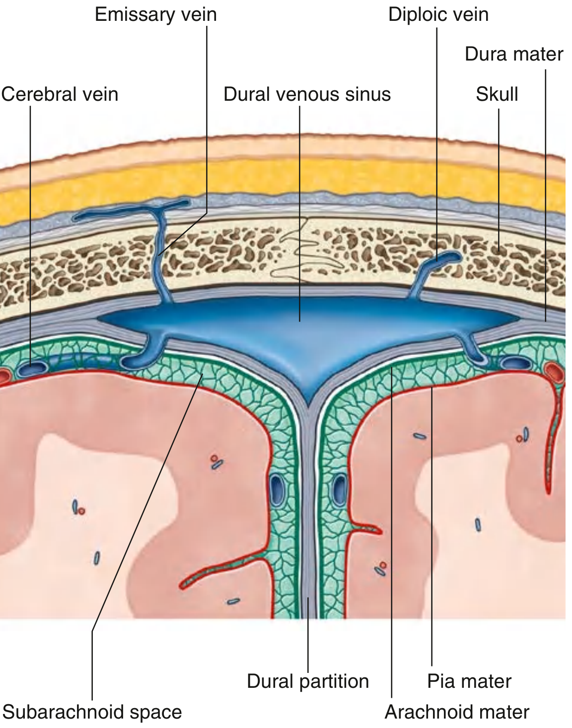
Dural venous sinuses are endothelial-lined venous spaces lying between the outer periosteal and inner meningeal layers of the dura mater. Think of them as rigid venous channels (unlike ordinary veins, their walls don't collapse). They are valveless, drain venous blood + CSF from the brain, and ultimately empty into the internal jugular veins.
Key tributaries feeding them:
- Cerebral veins (from brain parenchyma)
- Diploic veins (run between inner and outer tables of skull bones)
- Emissary veins (pass from outside the skull INTO the sinuses - no valves! clinically important)
- Arachnoid granulations (drain CSF)
Complete List with Locations and Tributaries
| Sinus | Location | Receives |
|---|---|---|
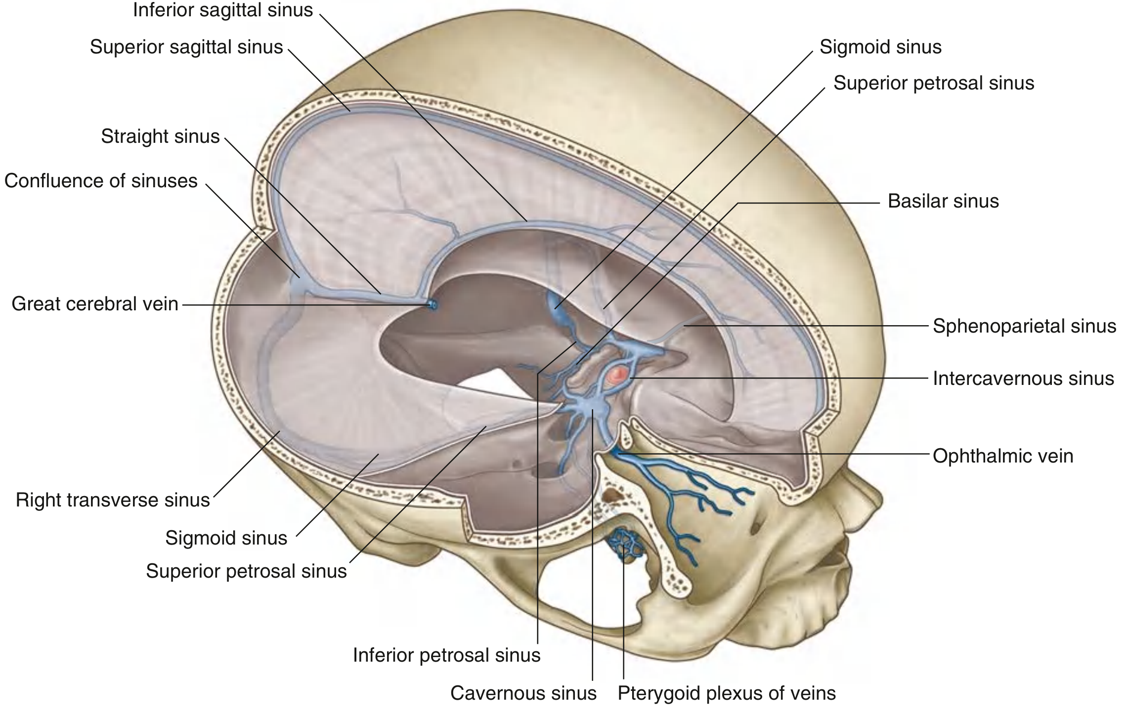
| Superior sagittal | Superior border of falx cerebri | Superior cerebral veins, diploic veins, emissary veins, CSF via arachnoid granulations |
| Inferior sagittal | Inferior margin of falx cerebri | A few cerebral veins + veins from falx cerebri |
| Straight | Junction of falx cerebri + tentorium cerebelli | Inferior sagittal sinus + great cerebral vein (of Galen) + posterior cerebral veins + superior cerebellar veins |
| Occipital | In falx cerebelli against occipital bone | Communicates with vertebral venous plexus |
| Confluence of sinuses | Dilated space at internal occipital protuberance | Superior sagittal + straight + occipital sinuses |
| Transverse (paired) | Horizontal along posterior/lateral tentorium cerebelli | From confluence; also superior petrosal sinus, inferior cerebral/cerebellar veins |
| Sigmoid (paired) | S-shaped continuation of transverse → internal jugular vein; grooves parietal, temporal, occipital bones | Transverse sinus + cerebral, cerebellar, diploic, emissary veins |
| Cavernous (paired) | Lateral aspect of body of sphenoid, each side of sella turcica | Cerebral + ophthalmic veins + sphenoparietal sinuses + emissary veins from pterygoid plexus |
| Intercavernous | Across sella turcica (anterior + posterior to pituitary stalk) | Interconnects the two cavernous sinuses |
| Sphenoparietal (paired) | Inferior surface of lesser wings of sphenoid | Diploic + meningeal veins |
| Superior petrosal (paired) | Superior margin of petrous temporal bone | Cavernous sinus + cerebral + cerebellar veins |
| Inferior petrosal (paired) | Groove between petrous temporal bone and occipital bone | Cavernous sinus + cerebellar veins + veins from internal ear and brainstem |
| Basilar | Clivus (posterior to sella turcica) | Connects bilateral inferior petrosal sinuses; communicates with vertebral plexus |
Flow Pathway (How Blood Gets Out)
Brain → Cerebral veins
↓
Superior sagittal sinus ──────────────┐
Straight sinus (← great cerebral vein + inf. sagittal sinus) ──→ CONFLUENCE OF SINUSES
Occipital sinus ───────────────────────┘
↓
Transverse sinuses (L + R)
↓
Sigmoid sinuses (L + R)
↓
Internal jugular veins
Cavernous sinus pathway:
Orbit (ophthalmic veins) → Cavernous sinus
Face (angular vein → sup. ophthalmic vein) → Cavernous sinus
Cavernous sinus → Superior petrosal sinus → Transverse/sigmoid sinus → IJV
Cavernous sinus → Inferior petrosal sinus → IJV directly
The Cavernous Sinus - HIGH YIELD for Exams
This is the most clinically tested sinus. Memorize the structures running through it.
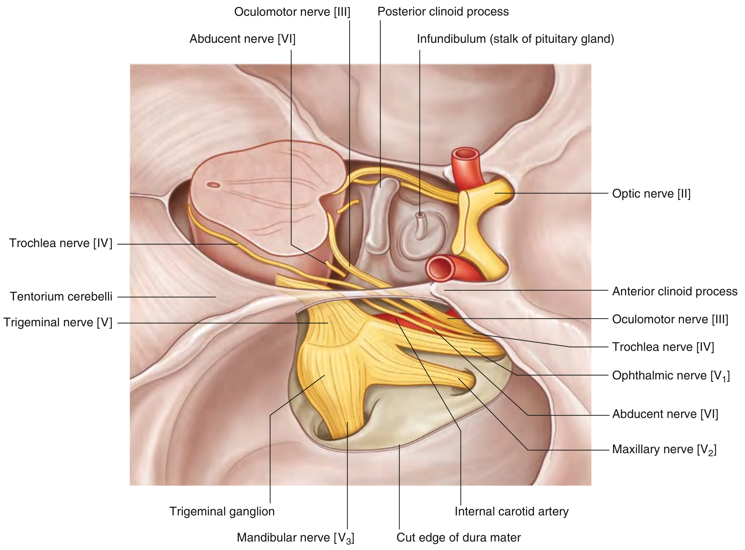
Location: Lateral to sella turcica / body of sphenoid, one on each side.
Structures INSIDE the sinus (in the lumen):
- Internal carotid artery (with sympathetic plexus)
- Abducent nerve (CN VI) - most medial, directly in the blood stream
Structures in the LATERAL WALL of the sinus (superior to inferior):
- Oculomotor nerve (CN III)
- Trochlear nerve (CN IV)
- Ophthalmic nerve (CN V1)
- Maxillary nerve (CN V2)
Mnemonic: "O TOM CAT" or remember O-T-O-M in the wall, ICA + VI inside.
Connections of the cavernous sinus:
- To fellow cavernous sinus via intercavernous sinuses (anterior + posterior)
- Receives from orbit via superior ophthalmic vein (and thus from facial vein/angular vein)
- Receives from infratemporal fossa via emissary veins from pterygoid plexus
- Drains to sigmoid sinus via superior petrosal sinus
- Drains to IJV via inferior petrosal sinus
Clinical High-Yield Points
1. Cavernous Sinus Thrombosis (CST)
- Source of infection: Most common - sphenoid/ethmoid sinusitis; also boils of the face (danger area), dental infections
- Why face infections reach here: Facial vein → superior ophthalmic vein → cavernous sinus (all valveless)
- Features (from nerve compression): Proptosis, chemosis, periorbital edema, painful ophthalmoplegia (III, IV, VI palsy), V1/V2 sensory loss, papilledema
- CN VI affected first - because it runs freely inside the sinus (not protected in the wall)
2. Emissary Veins - The "Danger Pathway"
- Emissary veins are valveless - they pass from outside the skull through foramina to the dural sinuses
- This means scalp infections can track intracranially - the reason the scalp's "loose connective tissue layer" is called the "danger area"
3. Superior Sagittal Sinus Thrombosis
- Can cause raised intracranial pressure (blocks CSF reabsorption at arachnoid granulations)
- Presents with headache, papilledema, seizures, bilateral leg weakness (parafalcine cortex affected)
- Associated with pregnancy, oral contraceptive pills, hypercoagulable states
4. Great Cerebral Vein (of Galen)
- Drains deep cerebral structures (basal ganglia, thalamus, internal capsule)
- Joins inferior sagittal sinus to form the straight sinus
- Vein of Galen malformation is a rare pediatric condition
5. Arachnoid Granulations
- Project into the lateral lacunae of the superior sagittal sinus
- CSF is absorbed here by bulk flow into the venous blood
- If blocked → communicating hydrocephalus
Quick Summary Table for Revision
| Feature | Detail |
|---|---|
| Wall layers | Between periosteal + meningeal dura |
| Valves | None (valveless) |
| Lining | Endothelium |
| Paired sinuses | Transverse, sigmoid, cavernous, sphenoparietal, superior/inferior petrosal, intercavernous |
| Unpaired sinuses | Superior sagittal, inferior sagittal, straight, occipital, confluence, basilar |
| Final drainage | Internal jugular vein (via sigmoid sinus) |
| CSF drainage site | Superior sagittal sinus (via arachnoid granulations) |
| Most clinically important | Cavernous sinus |
- Gray's Anatomy for Students, pp. 1024-1027
- Color Atlas of Human Anatomy, Vol. 2, pp. 133-134
This is a shared conversation. Sign in to Orris to start your own chat.