
Haemorrhage in animal
Reading File
Haemorrhage in animals is bleeding - the escape of blood from the vascular system. This is a broad pathology topic. Let me search the medical library for comprehensive coverage.
Searching sources...
Finding Sources
Finding Sources
Finding Sources
Finding Sources
The library has human medical textbooks. Let me search specifically for haemorrhage pathology content in Robbins (which covers general pathology applicable to animals too) and also look for veterinary-relevant content.
Finding Sources
Finding Sources
The Robbins Basic Pathology entry is the most relevant - it covers haemorrhage comprehensively. Let me read that section plus search for more specific animal/veterinary pathology content.
Reading File
Finding Sources
I have excellent content from Robbins. Let me read the continuation of the hemorrhage section and also fetch the image mentioned.
Reading File
Searching the Web
haemorrhage classification in animals veterinary pathology petechiae purpura ecchymosis
I now have excellent material for a comprehensive answer. Let me also fetch the petechiae image from Robbins for visual illustration.
Loading Image
Loading Image
I now have comprehensive information for a thorough answer. Let me compile the complete response on haemorrhage in animals.
Haemorrhage in Animals
Haemorrhage is defined as the extravasation (escape) of blood from blood vessels into surrounding tissues, body cavities, or to the exterior. It is a fundamental pathological process seen across all animal species.
Definition
Haemorrhage results from damage to blood vessel walls and may be exacerbated by defects in blood clotting. Even capillary bleeding can occur in chronically congested tissues. Trauma, atherosclerosis, inflammatory or neoplastic erosion of vessel walls are all common causes.
- Robbins & Kumar Basic Pathology, p. 82
Classification by Size / Morphology
| Type | Size | Description |
|---|---|---|
| Petechiae | 1-2 mm | Minute, pinpoint haemorrhages into skin, mucous membranes, or serosal surfaces |
| Purpura | 3-5 mm | Slightly larger haemorrhages |
| Ecchymoses | 1-2 cm | Larger subcutaneous hematomas (bruises) |
| Hematoma | >2 cm | Large accumulation of blood within tissue |
Petechial Haemorrhages (image below - colonic mucosa, thrombocytopenia):
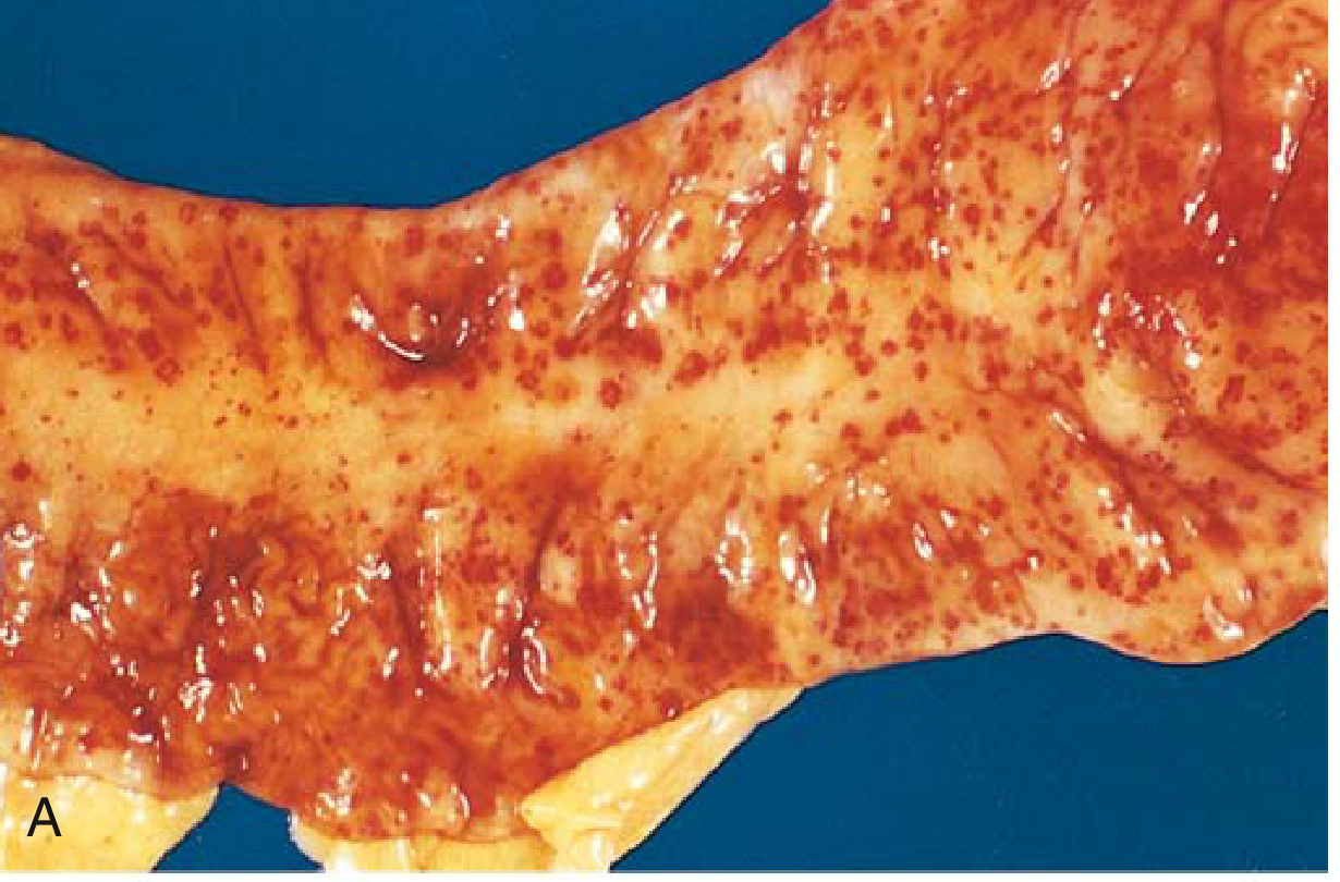
FIG 3.4A - Punctate petechial haemorrhages of the colonic mucosa, a consequence of thrombocytopenia.
Fatal Intracerebral Haemorrhage (image below):
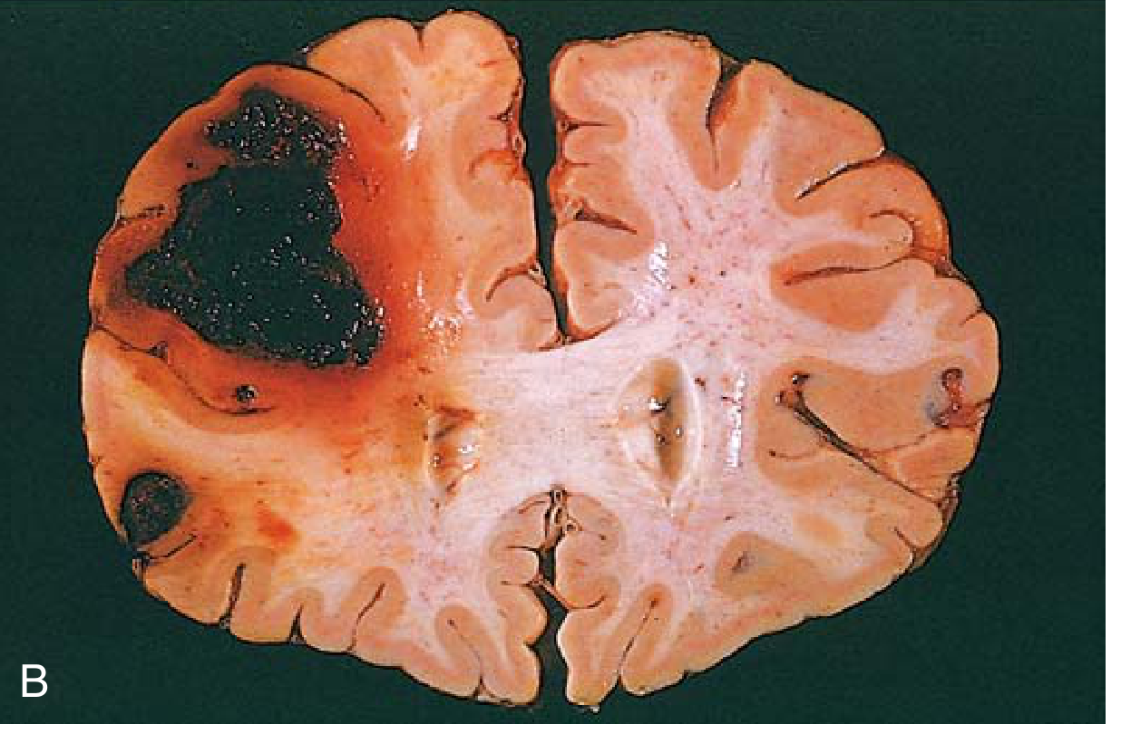
FIG 3.4B - Fatal intracerebral haemorrhage with large dark clot in the right hemisphere.
Classification by Location / Cavity
Large haemorrhages are named according to their location in body cavities:
- Hemothorax - blood in the thoracic (pleural) cavity
- Hemopericardium - blood in the pericardial sac
- Hemoperitoneum - blood in the peritoneal/abdominal cavity
- Hemarthrosis - blood in a joint
- Epistaxis - bleeding from the nose
- Hemoptysis - coughing up blood from the respiratory tract
- Hematemesis - vomiting blood
- Melena - blood in faeces (digested, tarry)
- Hematuria - blood in urine
Classification by Cause
1. Rhexis (Rexis) - Rupture of the Vessel Wall
- Traumatic laceration or rupture
- Erosion by inflammatory/neoplastic processes
- Aneurysm rupture
- Example: aortic rupture in horses, hepatic rupture in cattle with abscessation
2. Diapedesis
- Passive leakage of red cells through intact but damaged vessel walls (especially capillaries and venules)
- Seen in severe congestion, hypoxia, and septicaemia
- Common in bacterial infections causing endothelial damage (e.g., salmonellosis, hog cholera/Classical Swine Fever in pigs)
3. Per Rhexin (Haemorrhagic Diatheses)
A wide variety of disorders (collectively called haemorrhagic diatheses) increase the risk of haemorrhage, often after minimal injury. These involve:
- Inherited or acquired vascular wall defects (e.g., vitamin C deficiency/scurvy)
- Platelet disorders - thrombocytopenia or thrombocytopathy
- In dogs: immune-mediated thrombocytopenia (common in Cocker Spaniels, Toy Poodles, Old English Sheepdogs)
- Coagulation factor deficiencies
- von Willebrand disease (common in Doberman Pinschers, Scottish Terriers)
- Haemophilia A (Factor VIII deficiency) and B (Factor IX) in dogs and cats
Causes of Haemorrhage in Animals
Traumatic
- Road traffic accidents, bites, kicks, falls
- Surgical procedures
- Foreign body penetration (wire disease in cattle)
Vascular/Cardiovascular
- Aortic or vessel rupture
- Arteriovenous malformations
Inflammatory / Infectious
- Septicaemia - petechiae on serosal surfaces from endothelial damage
- Classical Swine Fever (Hog Cholera) - "paint brush" haemorrhages, petechiae on kidneys ("turkey egg kidney"), lymph node haemorrhages
- African Swine Fever - severe haemorrhages throughout the body
- Equine Infectious Anaemia - petechiae on mucous membranes
- Bovine Viral Diarrhoea (BVD) - petechiae, oral erosions
- Canine Parvovirus - haemorrhagic gastroenteritis
- Anthrax - bloody discharges from body orifices
- Warfarin/rodenticide poisoning - depletes Vitamin K-dependent clotting factors
Neoplastic
- Erosion of vessel walls by tumours (haemangiosarcoma in dogs - splenic rupture)
Nutritional/Toxic
- Vitamin K deficiency - clotting factor deficiency
- Bracken fern toxicosis in cattle - bone marrow suppression, thrombocytopenia, and massive haemorrhages
- Mycotoxins (aflatoxin, trichothecenes)
- Warfarin and rodenticide (brodifacoum) poisoning
Parasitic
- Heavy fluke infestation (liver flukes causing haemorrhagic hepatitis)
- Strongylus vulgaris in horses - verminous arteritis and haemorrhagic infarction
Pathological Consequences of Haemorrhage
The clinical impact depends on:
- Volume of blood lost - Rapid loss of up to 20% of blood volume may be well tolerated in healthy adults; greater losses cause haemorrhagic (hypovolemic) shock
- Rate of bleeding - Acute massive loss is more dangerous than chronic slow loss
- Location - Even a relatively small bleed in the brain (e.g., intracranial) can be fatal, while similar volume in subcutaneous tissue may be trivial
- Underlying health - Animals with pre-existing cardiac or respiratory disease are far less tolerant
Colour Changes in Resolving Haemorrhages (Ecchymoses/Bruises)
Extravasated red cells are phagocytosed and degraded by macrophages:
- Haemoglobin (red-blue) → Biliverdin/Bilirubin (blue-green) → Haemosiderin (golden-brown)
- Large internal haemorrhages may cause jaundice as haemoglobin breakdown products accumulate
Anaemia
- External haemorrhage - leads to iron deficiency anaemia over time (chronic loss, e.g., GI bleeding, heavy parasite burden)
- Internal haemorrhage (hematoma) - iron is recycled from phagocytosed red cells; iron deficiency anaemia does NOT typically result
Species-Specific Notes
| Species | Common Haemorrhagic Conditions |
|---|---|
| Dog | Haemangiosarcoma (splenic/hepatic), immune-mediated thrombocytopenia, von Willebrand disease, rodenticide toxicity |
| Cat | Hepatic lipidosis-associated coagulopathy, trauma |
| Horse | Exercise-induced pulmonary haemorrhage (EIPH), guttural pouch mycosis (carotid artery erosion), verminous arteritis |
| Cattle | Bracken fern toxicosis, BVD, haemorrhagic bowel syndrome |
| Pig | Classical/African Swine Fever (pathognomonic haemorrhagic lesions) |
| Sheep | Haemonchus contortus (barber's pole worm) - severe anaemia/haemorrhage from abomasal bleeding |
Summary: Key Terminology
| Term | Meaning |
|---|---|
| Petechiae | Pinpoint (1-2 mm) haemorrhages - platelet/endothelial defects |
| Purpura | 3-5 mm haemorrhages |
| Ecchymosis | Bruise (1-2 cm), subcutaneous |
| Hematoma | Blood accumulation in tissue |
| Haemorrhagic diathesis | Increased tendency to bleed |
| Rhexis | Vessel rupture |
| Diapedesis | RBC leakage through intact walls |
- Robbins & Kumar Basic Pathology (Robbins Pathology), pp. 81-82
- eClinPath - Clinical Signs of Haemorrhage
- Veterinary Petechiae & Ecchymoses - Veterian Key
This is a shared conversation. Sign in to Orris to start your own chat.