
Hi I'm a Dermatology resident,I'm preparing for the University md theory examination Can you explain my doubts and make notes
Welcome! I'm glad to help you prepare for your MD Dermatology theory examination. I'm ready to explain concepts and create structured notes on any topic you need.
Go ahead and ask your first doubt or topic - whether it's about skin diseases, histopathology, dermatopathology, pharmacology, procedures, or any clinical scenario. What would you like to start with?
Explain keratinisation in detailed
Reading File
Searching sources... - looking up keratinisation in dermatology textbooks now.
Finding Sources
Finding Sources
Finding Sources
Finding Sources
Reading File
Finding Sources
Reading File
Finding Sources
Reading File
Reading File
Reading File
Finding Sources
Now let me fetch the key diagrams to include in the notes:
Loading Image
Loading Image
Excellent - I now have comprehensive textbook content and diagrams. Here are your detailed MD-level notes on keratinisation:
KERATINISATION - Detailed Notes for MD Dermatology
DEFINITION
Keratinisation is the process by which keratinocytes undergo terminal differentiation - passing from proliferating basal cells through successive layers to become anucleate, protein-rich corneocytes (horn cells) that are ultimately shed (desquamation). It involves both a synthetic phase (building keratin filaments) and a degradative phase (losing organelles, forming the cornified envelope).
LAYERS OF THE EPIDERMIS (Zones of Keratinisation)
| Layer | Old Name | Key Features |
|---|---|---|
| Stratum Basale | Stratum Germinativum | Mitotically active; attached to BM; K5/K14 keratins |
| Stratum Spinosum | Malpighian / Prickle layer | Desmosomes (spines); K1/K10 appear; lamellar bodies form |
| Stratum Granulosum | Granular layer | Keratohyalin granules (profilaggrin); lamellar bodies exocytose |
| Stratum Lucidum | Clear layer | Only on palms and soles; appears pale/clear on H&E |
| Stratum Corneum | Horny layer | Anucleate corneocytes; cornified envelopes; final barrier |
On volar skin (palms/soles), the epidermis is thickest (~1.5 mm) and all five layers are visible. On eyelid skin it is <0.1 mm.
- Andrews' Diseases of the Skin, p. 12
THE TWO PHASES OF KERATINISATION
Phase 1 - SYNTHETIC Phase
- The keratinocyte accumulates intermediate filaments composed of fibrous protein (keratin) arranged in an α-helical coiled pattern
- These tonofilaments are bundled and converge on desmosomes at the plasma membrane (specialized attachment plates)
- Keratins produced: K5/K14 in basal layer → switch to K1/K10 in suprabasal layers
- This switch is a marker of commitment to terminal differentiation
Phase 2 - DEGRADATIVE Phase (Terminal Differentiation)
- Characterized by disappearance of cell organelles (nucleus, mitochondria, ribosomes)
- All contents consolidate into a mixture of filaments and amorphous cell envelopes
- Results in programmed death of the cell (distinct from apoptosis)
- Andrews' Diseases of the Skin, p. 13
KEY MOLECULAR PLAYERS IN KERATINISATION
1. Keratins
- Fibrous proteins; encoded by two subfamilies - acidic and basic
- One acidic + one basic gene product combine to form functional keratin pairs
- K5/K14 - basal layer (mutations → Epidermolysis Bullosa Simplex)
- K1/K10 - suprabasal differentiated layers (mutations → Epidermolytic Hyperkeratosis)
2. Keratohyalin Granules (KHG)
- Present in stratum granulosum cells
- Contain profilaggrin - a high-sulfur protein precursor
- Profilaggrin is converted to filaggrin in the granular layer
- Filaggrin = "Filament aggregating protein" - aggregates keratin tonofilaments
- Cleavage of profilaggrin to filaggrin is aided by Kallikrein-related peptidase 5 (a serine protease from lamellar granules)
- KHG are hygroscopic; repeated hydration/dehydration cycles aid normal desquamation
- Soft keratin (epidermis) = formed WITH keratohyalin granules
- Hard keratin (hair, nails) = formed WITHOUT keratohyalin granules
- Andrews' Diseases of the Skin, p. 13-14
3. Lamellar Granules (Odland Bodies / Membrane-Coating Granules)
- Produced in the Golgi apparatus of granular cells
- Contain: glycosphingolipids, phospholipids, ceramides, and proteases (kallikreins)
- Secreted by exocytosis at the interface of stratum granulosum and stratum corneum
- Their contents form the intercellular lipid envelope (water barrier)
- Abnormal in: Lamellar ichthyosis, Flegel hyperkeratosis
4. Cornified Cell Envelope (CE)
A 15-nm-thick insoluble protein layer deposited on the inner surface of the plasma membrane.
Key structural proteins (cross-linked by transglutaminase):
| Protein | Role |
|---|---|
| Loricrin | Major protein (~80% of CE mass); highest glycine content of any human protein; 26-kDa |
| Involucrin | Adjacent to cytoplasmic plasma membrane surface; tightly packed |
| Filaggrin | Anchors keratin filaments into CE |
| Envoplakin / Periplakin | Scaffold proteins |
| Cystatin α | Alongside involucrin |
| Elafin, SPR proteins | Cross-linkers |
| Desmoplakin | Desmosomal protein incorporated into CE |
- Histology: A Text and Atlas, p. 1296-1297
5. Lipid Envelope
- A 5-nm-thick layer of lipids attached to the outer cell surface by ester bonds
- Key lipids: ceramides, cholesterol, free fatty acids
- Most important component: monomolecular layer of acylglucosylceramide ("Teflon-like" non-stick layer)
- Ceramides are also found in topical barrier-repair products
EPIDERMAL BARRIER - Schematic
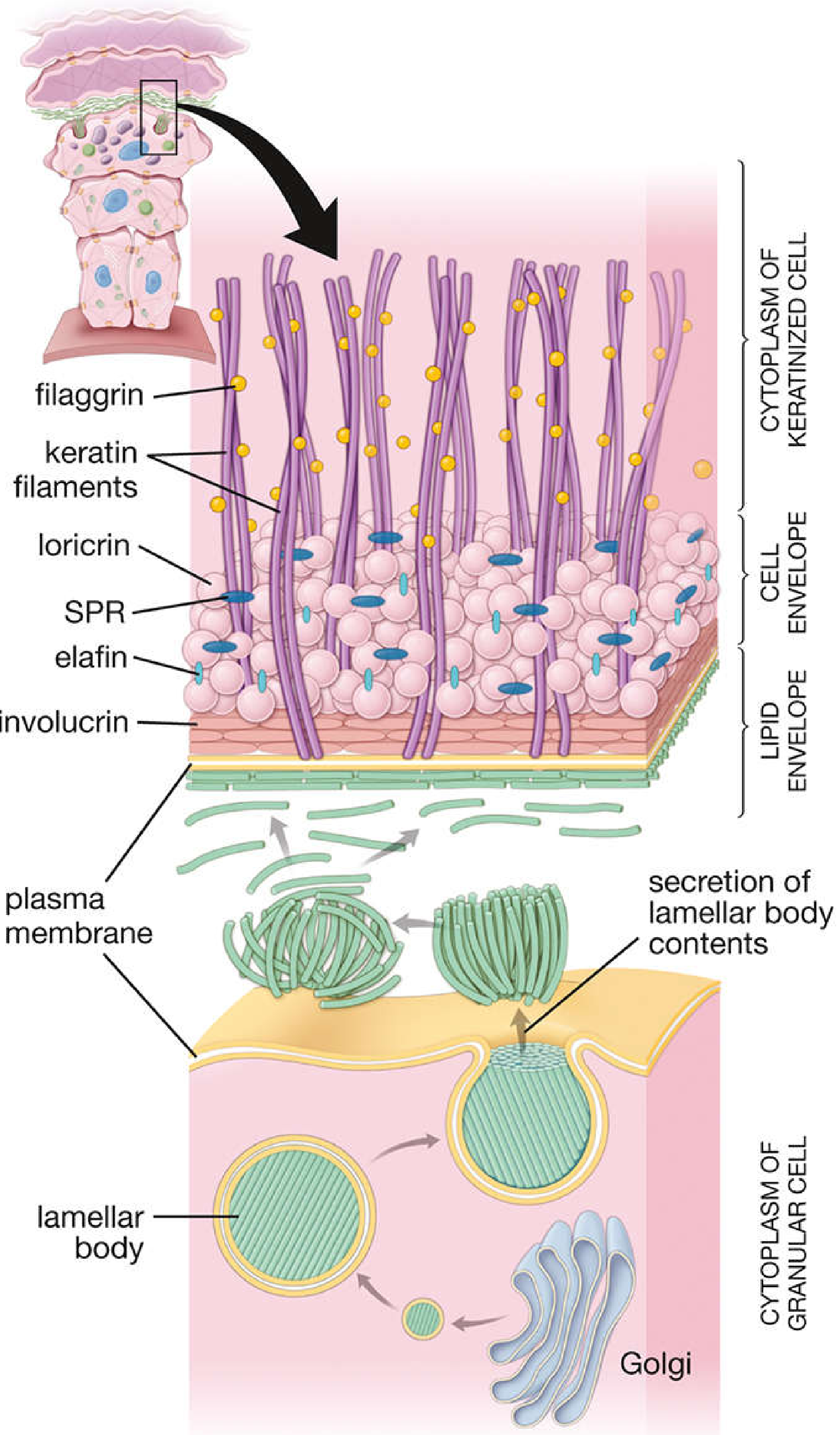
The lipid is secreted from lamellar bodies in granular cells (Golgi-derived), exocytosed into the intercellular space, forming the lamellar lipid barrier. Keratin filaments are bound by filaggrin and anchored into the CE.
EPIDERMAL TURNOVER TIME
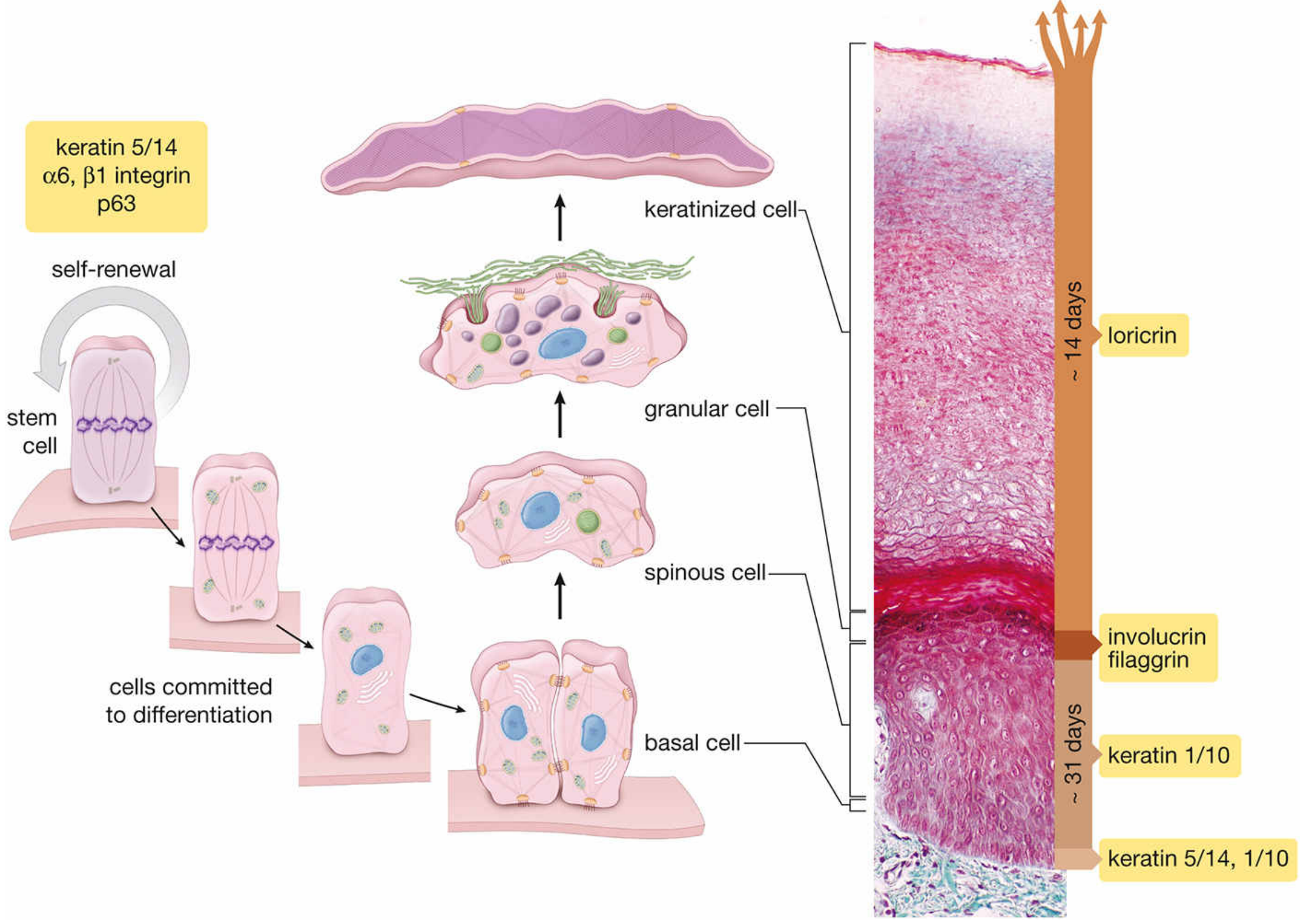
| Compartment | Time |
|---|---|
| Mitotic divisions (stratum basale) | ~1-2 days |
| Stratum spinosum + granulosum | ~31 days |
| Stratum corneum (16-20 cell layers) | ~14 days |
| Total epidermal turnover | ~47 days |
| In Psoriasis (accelerated) | ~8-10 days |
One cell layer of stratum corneum desquamates every 22.4 hours under normal conditions.
- Histology: A Text and Atlas, p. 1298-1299
STEM CELLS AND REGULATION
- Stem cells are slow-cycling, found in the deepest rete ridges (especially palmoplantar) and hair bulge
- Identified by: high β1-integrin expression, low desmosomal proteins (e.g., desmoglein 3), lack of terminal differentiation markers
- Histone deacetylation suppresses profilaggrin expression - histones regulate epidermal differentiation
- Desmoglein 1 cytoplasmic tail binds scaffolding protein Erbin → downregulates Ras-Raf pathway → promotes stratification and differentiation
- Andrews' Diseases of the Skin, p. 13
ABNORMAL KERATINISATION - Key Histological Patterns
| Pattern | Description | Example Disease |
|---|---|---|
| Parakeratosis | Retained nuclei in stratum corneum | Psoriasis, seborrhoeic dermatitis |
| Hyperkeratosis | Thickened stratum corneum | Ichthyosis vulgaris, callus |
| Corps ronds | Round, clear-to-pink, abnormally keratinized cells | Darier's disease |
| Grains | Elongated, basophilic, abnormally keratinized cells | Darier's disease |
| Dyskeratosis | Premature/abnormal individual cell keratinization | Various |
- In Ichthyosis vulgaris: diminished/absent granular layer → profilaggrin deficiency → retention hyperkeratosis
- In Psoriasis: accelerated turnover, parakeratosis, epidermal thickening
- Andrews' Diseases of the Skin, p. 13
DESQUAMATION (Normal Shedding)
- Mediated by protease activity (kallikreins/serine proteases) acting on desmosomal proteins (desmoglein, desmocollin)
- Lamellar granule-derived enzymes play a role
- The lipid barrier keeps proteases in check; disruption (e.g., Netherton syndrome - SPINK5 mutation blocking LEKTI inhibitor) leads to excessive desquamation
HIGH-YIELD SUMMARY TABLE FOR EXAMS
| Feature | Detail |
|---|---|
| Keratin gene pair rule | 1 acidic + 1 basic → functional keratin |
| Basal layer keratins | K5, K14 |
| Suprabasal keratins | K1, K10 |
| EBS mutation | K5 or K14 |
| Epidermolytic hyperkeratosis mutation | K1 or K10 |
| Profilaggrin → Filaggrin enzyme | Kallikrein-related peptidase 5 |
| Soft keratin | With keratohyalin (epidermis) |
| Hard keratin | Without keratohyalin (hair, nails) |
| Lamellar body origin | Golgi apparatus |
| Major CE protein | Loricrin (~80%) |
| CE enzyme | Transglutaminase |
| Total turnover time | ~47 days |
| Psoriasis turnover | ~8-10 days |
| Stratum lucidum location | Palms and soles only |
| Odland body = | Lamellar granule = membrane-coating granule |
These notes draw from Andrews' Diseases of the Skin (Clinical Dermatology) and Histology: A Text and Atlas with Correlated Cell and Molecular Biology. Let me know your next topic or doubt!
This is a shared conversation. Sign in to Orris to start your own chat.