
anatomy of forearm
I'll start by reading the medical library skill, then search for forearm anatomy content in parallel.
Reading File
Now let me search for forearm anatomy content in the library in parallel:
Finding Sources
Finding Sources
Finding Sources
Reading File
Gray's Anatomy for Students is the best source. Let me read the FOREARM section from it, plus the THIEME Atlas images, in parallel:
Reading File
Reading File
Reading File
Loading Image
Reading File
Loading Image
Reading File
Loading Image
Reading File
Loading Image
I now have a thorough picture of forearm anatomy. Let me fetch one more key image - the posterior compartment muscles, and get the muscles table data:
Reading File
I have comprehensive data. Now I'll compile the full anatomy of the forearm answer.
Anatomy of the Forearm
Source: Gray's Anatomy for Students, THIEME Atlas of Anatomy, Imaging Anatomy: Bones, Joints, Vessels and Nerves
Overview
The forearm extends between the elbow joint proximally and the wrist joint distally. Most major structures enter the forearm through the cubital fossa anteriorly. The ulnar nerve is the major exception - it passes posterior to the medial epicondyle of the humerus before entering the forearm.
Distally, most structures exit through or anterior to the carpal tunnel, with the radial artery being the exception - it passes dorsally around the wrist to enter the hand posteriorly.
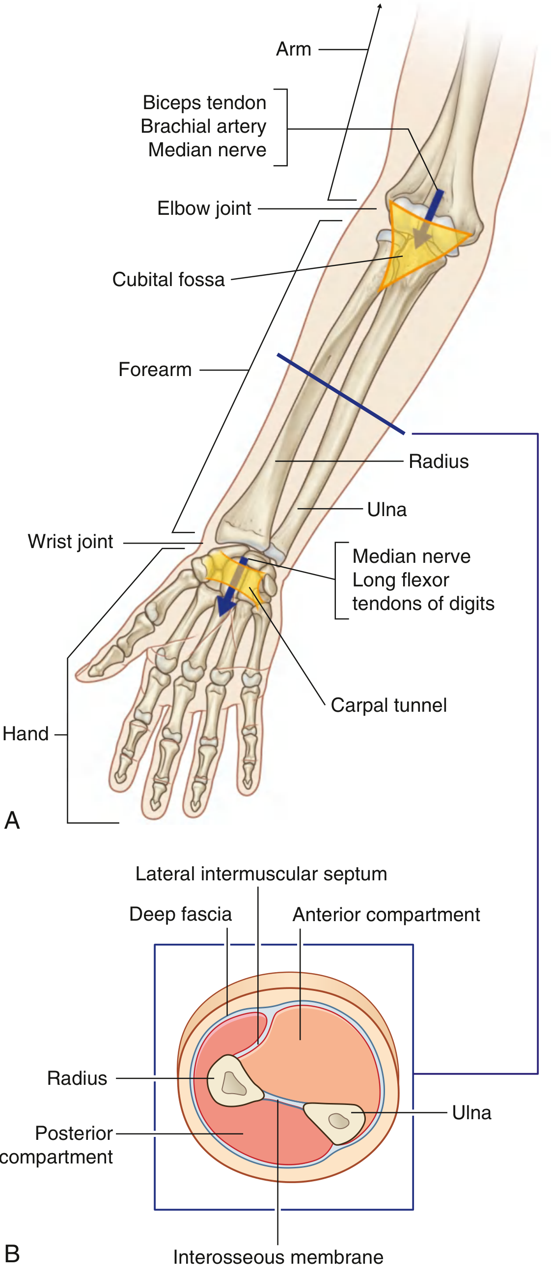
Fig. 7.79 - Forearm: (A) Proximal and distal relationships; (B) Transverse section through the middle of the forearm. (Gray's Anatomy for Students)
1. Bones
Radius (lateral bone)
The radius is small proximally and large distally. Its shaft is triangular in cross section with:
- Three borders: anterior (oblique line in superior third), posterior (distinct only in middle third), and interosseous (sharp; attachment for interosseous membrane)
- Three surfaces: anterior (smooth), posterior (smooth), and lateral (has oval roughening for pronator teres attachment midshaft)
The distal end is broad and flat. Key features:
- Dorsal tubercle - acts as a pulley for extensor pollicis longus tendon
- Ulnar notch - articulates with the head of the ulna
- Radial styloid process - projects distally on the lateral side
- Two carpal facets for articulation with the scaphoid and lunate bones
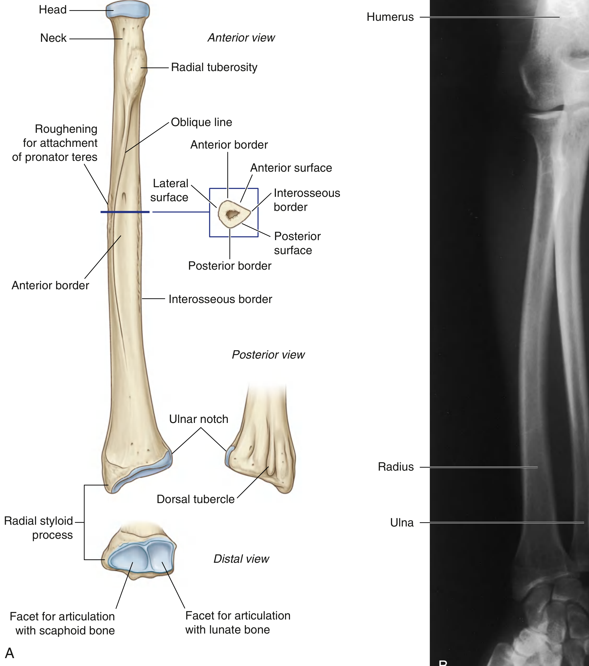
Shaft and distal end of the right radius showing surfaces, borders, and bony landmarks. (Gray's Anatomy for Students)
Ulna (medial bone)
The ulna is the opposite of the radius: large proximally and small distally. Its shaft is also triangular in cross section:
- Three borders: anterior (smooth/rounded), posterior (sharp and palpable along entire length), and interosseous
- Three surfaces: anterior (has roughening for pronator quadratus distally), posterior, and medial
The distal end features:
- A rounded head covered by articular cartilage anterolaterally
- Ulnar styloid process - originates from the posteromedial aspect
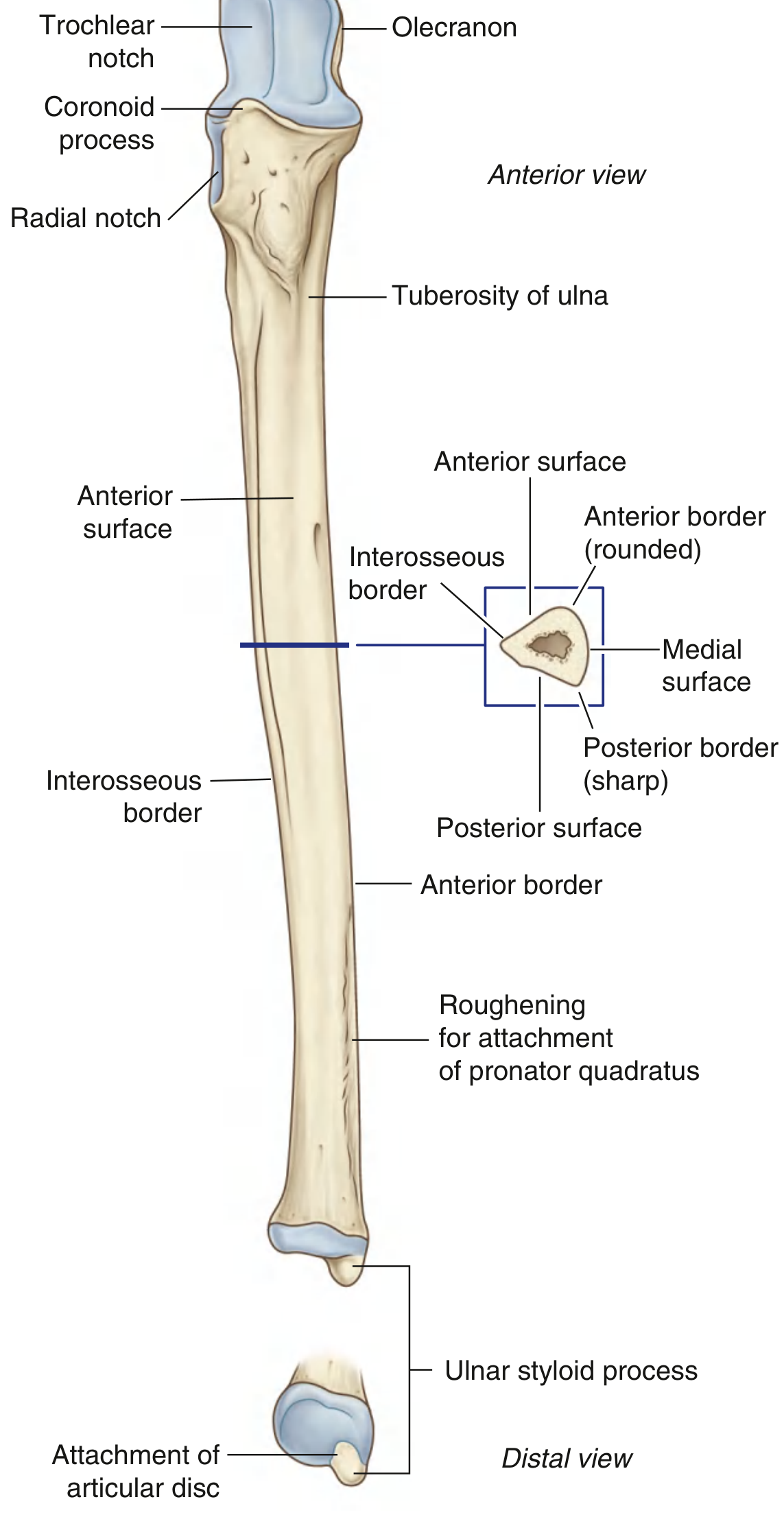
Shaft and distal end of the right ulna. (Gray's Anatomy for Students)
Interosseous Membrane
A fibrous sheet that links the interosseous borders of the radius and ulna along most of their length. It:
- Separates anterior from posterior compartments
- Transmits forces from radius to ulna
- Has an aperture proximally for the anterior interosseous artery to pass
2. Compartments
The forearm is divided into anterior (flexor) and posterior (extensor) compartments, separated by:
- A lateral intermuscular septum (from anterior border of radius to deep fascia)
- The interosseous membrane
- Deep fascia along the posterior border of the ulna
3. Anterior (Flexor) Compartment
Layers of muscles
Superficial layer (all originate from medial epicondyle of humerus):
| Muscle | Insertion | Innervation | Function |
|---|---|---|---|
| Flexor carpi ulnaris | Pisiform → hamate and metacarpal V | Ulnar nerve (C7, C8, T1) | Flexes and adducts wrist |
| Palmaris longus | Palmar aponeurosis | Median nerve (C7, C8) | Flexes wrist; resists shearing during grip |
| Flexor carpi radialis | Bases of metacarpals II & III | Median nerve (C6, C7) | Flexes and abducts wrist |
| Pronator teres | Midshaft lateral surface of radius | Median nerve (C6, C7) | Pronation |
Intermediate layer:
- Flexor digitorum superficialis (FDS) - originates from medial epicondyle, coronoid process, and proximal radius; splits into four tendons for the middle phalanges of fingers II-V; innervated by median nerve (C7, C8, T1).
Deep layer:
- Flexor digitorum profundus (FDP) - originates from the anterior and medial ulna and interosseous membrane; four tendons insert into distal phalanges. Medial half (ring + little finger) = ulnar nerve; lateral half (index + middle finger) = anterior interosseous nerve (branch of median).
- Flexor pollicis longus (FPL) - originates from anterior radius and interosseous membrane; inserts into the distal phalanx of the thumb; innervated by anterior interosseous nerve.
- Pronator quadratus - a flat, square muscle crossing the distal forearm; originates from the distal ulna and inserts on the distal radius; innervated by anterior interosseous nerve; primary pronator.
4. Posterior (Extensor) Compartment
Superficial layer (all arise from lateral epicondyle unless noted):
| Muscle | Insertion | Innervation | Function |
|---|---|---|---|
| Brachioradialis | Lateral distal radius | Radial nerve (C5, C6) | Elbow flexion (midproned) |
| Extensor carpi radialis longus | Base of metacarpal II | Radial nerve (C6, C7) | Extends and abducts wrist |
| Extensor carpi radialis brevis | Bases of metacarpals II & III | Deep branch of radial nerve (C7, C8) | Extends and abducts wrist |
| Extensor digitorum | Extensor hoods of fingers II-V | Posterior interosseous nerve (C7, C8) | Extends fingers II-V; assists wrist extension |
| Extensor digiti minimi | Extensor hood of little finger | Posterior interosseous nerve (C7, C8) | Extends little finger |
| Extensor carpi ulnaris | Base of metacarpal V | Posterior interosseous nerve (C7, C8) | Extends and adducts wrist |
| Anconeus | Olecranon + proximal posterior ulna | Radial nerve (C7, C8) | Assists elbow extension |
Deep layer:
- Supinator - wraps around the proximal radius; the deep branch of the radial nerve passes through it; supinates the forearm.
- Abductor pollicis longus (APL) - abducts the thumb and wrist.
- Extensor pollicis brevis (EPB) - extends the proximal phalanx of the thumb.
- Extensor pollicis longus (EPL) - extends the distal phalanx of the thumb; uses dorsal tubercle of radius as a pulley.
- Extensor indicis - extends the index finger independently.
5. Cross-Sectional Anatomy
The mid-forearm cross section is a key exam topic:
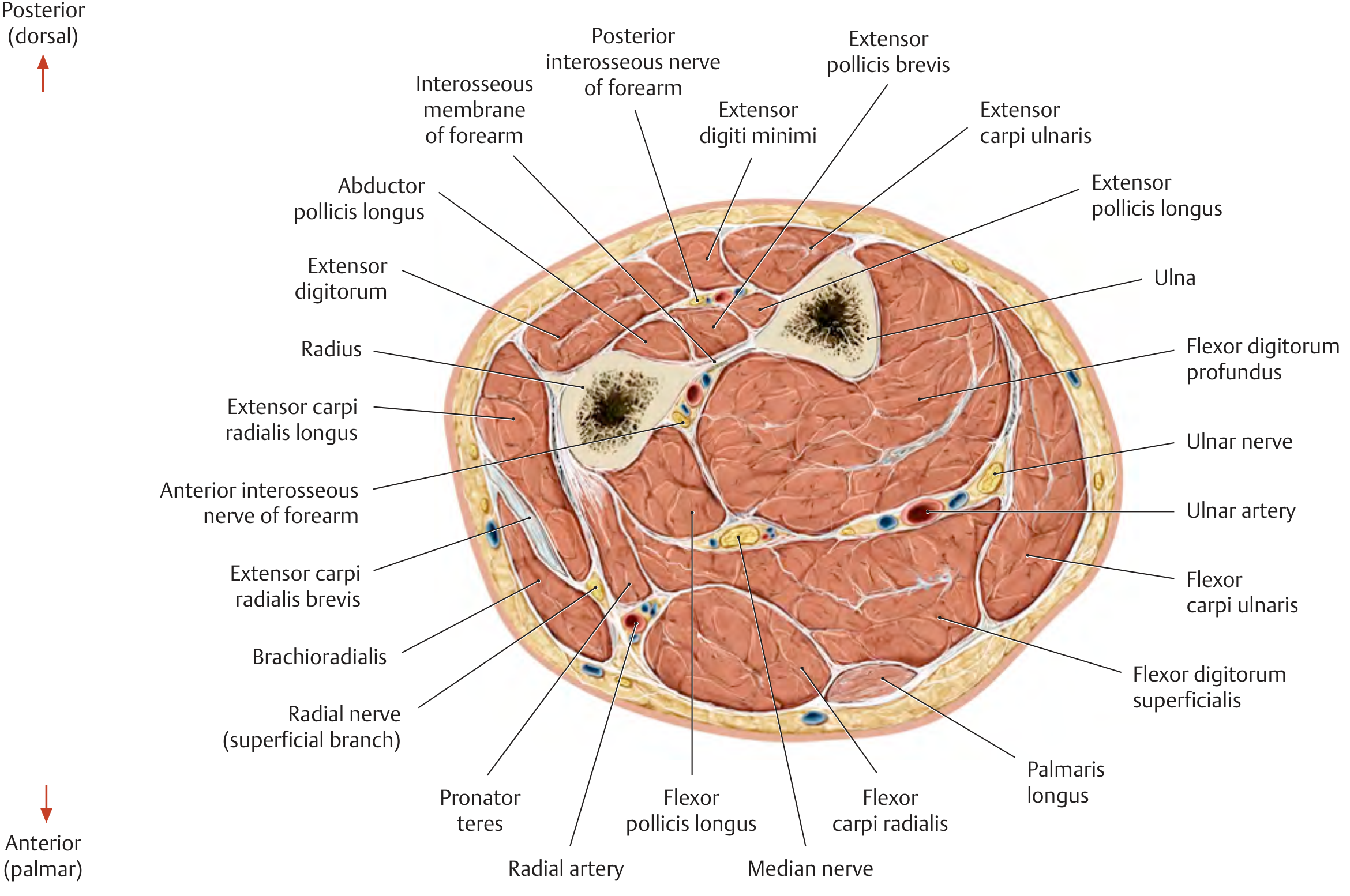
Cross section through the right forearm - THIEME Atlas of Anatomy. Note the positions of the radial artery and nerve (lateral/anterior), median nerve (central), ulnar artery and nerve (medial), and the interosseous membrane separating the compartments.
6. Arteries
Radial artery:
- Branch of the brachial artery; runs lateral in the forearm deep to the brachioradialis
- Passes between brachioradialis and flexor carpi radialis tendons distally - its pulse is palpable here (radial pulse)
- Exits by passing dorsally around the wrist into the hand
Ulnar artery:
- Larger branch; runs medially under the flexor carpi ulnaris
- Gives off the common interosseous artery which divides into:
- Anterior interosseous artery - travels along the anterior surface of the interosseous membrane supplying deep muscles
- Posterior interosseous artery - passes dorsally over the proximal membrane into the posterior compartment
- Leaves the forearm lateral to the pisiform, superficial to the flexor retinaculum
7. Nerves
Median Nerve
- Enters the forearm by passing between the two heads of pronator teres
- Then passes under the tendinous arch of flexor digitorum superficialis
- Runs deep to FDS but superficial to FDP throughout the forearm
- Gives off the anterior interosseous nerve (deep branch) supplying FDP (lateral half), FPL, and pronator quadratus
- Innervates all anterior compartment muscles except flexor carpi ulnaris and medial FDP
Ulnar Nerve
- Enters the forearm by passing posterior to the medial epicondyle through the cubital tunnel
- Runs between the two heads of flexor carpi ulnaris
- Descends on the medial side of the forearm deep to FCU, on the surface of FDP
- Innervates: flexor carpi ulnaris and medial half of FDP (ring and little fingers)
- Becomes palpable in the distal forearm medial to the ulnar artery
Radial Nerve (and branches)
- The superficial (sensory) branch runs deep to brachioradialis in the lateral forearm
- The deep branch enters the posterior compartment by passing through the supinator muscle, then becomes the posterior interosseous nerve which innervates all muscles of the posterior compartment (except brachioradialis, ECRL, and ECRB which are innervated before the split)
8. Joints
Proximal radio-ulnar joint - at the elbow; the head of the radius rotates within the radial notch of the ulna, held by the annular ligament.
Distal radio-ulnar joint - the articular surface of the ulnar head articulates with the ulnar notch of the radius and with a triangular fibrocartilage complex (TFCC) / articular disc that separates it from the wrist joint. Allows the distal radius to swing anteromedially over the ulna during pronation.
Together, these two joints allow pronation and supination (~180° range total).
9. Clinical Relevance
| Condition | Anatomy |
|---|---|
| Colles fracture | Distal radius fracture with posterior displacement - most common forearm fracture |
| Galeazzi fracture | Fracture of distal third of radius + subluxation of distal radio-ulnar joint |
| Monteggia fracture | Fracture of proximal ulna + anterior dislocation of radial head |
| Pronator syndrome | Median nerve compressed between heads of pronator teres |
| Anterior interosseous nerve palsy | Weakness of FDP (index/middle), FPL, pronator quadratus - cannot make "OK sign" |
| Cubital tunnel syndrome | Ulnar nerve compressed at medial epicondyle; causes ring/little finger weakness and sensory loss |
| Radial pulse | Palpated between brachioradialis and FCR tendons at the wrist |
Sources: Gray's Anatomy for Students (9780323934237), THIEME Atlas of Anatomy - General Anatomy and Musculoskeletal System (9781626237186), Imaging Anatomy: Bones, Joints, Vessels and Nerves Vol. 3 (9781626239845)
This is a shared conversation. Sign in to Orris to start your own chat.