
Juvenile angifibroma
I'll look this up in the medical library for you right away.
Searching sources...
Reading File
Finding Sources
Finding Sources
Searching PubMed
juvenile nasopharyngeal angiofibroma
Good - I found dedicated sections in Cummings Otolaryngology and Scott-Brown's. Let me read the most comprehensive ones now.
Reading File
Reading File
Let me read more of the Cummings section for staging, clinical features, and management:
Reading File
The offset drifted into the osteoma section. Let me read the correct JA section (lines 787-840):
Reading File
Reading File
Now let me also fetch the key endoscopic image and check the staging table more specifically:
Loading Image
Finding Sources
I now have comprehensive content to write a thorough answer. Let me compile everything.
Juvenile Angiofibroma (JA)
Also called Juvenile Nasopharyngeal Angiofibroma (JNA) - a benign but locally aggressive vascular tumor that almost exclusively affects adolescent males.
Definition & Pathology
JA is a benign vascular tumor histologically characterized by vascular endothelium-lined spaces embedded in a fibrous stroma. The vessels lack a smooth muscle layer (unlike normal arteries), sitting beside a conventional arteriocapillary tree that does have muscle layers. Immunohistochemistry and electron microscopy suggest it is better classified as a vascular malformation (hamartoma) rather than a true neoplasm.
- Cummings Otolaryngology, p. 988
Pathogenesis
The leading theory (Schick et al.) proposes that JA arises from incomplete regression of the first branchial arch artery, which forms during embryogenesis between days 22-24 as a temporary connection between the ventral and dorsal aorta. Remnants of this regression may form the nidus of JA, supported by the finding that JA vessels express laminin α2, a marker for early angiogenesis.
Molecular heterogeneity has been documented, with variable expression of: bFGF, H-Ras, IL-6, c-Kit, c-Myc, PDGFa, TP53, VEGFA mRNA, and β-catenin. Recent RNA sequencing studies support upregulation of VEGFA and activation of the FGFR pathway (implicated in angiogenesis).
Regarding hormonal influence: testosterone stimulation and oestrogen hypersensitivity at puberty are thought to cause nasal mucosal target cells to develop into muscularized vascular channels - explaining the male adolescent predilection.
- Cummings Otolaryngology, p. 988; Scott-Brown's Vol 1, p. 2899
Epidemiology
- Almost exclusively in adolescent males (10-25 years)
- Peak incidence around puberty (~14-17 years)
- Most involute after hormonal maturation
- Rare in females - if diagnosed, the diagnosis should be reconsidered
Site of Origin
The pathognomonic epicenter is the pterygopalatine fossa, specifically at the level of the sphenopalatine foramen or the base of the pterygoid process / superior choana (per recent imaging-based studies).
Patterns of spread (Fig. 50.7):
| Direction | Extension into |
|---|---|
| Medial | Nasopharynx, nasal cavity via sphenopalatine foramen |
| Superior | Sphenoid sinus floor, floor of middle cranial fossa |
| Lateral | Pterygopalatine fossa → pterygomaxillary fissure → infratemporal fossa |
| Anterior | Posterior wall of maxillary sinus (pushed forward) |
| Orbital | Via inferior orbital fissure → proptosis |
| Intracranial | Via middle cranial fossa erosion |
Bone involvement occurs via: (1) pressure resorption (subperiosteal growth), or (2) invasion of cancellous bone at the pterygoid root with expansion into the greater wing of sphenoid.
- Cummings Otolaryngology, p. 988
Clinical Features
Early/Small lesions:
- Unilateral nasal obstruction (most common)
- Epistaxis (often recurrent, may be severe enough to cause anaemia)
Advanced lesions:
- Swelling of the cheek (infratemporal fossa involvement)
- Proptosis + diplopia (orbital invasion)
- Headache (cranial fossa involvement)
- Conductive hearing loss (Eustachian tube obstruction)
Endoscopic appearance: A smooth, hypervascularized polypoid mass behind the middle turbinate (which is laterally displaced), completely obstructing the choana - in a teenage boy this is virtually diagnostic.
⚠️ Biopsy is contraindicated - carries high risk of severe hemorrhage.
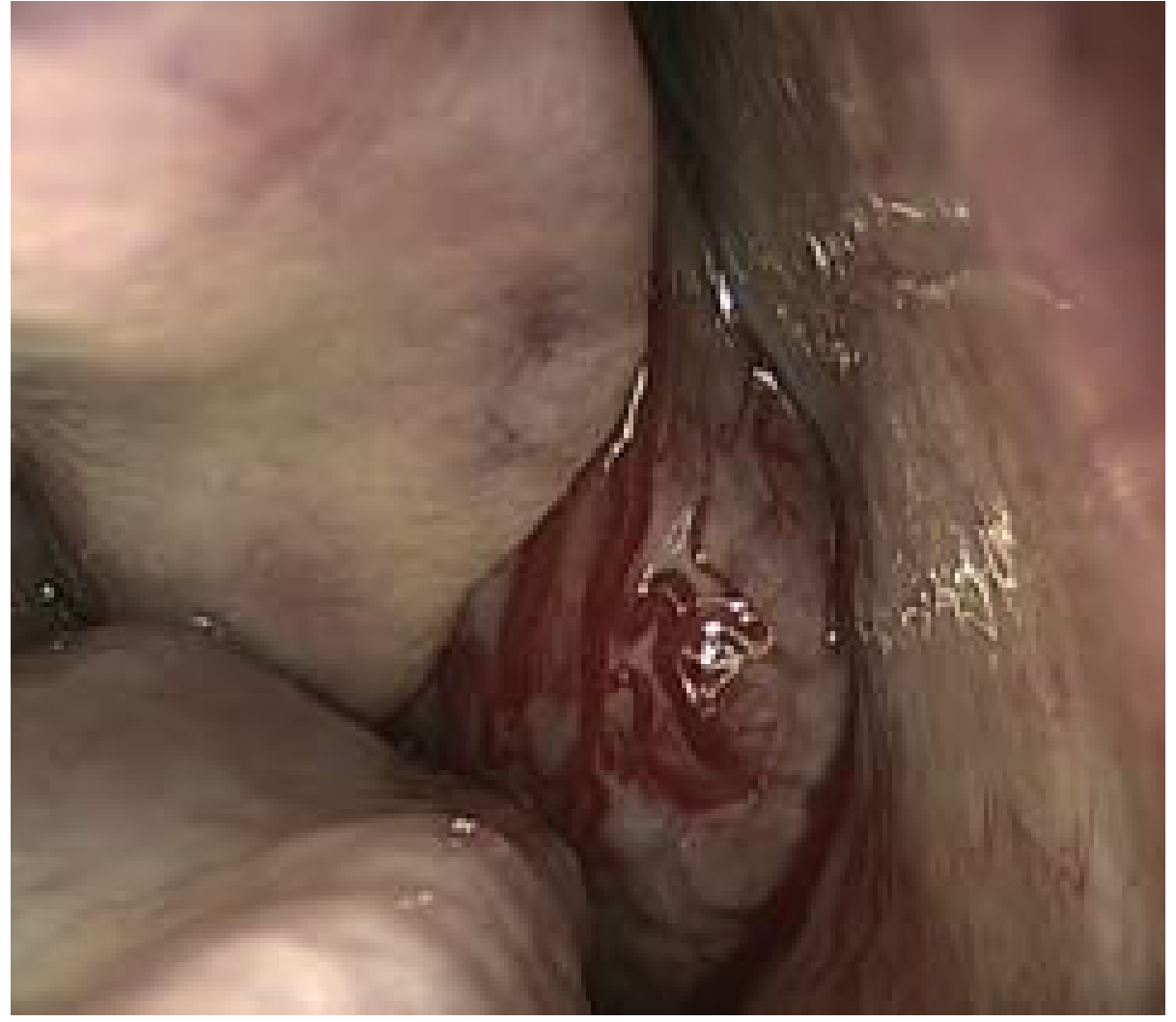
Endoscopic view: JA appearing as a polypoid hypervascularized mass with the choana completely obstructed (Cummings Otolaryngology, Fig. 50.8)
Imaging
CT findings:
- Mass centered in pterygopalatine fossa with erosion of the base of the medial pterygoid plate
- Intense contrast enhancement (hypervascular)
- Anterior bowing of posterior maxillary sinus wall (Holman-Miller sign / Antral sign)
MRI findings:
- Intense enhancement on contrast T1
- "Salt and pepper" appearance - multiple signal voids (flow voids) on T1 and T2 = major intralesional vessels (pathognomonic)
- Better delineates intracranial extension and orbital involvement
DSA (Digital Subtraction Angiography):
-
Enlarged intratumoral vessels + intense inhomogeneous tumor blush in venous phase
-
Maps vascular supply before embolization
-
Primary supply: branches of external carotid artery (internal maxillary, ascending pharyngeal)
-
Advanced lesions: internal carotid artery supply in ~35.6% of cases; bilateral supply in ~30.8%
-
Cummings Otolaryngology, p. 989; Scott-Brown's Vol 1, p. 2907
Staging
Several systems exist. The most widely used is based on extent of disease with special attention to:
- Infratemporal fossa extension
- Skull base involvement
- Intracranial invasion
The Snyderman/UPMC staging system (2010) emphasizes:
- Residual vascularization from internal carotid artery after embolization
- Route of intracranial growth - medial vs lateral to ICA and cavernous sinus - as key predictors for endoscopic resectability
- Cummings Otolaryngology, p. 990
Treatment
1. Preoperative Embolization
- Standard adjunct performed 48 hours before surgery
- Reduces intraoperative bleeding dramatically, improves tumor border visualization
- Uses: polyvinyl alcohol (PVA) particles, liquid agents (NBCA, Onyx), via superselective intra-arterial catheterization
- Direct puncture technique with Onyx injection improves devascularization, especially when ICA branches supply the tumor (transarterial approach risks neurological deficit in these cases)
- Some surgeons now consider embolization unnecessary for smaller tumors accessible endoscopically (maxillary artery can be clipped at start of dissection)
2. Surgery - Mainstay of Treatment
- Endoscopic resection has replaced open approaches for most cases (small, intermediate, and selected large tumors)
- Key surgical principles:
- Dissection in the subperiosteal plane
- Bipolar coagulation
- Extensive drilling of the basisphenoid (removes cancellous bone with tumor digitations - Howard et al. showed this dramatically reduces recurrence)
- Multiportal approaches (transnasal + transoral or transorbital) for advanced lesions
- Open approaches (midfacial degloving, infratemporal fossa) reserved for extensive ICA encasement or cavernous sinus involvement
3. Radiotherapy
- Used as an alternative or adjunct for intracranial involvement, unresectable disease, or recurrence
- Stereotactic radiosurgery also reported
4. Hormone therapy / Medical
- Antiandrogens and oestrogens have been tried (given hormonal dependence) but are not standard
- Flutamide (anti-androgen) has been used preoperatively to reduce tumor size in some series
Recurrence
- Residual disease is the most common reason for "recurrence" (incomplete initial resection)
- Systematic removal of involved sphenoid cancellous bone is the single most important step to reduce this
- Residual disease may spontaneously involute on serial MRI follow-up (particularly after puberty)
- MRI is the follow-up modality of choice
Key Points Summary
| Feature | Detail |
|---|---|
| Type | Benign, locally aggressive vascular hamartoma |
| Who | Adolescent males only |
| Origin | Pterygopalatine fossa / sphenopalatine foramen |
| Classic symptoms | Unilateral nasal obstruction + epistaxis |
| Biopsy | Contraindicated (hemorrhage risk) |
| Imaging hallmark | Salt-and-pepper on MRI; Holman-Miller sign on CT |
| Vascular supply | External carotid (IMA) ± ICA in advanced disease |
| Treatment | Surgery (endoscopic) ± preoperative embolization |
| Recurrence driver | Residual tumor in sphenoid cancellous bone |
Sources: Cummings Otolaryngology Head and Neck Surgery, pp. 988-990; Scott-Brown's Otorhinolaryngology Head & Neck Surgery Vol 1, pp. 2889-2940
This is a shared conversation. Sign in to Orris to start your own chat.