
Dentin bonding agents
dentin bonding agent hybrid layer adhesive
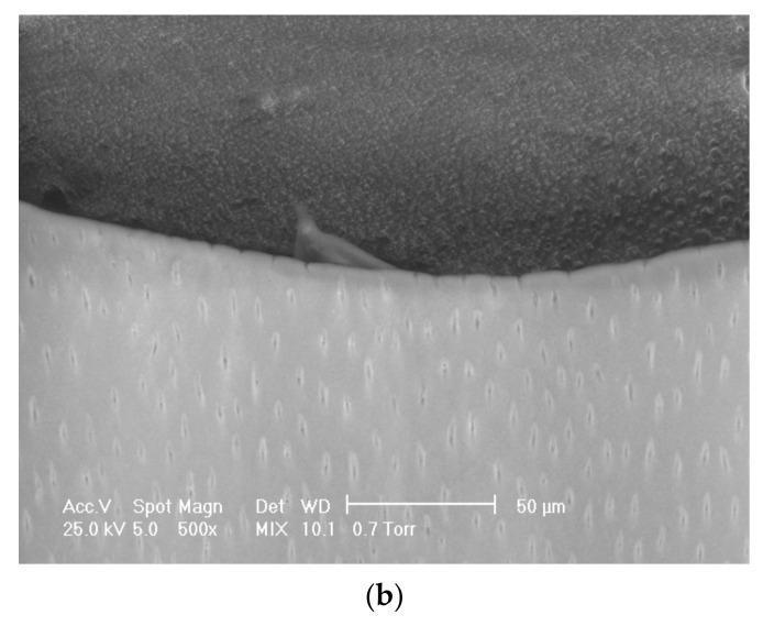
This Scanning Electron Microscopy (SEM) diagnostic image at 500x magnification illustrates the adhesive-dentin interface in a restorative dental specimen. The image is divided into two distinct regions. The lower portion (lighter tone) represents the dentin layer, characterized by a high density of evenly distributed, elongated dentinal tubules or canaliculi. These tubules appear as small, dark, vertically oriented ovals within a smooth calcified matrix. The upper portion (darker tone) depicts the dental adhesive material, showing a granular and homogeneous texture. The interface between the two materials is clearly defined, demonstrating clinical evidence of hybridization. At one point along the boundary, the adhesive material can be seen interpenetrating a dentinal tubule, indicating the formation of a resin tag for micromechanical retention. The image is critical for evaluating the quality of dental bonding, the presence of a hybrid layer, and the potential for microleakage in restorative dentistry.
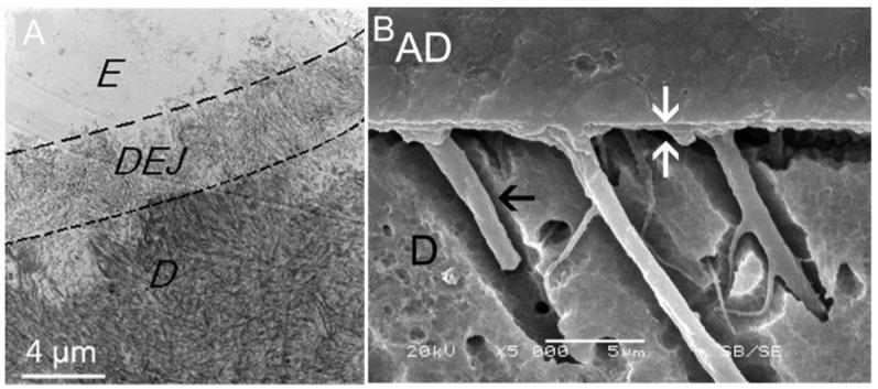
This figure presents two Scanning Electron Microscopy (SEM) microphotographs illustrating natural and artificial dental interfaces. Image A shows a cross-section of the natural dentinoenamel junction (DEJ), marked by dashed lines, which serves as a transitional interface between the highly mineralized enamel (E) and the more granular dentin (D). A scale bar of 4 µm is provided for reference. Image B demonstrates the resin-dentin interface achieved through dental bonding. The adhesive (AD) layer is visible at the top, forming a hybrid layer (indicated by white arrows) where the resin interdiffuses with the dentin (D). A prominent black arrow identifies a resin tag, where adhesive material has successfully penetrated into a dentinal tubule to provide micromechanical retention. The images compare the biomimetic integration of natural structures with the artificial adhesion used in restorative dentistry. Key educational concepts include dental histology, adhesive dentistry, and the morphology of the hybrid layer.
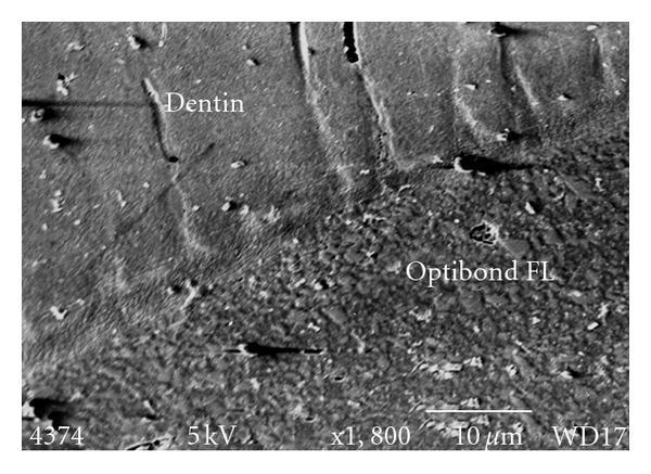
Scanning electron micrograph (SEM) at 1,800x magnification showing the cross-sectional interface between human dentin and the dental adhesive Optibond FL. The image illustrates the formation of a hybrid layer, a key educational concept in restorative dentistry and biomaterials. The dentin region occupies the upper portion of the frame, characterized by a relatively smooth, homogenous texture with subtle parallel striations and minor imperfections. The lower portion displays the Optibond FL material, which exhibits a highly granular and heterogeneous appearance due to the presence of numerous small, bright filler particles. A distinct transition zone is visible where the adhesive infiltrates the demineralized dentin substrate to form the resin-dentin interface. The micrograph includes technical metadata such as a 10 μm scale bar, 5kV accelerating voltage, and WD17 working distance, providing context for the microscopic evaluation of bond integrity and failure modes in dental luting cements.
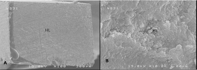
This diagnostic image consists of two Scanning Electron Microscopy (SEM) micrographs showing the microstructural analysis of a dental fractured surface. The visuals demonstrate a failure in the adhesive bond between restorative material and tooth structure. Image A, at 100x magnification, provides a macro-view of the fractured surface, labeled 'HL' for Hybrid Layer, which appears as a relatively uniform and smooth region with subtle linear striations. Image B, at a much higher 10000x magnification, reveals the topographical details of the same hybrid layer, showcasing a granular, rough, and porous micro-architecture. These images are clinically significant in dentistry for evaluating the quality of dentin bonding and characterizing the mode of failure—in this case, an adhesive failure predominantly involving the hybrid layer. The comparison between the two magnifications illustrates the difference between gross surface continuity and microscopic irregularities essential for assessing dental material performance and bond integrity.
Dentin Bonding Agents (DBAs)
Definition
Composition of a DBA
| Component | Function | Key Contents |
|---|---|---|
| Etchant | Removes smear layer, demineralizes dentin 4-8 µm, opens tubules | 30-40% phosphoric acid |
| Primer | Hydrophilic monomer; wets and penetrates moist dentin collagen | HEMA, acetone/ethanol/water solvent, bi-functional monomers |
| Adhesive resin (bond) | Hydrophobic resin; seals the surface, provides mechanical bridge | Bis-GMA, UDMA, unfilled or lightly filled resin |
Classification by Generation
1st Generation (1950s-60s)
- Used NPG-GMA (N-phenyl glycine + glycidyl methacrylate)
- Bond strength very low (~1-3 MPa); no clinical significance today
- Bonded only to enamel, not dentin
2nd Generation (1970s-early 80s)
- Halophosphorous esters (e.g., Bis-GMA derivatives)
- Bonded to calcium in smear layer; unreliable (~5 MPa)
- Smear layer left intact
3rd Generation (mid-1980s)
- First to remove smear layer with mild acids
- Separate etch, prime, and bond steps
- Bond strength ~15-20 MPa
- Examples: Scotchbond 2, Clearfil New Bond
4th Generation (early 1990s) - GOLD STANDARD
- Total-etch technique: 35-37% phosphoric acid applied to both enamel and dentin
- Three-bottle system: etch + primer + bond
- Bond strength: 20-25 MPa to dentin
- Examples: Optibond FL, All-Bond 2, Scotchbond Multi-Purpose
- Forms true hybrid layer with long resin tags into dentinal tubules
5th Generation (mid-1990s)
- Total-etch, but two-step: primer and adhesive combined into one bottle
- Equal or slightly less bond strength vs. 4th gen
- Examples: Single Bond (3M), Prime & Bond NT, One-Step
6th Generation (early 2000s) - Self-Etch
- Two-step self-etch: acidic primer etches and primes simultaneously; separate adhesive applied after
- Does NOT require separate acid etching
- Less post-operative sensitivity; milder demineralization
- Examples: Clearfil SE Bond, AdheSE
- Mild self-etching primer: pH ~2; aggressive: pH <1
7th Generation (2005 onwards) - All-in-One / Universal
- All three steps (etch + prime + bond) in a single bottle
- Convenient but somewhat lower bond strength
- Examples: Xeno V, G-Bond, iBond, Adper Prompt-L-Pop
- Hydrophilic nature makes the bond layer act like a semi-permeable membrane
8th Generation (Universal Adhesives, ~2012-present)
- Can be used in total-etch, self-etch, or selective-etch mode
- Contain 10-MDP monomer (strong chemical bond to hydroxyapatite)
- Examples: Scotchbond Universal, All-Bond Universal, Clearfil Universal Bond
- Bond strength comparable to 4th gen in total-etch mode
Mechanism of Bonding
Enamel Bonding
- Phosphoric acid etching creates micro-porosities (10-20 µm deep)
- Resin penetrates and polymerizes = micromechanical interlocking
- Very reliable; bond strength 20-30 MPa
Dentin Bonding (More Complex)
- Acid etching removes smear layer and demineralizes superficial dentin
- Exposed collagen network remains (intertubular dentin scaffold)
- Hydrophilic primer infiltrates the wet collagen mesh
- Adhesive resin encapsulates the collagen fibers
- Light-curing polymerizes the resin = hybrid layer (0.5-8 µm thick)
- Resin also extends into tubule orifices = resin tags
Smear Layer
| Strategy | What happens | Example systems |
|---|---|---|
| Remove (Total-etch) | Acid dissolves it completely; collagen exposed | 4th, 5th gen |
| Modify (mild self-etch) | Partially dissolves; incorporated into hybrid layer | 6th gen (mild) |
| Preserve (no-etch older) | Bond to smear layer only (weak) | 2nd gen |
Total-Etch vs. Self-Etch Technique
| Feature | Total-Etch | Self-Etch |
|---|---|---|
| Acid step | Separate phosphoric acid | Built into primer |
| Demineralization depth | 4-8 µm | 0.5-1 µm (mild) |
| Bond strength to enamel | Excellent | Good (mild SE less effective) |
| Bond strength to dentin | Excellent | Comparable |
| Post-op sensitivity | Higher risk | Lower risk |
| Technique sensitivity | High (over-drying kills bond) | Lower |
| Smear layer | Removed | Incorporated |
Key Monomers in Modern DBAs
| Monomer | Full Name | Role |
|---|---|---|
| HEMA | 2-Hydroxyethyl methacrylate | Hydrophilic; wets dentin |
| Bis-GMA | Bisphenol A glycidyl dimethacrylate | Hydrophobic adhesive resin |
| UDMA | Urethane dimethacrylate | Adhesive resin (less brittle) |
| 10-MDP | 10-methacryloyloxydecyl dihydrogen phosphate | Chemical bonding to Ca in hydroxyapatite; key in 8th gen |
| 4-META | 4-methacryloxyethyl trimellitate anhydride | Chemical adhesion; used in Superbond C&B |
| Glutaraldehyde | - | Collagen cross-linker; used in 5th gen (e.g., Gluma) |
Failure Modes
- Adhesive failure: at the resin-dentin interface (between hybrid layer and unaffected dentin)
- Cohesive failure: within the dentin or within the adhesive/composite itself
Factors Affecting Bond Strength
- Moisture - dentin must be moist (not wet or dry) for total-etch; smear layer washed away but collagen must not collapse
- Dentinal tubule orientation - deeper dentin (more tubules, more fluid) = lower bond strength
- Smear layer thickness - thicker smear after fine burs vs. coarse
- Remaining dentin thickness (RDT) - thin dentin near pulp has more tubules and dentinal fluid pressure
- Caries-affected dentin - partially demineralized; more tubule occlusion; bond may be weaker
- Sclerotic dentin - tubules occluded with mineral; reduced resin tag penetration
- C-factor (configuration factor) - ratio of bonded to unbonded surfaces; high C-factor increases polymerization stress and debonding risk
Clinical Considerations
- Post-operative sensitivity is mainly due to incomplete resin infiltration of demineralized dentin, leaving unprotected collagen exposed to fluid flow (hydrodynamic theory of pain)
- Nanoleakage: water/ions permeate the hybrid layer even without visible gaps - major cause of secondary caries and bond degradation over time
- Over-etching desiccates collagen fibers, causing them to collapse and preventing resin infiltration
- Self-etch systems tend to have lower post-op sensitivity because they do not over-demineralize dentin
- Shelf life and solvent evaporation - acetone-based primers evaporate quickly; must be capped between applications
Bond Strength Values (Approximate)
| System Generation | Enamel Bond (MPa) | Dentin Bond (MPa) |
|---|---|---|
| 1st gen | <3 | <3 |
| 2nd gen | 8-12 | 4-8 |
| 3rd gen | 15-20 | 12-18 |
| 4th gen | 20-30 | 18-25 |
| 5th gen | 20-28 | 18-24 |
| 6th gen | 15-22 | 18-25 |
| 7th gen | 15-20 | 15-20 |
| 8th gen (universal) | 22-28 | 20-26 |
Summary: Choosing the Right System
- Best bond strength overall: 4th gen (Optibond FL) or 8th gen Universal in total-etch mode
- Least technique sensitive / least sensitivity: 6th gen self-etch (Clearfil SE Bond)
- Most convenient: 7th/8th gen universal adhesives
- Deep dentin / near-pulp cavities: Self-etch preferred to avoid sensitivity
- Enamel margins: Total-etch or selective-etch preferred for better enamel bond