
tilting towards one side after dizzniess age 80 what could be thecause
dizziness vertigo elderly causes
BPPV benign paroxysmal positional vertigo mechanism inner ear
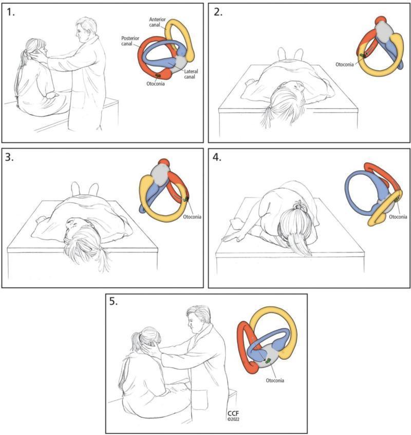
A five-panel medical illustration sequence detailing the Epley maneuver for treating Benign Paroxysmal Positional Vertigo (BPPV). Each panel pairs a clinical illustration of a patient and clinician with a corresponding anatomical diagram of the right inner ear's semicircular canals (anterior in yellow, posterior in red, and lateral in blue). Step 1 shows the patient seated with the head rotated 45° toward the affected side, with otoconia located in the posterior canal. Step 2 depicts the patient moved into a supine position with the neck slightly extended. Step 3 illustrates the head being rotated 90° toward the opposite (healthy) side. Step 4 shows the patient rolling onto their side while maintaining head rotation, facilitating the movement of otoconia through the canal toward the utricle. Step 5 shows the patient returning to a seated position. The anatomical insets track the gravitational migration of otoconia out of the posterior semicircular canal, demonstrating the mechanical rationale for this canalith repositioning procedure.
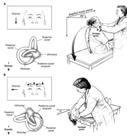
Summary : This figure illustrates the steps and anatomical basis of a clinical maneuver for diagnosing or treating posterior canal benign paroxysmal positional vertigo (BPPV), showing both the head/body positions and the orientation of the inner ear semicircular canals relative to gravity. illustration: # Step A : Initial Position and Head Turn • Patient is seated upright on an examination table. • Head is turned 45° to the side (sagittal body plane indicated). • Gravity direction is shown as downward. • Anatomical diagram of the inner ear highlights the superior canal, posterior canal, utriculus, and posterior-canal ampulla. • Particles are depicted within the posterior canal, indicating the suspected location of otoconia in BPPV. # Step B : Reclining Position and Head Extension • Patient is moved from sitting to a supine position with the head extended backward at a 20° angle. • Examiner supports the patient’s head at the vantage point. • Gravity direction is now shown as leftward. • Anatomical diagram again highlights the superior canal, posterior canal, utriculus, and posterior-canal ampulla. • Particles remain in the posterior canal, now oriented differently due to the change in gravity direction. # Spatial Relationships : • The examiner is positioned behind the patient in both steps, guiding the head movement. • The vantage point for head support is indicated in both illustrations. • The anatomical diagrams are aligned with the patient’s head orientation in each step. # Technical Details : • No scale bar or magnification is present. • The angle of head extension (20°) is explicitly labeled. • The sagittal body plane and gravity vectors are clearly marked. # Analysis : • The figure demonstrates how head positioning relative to gravity affects the orientation of the semicircular canals and the movement of particles within the posterior canal. • The maneuver is designed to provoke or treat vertigo by repositioning otoconia, with each step altering the gravitational vector acting on the inner ear structures. • The anatomical diagrams clarify the rationale for the head movements in the clinical procedure.
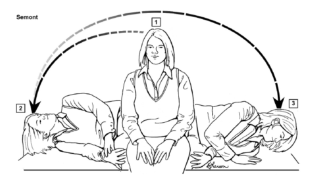
Summary : This illustration demonstrates the Semont maneuver, a physical therapy technique used to treat benign paroxysmal positional vertigo (BPPV) by repositioning canaliths in the inner ear. illustration: # Scene Overview : • The sequence shows a person sitting on an examination table, then quickly moving to lie on one side, and then to the opposite side. • Three positions are depicted: 1. Sitting upright in the center of the table. 2. Lying on the left side with the head turned upward. 3. Lying on the right side with the head turned downward. • Arrows indicate the direction of movement from the sitting position to each side. # Technical Details : • The illustration is labeled "Semont" in the upper left. • The person is wearing a vest over a shirt and pants. • No scale bar or measurement units are present. • The sequence is shown with dashed and solid arrows to indicate the order of movements. # Spatial Relationships : • The person starts in the center, moves rapidly to one side, then to the other, maintaining a consistent orientation of the torso and head as indicated by the arrows. • The table is depicted horizontally, with the person’s movements spanning from left to right. # Analysis : • The illustration clearly outlines the three-step Semont maneuver, emphasizing the rapid, sequential movement from sitting to lying on one side, then quickly to the other. • The arrows and body positions visually guide the viewer through the maneuver, making it suitable for patient or clinician education.
Dizziness + Tilting to One Side in an 80-Year-Old: Possible Causes
1. BPPV - Benign Paroxysmal Positional Vertigo (MOST COMMON)
- Key features: Brief spinning (seconds to <1 min) triggered by head position changes (lying down, rolling over, looking up). Patient may stumble or lean toward one side when rising.
- Diagnosis: Dix-Hallpike maneuver
- Treatment: Epley maneuver (canalith repositioning) - very effective
2. Wallenberg Syndrome (Lateral Medullary Infarction) - URGENT
- Key features:
- Vertigo + illusion of environmental tilting
- Tendency to fall toward the affected side (lateropulsion)
- Ipsilateral facial numbness, Horner syndrome (drooping eyelid, small pupil)
- Contralateral loss of pain/temperature sensation in body
- Difficulty swallowing (dysphagia)
- This is a stroke - requires immediate emergency care
3. Cerebellar Stroke or Hemorrhage - URGENT
- Key features: Severe vomiting, vertigo, truncal ataxia (inability to walk straight), gait unsteadiness, often leaning or falling to one side
- Cerebellar hemorrhage: dizziness + marked truncal ataxia + gaze palsies + depressed consciousness - rapidly progressive
- Cerebellar infarction can mimic vestibular neuritis and be missed - >34% of cerebellar strokes are initially misdiagnosed
4. Vertebrobasilar TIA / Stroke
- Key features: Abrupt onset, lasting minutes to hours; may include blurred vision, diplopia, dysarthria, or unilateral weakness
- Particularly common in elderly with atherosclerosis, atrial fibrillation (cardioembolic), or hypertension
5. Vestibular Neuritis / Labyrinthitis
- Key features: Acute onset prolonged vertigo (days), nausea/vomiting, patient leans/falls toward the affected ear
- In older adults, this more often results from infarction (labyrinthine artery occlusion) than a virus
- Recovery is slower in the elderly (can take months)
- Labyrinthitis includes hearing loss; vestibular neuritis does not
6. Orthostatic Hypotension
- Key features: Dizziness/lightheadedness on standing from sitting or lying; can cause falls and leaning
- Common in patients on antihypertensives, diuretics, alpha-blockers, antidepressants
- Diagnosed by measuring BP lying vs. standing (drop >20 mmHg systolic)
7. Cervicogenic Dizziness
- Key features: Dizziness triggered by turning the head or looking up; can cause "drop attacks"
- Common in elderly with degenerative cervical spine disease
8. Meniere's Disease
- Less common at age 80 but possible, especially if episodes are recurring
9. Multiple Sensory Deficits (Presbystasis)
- This is the cause in ~25% of elderly dizzy patients
- Not a single disease but accumulation of age-related sensory decline
10. Medications
| Drug Class | Example |
|---|---|
| Antihypertensives | Amlodipine, metoprolol |
| Diuretics | Furosemide |
| Sedatives/benzodiazepines | Diazepam |
| Anticonvulsants | Phenytoin |
| Aminoglycoside antibiotics | Gentamicin (ototoxic) |
| Antidepressants (SSRIs, TCAs) | Amitriptyline |
Red Flag Signs - Seek Emergency Care Immediately
- Sudden onset, severe headache
- Persistent (not brief) vertigo
- Difficulty walking, inability to stand at all
- Double vision, facial drooping, speech difficulty
- New hearing loss on one side
- Horner syndrome (ptosis + miosis)
- Weakness or numbness of face/limbs
Summary Table
| Cause | Onset | Duration | Key Clue |
|---|---|---|---|
| BPPV | Position change | Seconds | Dix-Hallpike positive |
| Wallenberg syndrome | Sudden | Persistent | Ipsilateral face + Horner, tilting |
| Cerebellar stroke | Sudden | Persistent | Ataxia, vomiting, headache |
| Vertebrobasilar TIA | Sudden | Minutes-hours | Resolves, risk factors |
| Vestibular neuritis | Sudden | Days | No hearing loss |
| Orthostatic hypotension | On standing | Seconds | BP drop on standing |
| Cervicogenic | Head turning | Seconds | Neck movement triggered |
| Multisensory deficit | Chronic | Ongoing | Multiple impairments |