
Opthalmology
What ophthalmology topic would you like to explore?
Which disease or condition would you like to focus on?
How deep would you like to go?
"age-related macular degeneration"[MeSH Terms] AND treatment
"age-related macular degeneration" AND "anti-VEGF"
Age-Related Macular Degeneration (AMD) - Full Deep-Dive
1. Overview & Epidemiology
- Affects >1.75 million people in the United States
- Prevalence increases sharply after age 55; cumulative incidence in those >75 years is approximately 8%
- Both eyes are typically affected, though often asymmetrically
- Peripheral vision is spared - only central/macular function is lost
2. Pathophysiology
The RPE-Bruch Membrane-Choriocapillaris Unit
- Retinal Pigment Epithelium (RPE) - nourishes photoreceptors, phagocytoses spent outer segments
- Bruch's Membrane - basement membrane of the RPE
- Choriocapillaris - the innermost choroidal vasculature supplying oxygen/nutrients
Pathogenesis Diagram (Robbins)
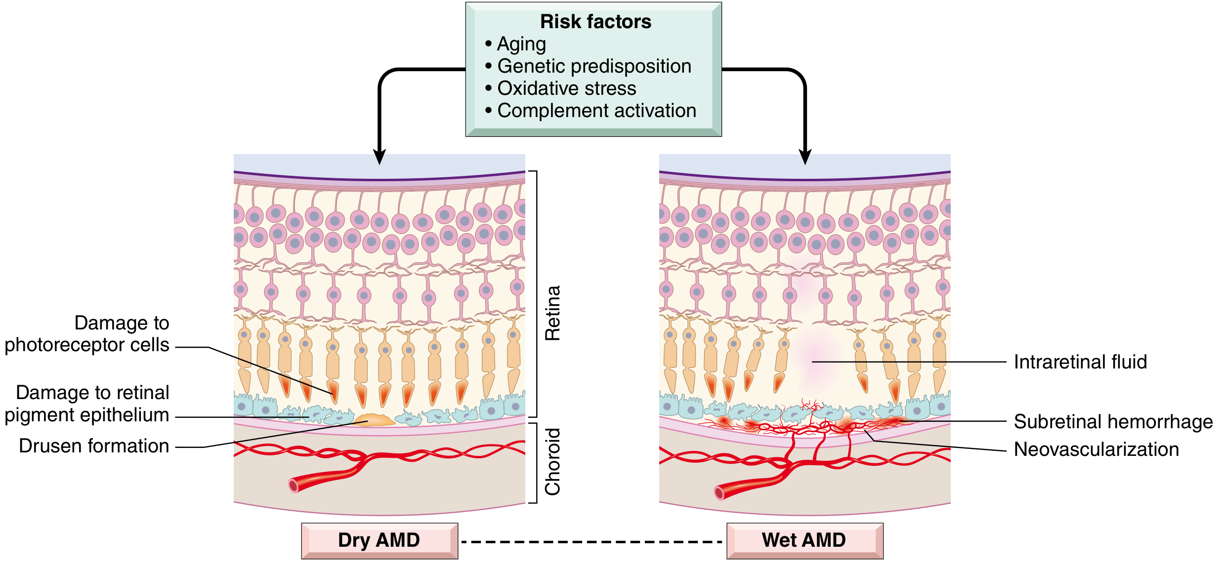
Dry (Atrophic) AMD
- Drusen form as focal thickenings/abnormal lipoprotein deposits within Bruch's membrane
- Lipofuscin (oxidation products of polyunsaturated fatty acids + vitamin A dimers) accumulates in stressed RPE cells
- Chronic complement system activation leads to RPE geographic atrophy and choriocapillaris loss
- Photoreceptors are secondarily lost overlying the atrophic RPE
Wet (Neovascular/Exudative) AMD
- Choroidal neovascularization (CNV): new vessels from the choriocapillaris penetrate through a breach in Bruch's membrane into the subretinal space
- These new vessels are fragile and leaky - they produce subretinal fluid (SRF), hemorrhage, and exudates
- VEGF (vascular endothelial growth factor) is the principal driver of neovascularization
- Blood may organize into a disciform scar (subretinal fibrosis), causing permanent photoreceptor loss
- Wet AMD is a complication of dry AMD, though both can coexist as parallel processes
3. Genetics & Risk Factors
Genetic
| Gene | Role |
|---|---|
| CFH (Complement Factor H) | Most strongly associated; abnormal CFH → unchecked complement activation |
| CFI (Complement Factor I) | Complement regulatory gene |
| Other complement regulatory genes | Polymorphisms increase risk |
Environmental & Lifestyle Risk Factors
| Factor | Notes |
|---|---|
| Smoking | Strongest modifiable risk factor; accelerates degeneration |
| Advanced age | Risk doubles each decade after 55 |
| Family history | Strong genetic component |
| Hyperopia | Associated risk |
| Blue eyes/fair skin | Increased susceptibility |
| Systemic hypertension | Vascular risk factor |
| High lipid levels | Accelerates disease |
| Uncontrolled blood sugar | Metabolic risk |
| UV exposure | Environmental oxidative stress |
4. Clinical Features
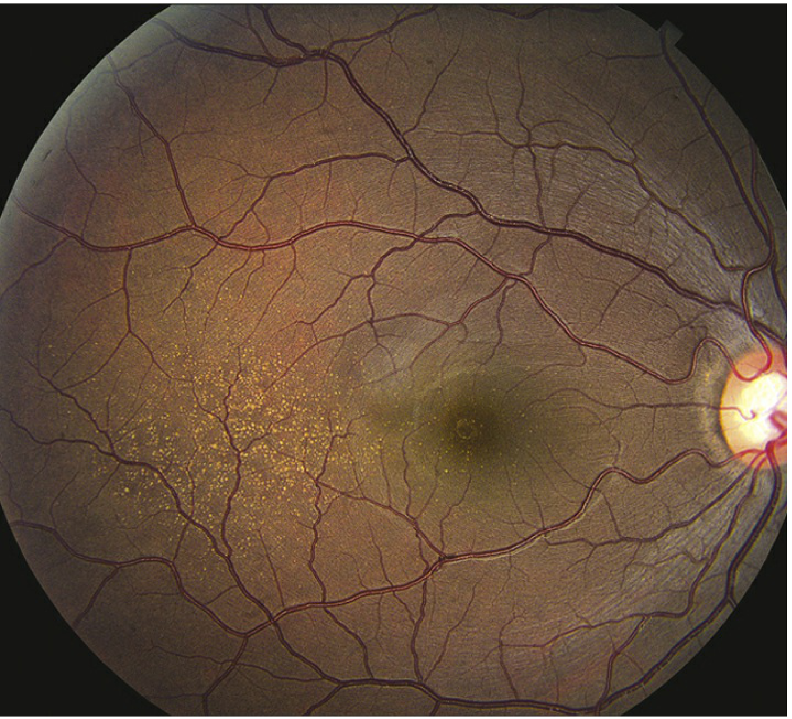
Dry AMD
- Gradual, insidious loss of central vision (may be asymptomatic for years)
- Amsler grid distortion (metamorphopsia may appear as the disease progresses)
- Difficulty reading, recognizing faces
- Drusen - soft drusen >60 µm are most significant (deep, hypopigmented spots)
- RPE mottling, depigmentation, hyperpigmentation clumps
- Geographic atrophy - well-defined areas of RPE and photoreceptor loss in advanced disease
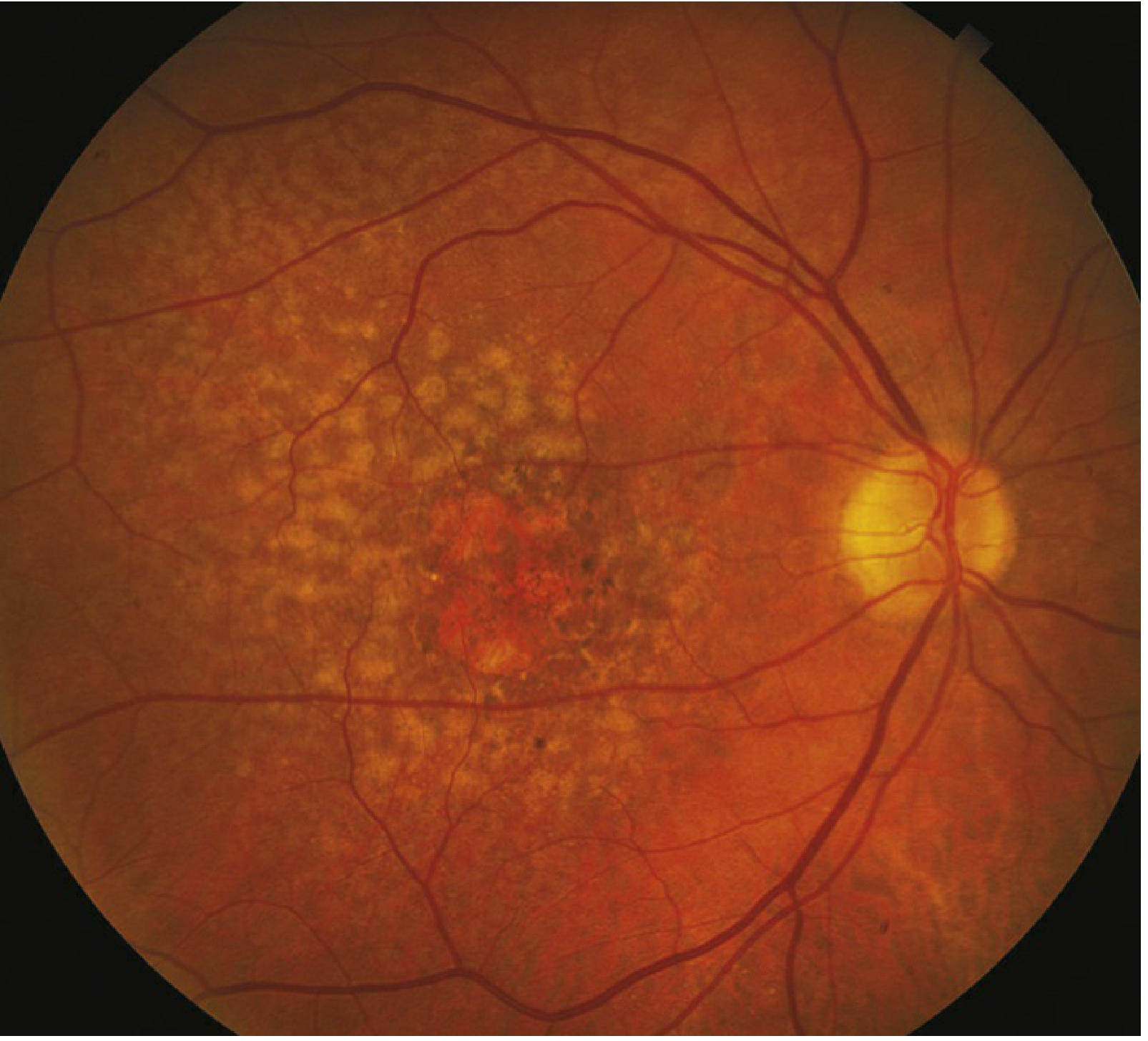
Wet AMD
- Sudden or rapid onset of central visual loss
- Central or paracentral scotoma (blind spot)
- Metamorphopsia (straight lines appear wavy) - hallmark symptom
- Photopsias in the central visual field
- Subretinal fluid (SRF), macular edema (ME), or RPE detachment associated with CNV
- Subretinal or intraretinal hemorrhage
- Retinal exudates
- Subretinal fibrosis / disciform scar (end-stage)
- Retinal angiomatous proliferation (RAP) - intraretinal variant with telangiectatic vessels
A Key Clinical Pearl
5. Diagnosis & Investigations
At the Slit-Lamp / Fundus Exam
- Dilated fundus exam with indirect ophthalmoscopy
- Soft drusen, RPE changes, CNV membranes, hemorrhage
Amsler Grid Testing
- Home monitoring tool for detecting metamorphopsia
- Patients with dry AMD at risk for CNV should perform daily Amsler grid testing and seek urgent attention if new distortion appears
Optical Coherence Tomography (OCT) - Gold Standard for Wet AMD
- Identifies subretinal fluid, intraretinal fluid, RPE detachments, CNV membranes
- Used for monitoring treatment response to anti-VEGF therapy
Fluorescein Angiography (FA)
- Identifies classic vs. occult CNV
- Classic CNV: well-defined, leaks early and progressively
- Occult CNV: poorly defined, late leakage
Indocyanine Green Angiography (ICGA)
- Better delineates occult CNV, polypoidal choroidal vasculopathy (PCV)
OCT Angiography (OCTA)
- Non-invasive visualization of CNV without dye injection
- Increasingly used in diagnosis and follow-up
6. Staging (AMD Classification)
| Stage | Features |
|---|---|
| Early AMD | Small to medium drusen (<125 µm), no pigmentary changes |
| Intermediate AMD | Large drusen (>125 µm) or medium drusen with pigmentary changes |
| Advanced Dry AMD | Geographic atrophy of RPE ± photoreceptors, not involving the fovea |
| Advanced Wet AMD | CNV with subretinal fluid/hemorrhage, disciform scar |
7. Differential Diagnosis
| Condition | Key Distinguishing Feature |
|---|---|
| Myopic degeneration | Peripapillary changes, macular changes without drusen, high myopia |
| Central serous chorioretinopathy (CSCR) | Serous retinal elevation, usually <50 years, no drusen |
| Ocular histoplasmosis | Small white-yellow chorioretinal scars, peripapillary atrophy |
| Inherited macular dystrophies (Stargardt, Best disease) | Usually <50 years, familial, no drusen |
| Chloroquine toxicity | Bull's eye maculopathy, drug history |
| Angioid streaks | Sub-retinal red-brown bands radiating from optic disc |
| IPCV (polypoidal choroidal vasculopathy) | Polyp-like aneurysmal choroidal dilations on ICGA; common in Asian patients |
8. Management
Dry AMD - No Proven Curative Treatment
- Vitamin C (500 mg)
- Vitamin E (400 IU)
- Lutein (10 mg) + Zeaxanthin (2 mg) - replaces beta-carotene
- Zinc (80 mg)
- Copper (2 mg) - to prevent zinc-induced copper deficiency
Note: Beta-carotene is NOT recommended in smokers due to increased risk of lung cancer.
- Cessation of smoking (most important)
- Control of blood pressure, blood sugar, and lipids
- Dietary changes (green leafy vegetables rich in lutein/zeaxanthin)
- Complement inhibitors are being investigated (target C3 or C5 pathway)
- Special magnifying glasses
- Video-based magnification aids
- Allow patients to continue independent living
Wet AMD - Anti-VEGF Therapy (Mainstay)
| Drug | Class | Notes |
|---|---|---|
| Ranibizumab (Lucentis) | Anti-VEGF-A fragment (Fab) | FDA-approved, gold standard |
| Bevacizumab (Avastin) | Full anti-VEGF antibody | Off-label, widely used; comparable efficacy in CATT trial |
| Aflibercept (Eylea) | VEGF trap (VEGF-A + PlGF) | Longer dosing intervals possible |
| Faricimab (Vabysmo) | Dual inhibitor of VEGF-A + Angiopoietin-2 | Newest agent; longest dosing intervals (up to 16 weeks) |
Recent Evidence Update (2025): A network meta-analysis (PMID: 40241463) comparing all intravitreal anti-VEGF therapies for neovascular AMD showed comparable efficacy across agents. A systematic review of faricimab (PMID: 39043575) confirmed non-inferiority to ranibizumab and aflibercept with extended dosing intervals.
- Loading phase: Monthly injections for 3 months
- Maintenance: Pro re nata (PRN), treat-and-extend (T&E), or fixed dosing depending on agent and response
Other Treatments for Wet AMD
- Photodynamic therapy (PDT) with verteporfin - used in classic subfoveal CNV; less common since anti-VEGF era
- Laser photocoagulation - for extrafoveal CNV lesions; risk of scotoma
- Macular translocation surgery - retina detached, translocated away from CNV; then laser applied to destroy pathological vessels (advanced/salvage cases)
- Vitrectomy - for advanced cases with vitreous hemorrhage or large subretinal hemorrhage
9. Prognosis & Follow-up
- Dry AMD progresses slowly over months to decades; most patients retain some functional vision
- Wet AMD without treatment: rapid central vision loss within months
- With anti-VEGF treatment: ~33% gain >15 letters vision; 90%+ maintain or improve
- Patients with wet AMD in one eye have 10-12% annual risk of CNV developing in the fellow eye
- High-risk features for fellow eye: multiple/confluent soft drusen, RPE clumping
10. Summary Table
| Feature | Dry AMD | Wet AMD |
|---|---|---|
| Frequency | 90% of cases | 10% of cases |
| Vision loss | Gradual, over years | Sudden/rapid |
| Key finding | Drusen, geographic atrophy | CNV, subretinal fluid/hemorrhage |
| VEGF role | Not primary driver | Central driver (→ CNV) |
| Treatment | AREDS2 supplements, lifestyle | Anti-VEGF injections |
| Prognosis | Variable; can be severe | Poor if untreated; treatable if caught early |
- Goldman-Cecil Medicine International Edition (2024)
- The Wills Eye Manual, 8th Edition
- Robbins, Cotran & Kumar Pathologic Basis of Disease
- Histology: A Text and Atlas with Correlated Cell and Molecular Biology
- Katzung's Basic and Clinical Pharmacology, 16th Edition
- Butler et al., Acta Ophthalmol, 2025 - Network Meta-Analysis PMID 40241463
- Samacá-Samacá et al., BMJ Open Ophthalmol, 2024 - Faricimab Systematic Review PMID 39043575
Make a ppt
mkdir -p /home/daytona/workspace/amd-ppt && node /home/daytona/skills/shared/scripts/fetch_images.js "https://cdn.orris.care/cdss_images/ff3b1658063ecd3f735cbb8212922fb6522032a38c526619243c37b6d5b8216d.png" "https://cdn.orris.care/cdss_images/3c9867ffbbcd1c46bffbf8705e439310d97ac977f310d159bd552d823398adad.png" "https://cdn.orris.care/cdss_images/e1fc92fd9a06475763eddd40ec36bbabc56feeca5f57d02f69144b0dfd9c5389.png" 2>/dev/null | node -e "const d=require('fs');const j=JSON.parse(require('fs').readFileSync('/dev/stdin','utf8'));j.forEach((img,i)=>{const b=img.base64.split(',')[1];d.writeFileSync('/home/daytona/workspace/amd-ppt/img'+i+'.txt',img.base64);});console.log('done',j.map(x=>x.error||'ok'))"
~/amd-ppt/amd.js
const pptxgen = require("pptxgenjs");
const fs = require("fs");

const pres = new pptxgen();
pres.layout = "LAYOUT_16x9";
pres.title = "Age-Related Macular Degeneration (AMD)";
pres.author = "Orris Medical";

// Load images
const img0 = fs.readFileSync("/home/daytona/workspace/amd-ppt/img0.txt", "utf8"); // pathogenesis diagram dry vs wet
const img1 = fs.readFileSync("/home/daytona/workspace/amd-ppt/img1.txt", "utf8"); // fundus dry AMD
const img2 = fs.readFileSync("/home/daytona/workspace/amd-ppt/img2.txt", "utf8"); // fundus wet AMD

// ── Color Palette ──────────────────────────────────────────────
const DARK_BG   = "0D1B2A"; // deep navy
const MID_BG    = "1A2E42"; // slate navy
const ACCENT    = "1E90FF"; // clinical blue
const ACCENT2   = "E83030"; // alert red (wet AMD)
const ACCENT3   = "2ECC71"; // treatment green
const WHITE     = "FFFFFF";
const LIGHT_GRAY= "D0D8E4";
const GOLD      = "F4C430"; // soft gold for headers

// ── Helper: dark slide background ──────────────────────────────
function darkBg(slide) {
  slide.addShape(pres.shapes.RECTANGLE, {
    x: 0, y: 0, w: 10, h: 5.625,
    fill: { color: DARK_BG }, line: { type: "none" }
  });
}

function midBg(slide) {
  slide.addShape(pres.shapes.RECTANGLE, {
    x: 0, y: 0, w: 10, h: 5.625,
    fill: { color: "F0F4F8" }, line: { type: "none" }
  });
}

// ── Slide 1: Title ─────────────────────────────────────────────
{
  const s = pres.addSlide();
  darkBg(s);

  // Top accent bar
  s.addShape(pres.shapes.RECTANGLE, {
    x: 0, y: 0, w: 10, h: 0.08,
    fill: { color: ACCENT }, line: { type: "none" }
  });

  // Eye cross-section decorative shape on right
  s.addShape(pres.shapes.OVAL, {
    x: 6.5, y: 0.8, w: 3.2, h: 3.2,
    fill: { color: MID_BG, transparency: 30 },
    line: { color: ACCENT, width: 1.5 }
  });
  s.addShape(pres.shapes.OVAL, {
    x: 7.5, y: 1.8, w: 1.2, h: 1.2,
    fill: { color: ACCENT, transparency: 50 },
    line: { color: ACCENT, width: 0.5 }
  });

  // Title text
  s.addText("Age-Related", {
    x: 0.5, y: 0.9, w: 6.5, h: 0.8,
    fontSize: 36, bold: true, color: WHITE, fontFace: "Calibri",
    charSpacing: 2, margin: 0
  });
  s.addText("Macular Degeneration", {
    x: 0.5, y: 1.65, w: 6.5, h: 0.8,
    fontSize: 36, bold: true, color: ACCENT, fontFace: "Calibri",
    charSpacing: 2, margin: 0
  });
  s.addText("(AMD)", {
    x: 0.5, y: 2.4, w: 6.5, h: 0.6,
    fontSize: 28, bold: false, color: LIGHT_GRAY, fontFace: "Calibri",
    margin: 0
  });

  // Divider line
  s.addShape(pres.shapes.RECTANGLE, {
    x: 0.5, y: 3.1, w: 4.5, h: 0.04,
    fill: { color: GOLD }, line: { type: "none" }
  });

  s.addText([
    { text: "Pathophysiology  •  Diagnosis  •  Management", options: { color: LIGHT_GRAY } }
  ], {
    x: 0.5, y: 3.25, w: 8, h: 0.4, fontSize: 13, fontFace: "Calibri", margin: 0
  });

  s.addText([
    { text: "Sources: Goldman-Cecil Medicine | Wills Eye Manual | Robbins Pathology", options: { color: "7A8FA6", italic: true } }
  ], {
    x: 0.5, y: 5.1, w: 9, h: 0.35, fontSize: 9.5, fontFace: "Calibri", margin: 0
  });

  // Bottom accent bar
  s.addShape(pres.shapes.RECTANGLE, {
    x: 0, y: 5.545, w: 10, h: 0.08,
    fill: { color: ACCENT2 }, line: { type: "none" }
  });
}

// ── Slide 2: Overview & Epidemiology ──────────────────────────
{
  const s = pres.addSlide();
  midBg(s);

  // Left accent bar
  s.addShape(pres.shapes.RECTANGLE, {
    x: 0, y: 0, w: 0.08, h: 5.625,
    fill: { color: ACCENT }, line: { type: "none" }
  });

  // Header band
  s.addShape(pres.shapes.RECTANGLE, {
    x: 0, y: 0, w: 10, h: 0.85,
    fill: { color: DARK_BG }, line: { type: "none" }
  });
  s.addText("Overview & Epidemiology", {
    x: 0.3, y: 0.1, w: 9, h: 0.65,
    fontSize: 22, bold: true, color: WHITE, fontFace: "Calibri", margin: 0
  });

  // Stat boxes
  const stats = [
    { val: "#1", label: "Cause of irreversible\ncentral vision loss in\nelderly (W. countries)", color: ACCENT },
    { val: "8.5%", label: "Of global blindness\ncaused by AMD", color: "8B5E3C" },
    { val: "1.75M+", label: "Americans affected", color: ACCENT2 },
    { val: "8%", label: "Cumulative incidence\nin those aged 75+", color: "2E8B57" }
  ];
  stats.forEach((st, i) => {
    const x = 0.4 + i * 2.35;
    s.addShape(pres.shapes.ROUNDED_RECTANGLE, {
      x, y: 1.0, w: 2.1, h: 1.8,
      fill: { color: WHITE },
      line: { color: st.color, width: 2 },
      shadow: { type: "outer", color: "000000", blur: 5, offset: 2, angle: 135, opacity: 0.1 },
      rectRadius: 0.1
    });
    s.addText(st.val, {
      x: x + 0.05, y: 1.1, w: 2.0, h: 0.7,
      fontSize: 28, bold: true, color: st.color, align: "center", fontFace: "Calibri", margin: 0
    });
    s.addText(st.label, {
      x: x + 0.05, y: 1.8, w: 2.0, h: 0.9,
      fontSize: 10, color: "333333", align: "center", fontFace: "Calibri", margin: 0
    });
  });

  // Key points
  s.addShape(pres.shapes.RECTANGLE, {
    x: 0.15, y: 3.0, w: 9.7, h: 2.4,
    fill: { color: MID_BG }, line: { type: "none" },
    shadow: { type: "outer", color: "000000", blur: 4, offset: 1, angle: 135, opacity: 0.2 }
  });
  s.addText([
    { text: "Key Facts", options: { bold: true, color: GOLD, breakLine: true, fontSize: 13 } },
    { text: "• Neurodegenerative disease of the retinal pigment epithelium (RPE) in the macula\n", options: { color: WHITE, fontSize: 11 } },
    { text: "• Affects both eyes; only central vision is lost — peripheral vision is spared\n", options: { color: WHITE, fontSize: 11 } },
    { text: "• Prevalence doubles each decade after age 55  |  Risk: smoking, genetics (CFH gene), hypertension\n", options: { color: WHITE, fontSize: 11 } },
    { text: "• Two forms: Dry (90%) and Wet (10%) — wet causes most severe vision loss", options: { color: WHITE, fontSize: 11 } }
  ], {
    x: 0.4, y: 3.05, w: 9.3, h: 2.3, fontFace: "Calibri", margin: 5
  });
}

// ── Slide 3: Pathophysiology ───────────────────────────────────
{
  const s = pres.addSlide();
  darkBg(s);

  s.addShape(pres.shapes.RECTANGLE, {
    x: 0, y: 0, w: 10, h: 0.85,
    fill: { color: MID_BG }, line: { type: "none" }
  });
  s.addText("Pathophysiology", {
    x: 0.3, y: 0.12, w: 9, h: 0.65,
    fontSize: 22, bold: true, color: WHITE, fontFace: "Calibri", margin: 0
  });
  s.addShape(pres.shapes.RECTANGLE, {
    x: 9.2, y: 0, w: 0.8, h: 0.85,
    fill: { color: ACCENT }, line: { type: "none" }
  });

  // Diagram image - Robbins
  s.addImage({ data: img0, x: 0.15, y: 0.95, w: 5.6, h: 3.4 });

  // Caption
  s.addText("Fig: Dry vs Wet AMD — Robbins Pathologic Basis of Disease", {
    x: 0.15, y: 4.35, w: 5.6, h: 0.3,
    fontSize: 8, color: "7A8FA6", italic: true, fontFace: "Calibri", margin: 0
  });

  // Right: annotation boxes
  const items = [
    { label: "RPE-Bruch's-Choriocapillaris Unit", body: "Functional triad. Disruption of any one component → photoreceptor loss", color: ACCENT },
    { label: "Dry AMD", body: "Drusen (lipoprotein deposits in Bruch's membrane) → geographic atrophy via complement activation (CFH gene)", color: "2ECC71" },
    { label: "Wet AMD", body: "VEGF-driven choroidal neovascularization (CNV) penetrates Bruch's → leakage, hemorrhage, disciform scar", color: ACCENT2 }
  ];
  items.forEach((it, i) => {
    const y = 0.95 + i * 1.5;
    s.addShape(pres.shapes.RECTANGLE, {
      x: 6.0, y, w: 3.8, h: 1.35,
      fill: { color: MID_BG }, line: { color: it.color, width: 1.5 }
    });
    s.addShape(pres.shapes.RECTANGLE, {
      x: 6.0, y, w: 3.8, h: 0.3,
      fill: { color: it.color }, line: { type: "none" }
    });
    s.addText(it.label, {
      x: 6.05, y: y + 0.02, w: 3.7, h: 0.28,
      fontSize: 9.5, bold: true, color: WHITE, fontFace: "Calibri", margin: 2
    });
    s.addText(it.body, {
      x: 6.05, y: y + 0.32, w: 3.7, h: 0.95,
      fontSize: 10, color: LIGHT_GRAY, fontFace: "Calibri", margin: 4
    });
  });
}

// ── Slide 4: Types & Clinical Features ────────────────────────
{
  const s = pres.addSlide();
  midBg(s);

  s.addShape(pres.shapes.RECTANGLE, {
    x: 0, y: 0, w: 10, h: 0.85,
    fill: { color: DARK_BG }, line: { type: "none" }
  });
  s.addText("Types & Clinical Features", {
    x: 0.3, y: 0.12, w: 9, h: 0.65,
    fontSize: 22, bold: true, color: WHITE, fontFace: "Calibri", margin: 0
  });

  // Dry AMD column
  s.addShape(pres.shapes.RECTANGLE, {
    x: 0.2, y: 0.95, w: 4.5, h: 4.5,
    fill: { color: WHITE },
    line: { color: "2ECC71", width: 2 },
    shadow: { type: "outer", color: "000000", blur: 6, offset: 2, angle: 135, opacity: 0.1 }
  });
  s.addShape(pres.shapes.RECTANGLE, {
    x: 0.2, y: 0.95, w: 4.5, h: 0.38,
    fill: { color: "2ECC71" }, line: { type: "none" }
  });
  s.addText("DRY AMD  (90%)", {
    x: 0.3, y: 0.97, w: 4.3, h: 0.35,
    fontSize: 12, bold: true, color: WHITE, fontFace: "Calibri", margin: 2
  });

  s.addImage({ data: img1, x: 0.35, y: 1.4, w: 2.5, h: 1.6 });
  s.addText("Fundus: fine macular drusen", {
    x: 0.35, y: 3.0, w: 2.5, h: 0.25,
    fontSize: 7.5, color: "555555", italic: true, fontFace: "Calibri", margin: 0, align: "center"
  });

  s.addText([
    { text: "Symptoms\n", options: { bold: true, color: "2E8B57", breakLine: false } },
    { text: "• Gradual central vision loss\n• Metamorphopsia (Amsler grid)\n• May be asymptomatic for years\n\n", options: { color: "333333" } },
    { text: "Key Signs\n", options: { bold: true, color: "2E8B57", breakLine: false } },
    { text: "• Soft drusen >60 μm\n• RPE mottling / depigmentation\n• Geographic atrophy (advanced)", options: { color: "333333" } }
  ], {
    x: 2.9, y: 1.38, w: 1.7, h: 3.0,
    fontSize: 9.5, fontFace: "Calibri", margin: 3
  });

  // Wet AMD column
  s.addShape(pres.shapes.RECTANGLE, {
    x: 5.2, y: 0.95, w: 4.5, h: 4.5,
    fill: { color: WHITE },
    line: { color: ACCENT2, width: 2 },
    shadow: { type: "outer", color: "000000", blur: 6, offset: 2, angle: 135, opacity: 0.1 }
  });
  s.addShape(pres.shapes.RECTANGLE, {
    x: 5.2, y: 0.95, w: 4.5, h: 0.38,
    fill: { color: ACCENT2 }, line: { type: "none" }
  });
  s.addText("WET AMD  (10%)", {
    x: 5.3, y: 0.97, w: 4.3, h: 0.35,
    fontSize: 12, bold: true, color: WHITE, fontFace: "Calibri", margin: 2
  });

  s.addImage({ data: img2, x: 5.35, y: 1.4, w: 2.5, h: 1.6 });
  s.addText("Fundus: CNV with hemorrhage & drusen", {
    x: 5.35, y: 3.0, w: 2.5, h: 0.25,
    fontSize: 7.5, color: "555555", italic: true, fontFace: "Calibri", margin: 0, align: "center"
  });

  s.addText([
    { text: "Symptoms\n", options: { bold: true, color: ACCENT2, breakLine: false } },
    { text: "• Sudden central vision loss\n• Metamorphopsia (wavy lines)\n• Central/paracentral scotoma\n\n", options: { color: "333333" } },
    { text: "Key Signs\n", options: { bold: true, color: ACCENT2, breakLine: false } },
    { text: "• Subretinal fluid / CNV\n• Subretinal hemorrhage\n• Disciform scar (end-stage)", options: { color: "333333" } }
  ], {
    x: 7.85, y: 1.38, w: 1.75, h: 3.0,
    fontSize: 9.5, fontFace: "Calibri", margin: 3
  });
}

// ── Slide 5: Risk Factors & Genetics ─────────────────────────
{
  const s = pres.addSlide();
  darkBg(s);

  s.addShape(pres.shapes.RECTANGLE, {
    x: 0, y: 0, w: 10, h: 0.85,
    fill: { color: MID_BG }, line: { type: "none" }
  });
  s.addText("Risk Factors & Genetics", {
    x: 0.3, y: 0.12, w: 9, h: 0.65,
    fontSize: 22, bold: true, color: WHITE, fontFace: "Calibri", margin: 0
  });

  // Genetic factors
  s.addShape(pres.shapes.ROUNDED_RECTANGLE, {
    x: 0.2, y: 0.98, w: 4.5, h: 2.5,
    fill: { color: "0A1628" }, line: { color: ACCENT, width: 1.5 }, rectRadius: 0.08
  });
  s.addText("🧬  Genetic Factors", {
    x: 0.35, y: 1.05, w: 4.2, h: 0.4,
    fontSize: 13, bold: true, color: ACCENT, fontFace: "Calibri", margin: 0
  });
  s.addText([
    { text: "CFH  ", options: { bold: true, color: GOLD } },
    { text: "(Complement Factor H) — strongest association\n", options: { color: LIGHT_GRAY } },
    { text: "CFI  ", options: { bold: true, color: GOLD } },
    { text: "(Complement Factor I) — regulatory gene\n", options: { color: LIGHT_GRAY } },
    { text: "Other complement regulatory gene polymorphisms\n", options: { color: LIGHT_GRAY } },
    { text: "→ Excess complement activity → RPE injury", options: { color: ACCENT, italic: true } }
  ], {
    x: 0.35, y: 1.45, w: 4.2, h: 1.9,
    fontSize: 10.5, fontFace: "Calibri", margin: 4
  });

  // Modifiable risk factors
  s.addShape(pres.shapes.ROUNDED_RECTANGLE, {
    x: 0.2, y: 3.6, w: 4.5, h: 1.8,
    fill: { color: "0A1628" }, line: { color: ACCENT2, width: 1.5 }, rectRadius: 0.08
  });
  s.addText("🚬  Modifiable Risk Factors", {
    x: 0.35, y: 3.67, w: 4.2, h: 0.4,
    fontSize: 13, bold: true, color: ACCENT2, fontFace: "Calibri", margin: 0
  });
  s.addText([
    { text: "• Smoking (strongest modifiable RF)\n", options: { bold: true, color: ACCENT2 } },
    { text: "• Hypertension  •  Dyslipidaemia\n• Poor glycaemic control\n• UV exposure", options: { color: LIGHT_GRAY } }
  ], {
    x: 0.35, y: 4.07, w: 4.2, h: 1.3,
    fontSize: 10.5, fontFace: "Calibri", margin: 4
  });

  // Non-modifiable risk factors
  s.addShape(pres.shapes.ROUNDED_RECTANGLE, {
    x: 5.2, y: 0.98, w: 4.5, h: 4.42,
    fill: { color: "0A1628" }, line: { color: GOLD, width: 1.5 }, rectRadius: 0.08
  });
  s.addText("Non-Modifiable Risk Factors", {
    x: 5.35, y: 1.05, w: 4.2, h: 0.4,
    fontSize: 13, bold: true, color: GOLD, fontFace: "Calibri", margin: 0
  });

  const nrfs = [
    ["Age", "Risk doubles every decade after 55; cumulative 8% at 75+"],
    ["Family history", "Strong genetic heritability"],
    ["Hyperopia", "Long-sighted individuals at higher risk"],
    ["Blue eyes / fair skin", "Lower macular pigment density"],
    ["Female sex", "Slightly higher prevalence in women"],
    ["Fellow eye involvement", "10–12% annual risk of CNV in second eye if first eye affected"]
  ];
  nrfs.forEach((r, i) => {
    s.addText([
      { text: r[0] + ": ", options: { bold: true, color: GOLD } },
      { text: r[1], options: { color: LIGHT_GRAY } }
    ], {
      x: 5.35, y: 1.5 + i * 0.63, w: 4.2, h: 0.55,
      fontSize: 10, fontFace: "Calibri", margin: 3
    });
    if (i < nrfs.length - 1) {
      s.addShape(pres.shapes.RECTANGLE, {
        x: 5.35, y: 2.03 + i * 0.63, w: 4.2, h: 0.01,
        fill: { color: "2A3D55" }, line: { type: "none" }
      });
    }
  });
}

// ── Slide 6: Diagnosis & Investigations ───────────────────────
{
  const s = pres.addSlide();
  midBg(s);

  s.addShape(pres.shapes.RECTANGLE, {
    x: 0, y: 0, w: 10, h: 0.85,
    fill: { color: DARK_BG }, line: { type: "none" }
  });
  s.addText("Diagnosis & Investigations", {
    x: 0.3, y: 0.12, w: 9, h: 0.65,
    fontSize: 22, bold: true, color: WHITE, fontFace: "Calibri", margin: 0
  });

  const investigations = [
    {
      title: "Amsler Grid",
      body: "Home monitoring tool\nDetects metamorphopsia (wavy/distorted lines)\nDaily use mandatory for at-risk patients",
      color: "2ECC71", icon: "📊"
    },
    {
      title: "Dilated Fundus Exam",
      body: "First-line clinical exam\nIdentifies drusen, RPE changes, CNV, hemorrhage\nBilateral evaluation essential",
      color: ACCENT, icon: "👁"
    },
    {
      title: "OCT (Gold Standard)",
      body: "Detects SRF, IRF, RPE detachments, CNV\nQuantifies fluid for treatment monitoring\nOCT-A: non-invasive CNV mapping (no dye)",
      color: GOLD, icon: "🔬"
    },
    {
      title: "Fluorescein Angiography",
      body: "Classic CNV: early leakage, well-defined\nOccult CNV: late, poorly defined leakage\nStill gold standard for CNV characterisation",
      color: ACCENT2, icon: "💉"
    },
    {
      title: "ICGA",
      body: "Indocyanine green angiography\nBetter for occult CNV & polypoidal choroidal vasculopathy (PCV)\nEssential in Asian patients",
      color: "9B59B6", icon: "🩺"
    },
    {
      title: "AMD Staging (AREDS)",
      body: "Early: small/medium drusen\nIntermediate: large drusen ±pigment change\nAdvanced: geographic atrophy or wet AMD",
      color: "E67E22", icon: "📋"
    }
  ];

  investigations.forEach((inv, i) => {
    const col = i % 3;
    const row = Math.floor(i / 3);
    const x = 0.15 + col * 3.3;
    const y = 0.98 + row * 2.25;

    s.addShape(pres.shapes.RECTANGLE, {
      x, y, w: 3.1, h: 2.1,
      fill: { color: WHITE },
      line: { color: inv.color, width: 1.5 },
      shadow: { type: "outer", color: "000000", blur: 4, offset: 1, angle: 135, opacity: 0.08 }
    });
    s.addShape(pres.shapes.RECTANGLE, {
      x, y, w: 3.1, h: 0.38,
      fill: { color: inv.color }, line: { type: "none" }
    });
    s.addText(inv.title, {
      x: x + 0.05, y: y + 0.03, w: 3.0, h: 0.33,
      fontSize: 11, bold: true, color: WHITE, fontFace: "Calibri", margin: 3
    });
    s.addText(inv.body, {
      x: x + 0.08, y: y + 0.42, w: 2.95, h: 1.6,
      fontSize: 9.5, color: "333333", fontFace: "Calibri", margin: 3
    });
  });
}

// ── Slide 7: Management — Dry AMD ─────────────────────────────
{
  const s = pres.addSlide();
  darkBg(s);

  s.addShape(pres.shapes.RECTANGLE, {
    x: 0, y: 0, w: 10, h: 0.85,
    fill: { color: "0F2D1A" }, line: { type: "none" }
  });
  s.addShape(pres.shapes.RECTANGLE, {
    x: 0, y: 0, w: 0.08, h: 5.625,
    fill: { color: ACCENT3 }, line: { type: "none" }
  });
  s.addText("Management — Dry AMD", {
    x: 0.3, y: 0.12, w: 9, h: 0.65,
    fontSize: 22, bold: true, color: WHITE, fontFace: "Calibri", margin: 0
  });

  // AREDS2 box
  s.addShape(pres.shapes.RECTANGLE, {
    x: 0.2, y: 1.0, w: 5.8, h: 3.2,
    fill: { color: "0A2012" }, line: { color: ACCENT3, width: 2 }
  });
  s.addShape(pres.shapes.RECTANGLE, {
    x: 0.2, y: 1.0, w: 5.8, h: 0.42,
    fill: { color: ACCENT3 }, line: { type: "none" }
  });
  s.addText("AREDS2 Supplementation Formula", {
    x: 0.3, y: 1.03, w: 5.6, h: 0.38,
    fontSize: 13, bold: true, color: WHITE, fontFace: "Calibri", margin: 2
  });

  const areds = [
    ["Vitamin C", "500 mg"],
    ["Vitamin E", "400 IU"],
    ["Lutein", "10 mg"],
    ["Zeaxanthin", "2 mg"],
    ["Zinc", "80 mg"],
    ["Copper", "2 mg (prevents zinc-induced deficiency)"]
  ];
  areds.forEach((a, i) => {
    s.addText([
      { text: a[0] + "  ", options: { bold: true, color: ACCENT3 } },
      { text: a[1], options: { color: LIGHT_GRAY } }
    ], {
      x: 0.4, y: 1.5 + i * 0.37, w: 5.5, h: 0.35,
      fontSize: 10.5, fontFace: "Calibri", margin: 2
    });
  });

  s.addShape(pres.shapes.RECTANGLE, {
    x: 0.25, y: 3.9, w: 5.7, h: 0.02,
    fill: { color: ACCENT3 }, line: { type: "none" }
  });
  s.addText([
    { text: "⚠ Beta-carotene is NOT recommended in smokers", options: { bold: true, color: ACCENT2 } },
    { text: " — increased lung cancer risk", options: { color: LIGHT_GRAY } }
  ], {
    x: 0.3, y: 3.95, w: 5.7, h: 0.45,
    fontSize: 10, fontFace: "Calibri", margin: 3
  });

  // Lifestyle column
  s.addShape(pres.shapes.RECTANGLE, {
    x: 6.2, y: 1.0, w: 3.6, h: 4.4,
    fill: { color: "0A2012" }, line: { color: GOLD, width: 1.5 }
  });
  s.addShape(pres.shapes.RECTANGLE, {
    x: 6.2, y: 1.0, w: 3.6, h: 0.42,
    fill: { color: GOLD }, line: { type: "none" }
  });
  s.addText("Lifestyle & Monitoring", {
    x: 6.3, y: 1.03, w: 3.4, h: 0.38,
    fontSize: 13, bold: true, color: DARK_BG, fontFace: "Calibri", margin: 2
  });

  const lifestyle = [
    ["Stop smoking", "Most important modifiable action"],
    ["Control BP", "Reduces vascular stress on RPE"],
    ["Control lipids", "Limit drusen progression"],
    ["Glycaemic control", "Prevents metabolic damage"],
    ["Diet", "Green leafy veg (lutein/zeaxanthin)"],
    ["Amsler grid", "Daily home monitoring for CNV onset"],
    ["Low vision aids", "Magnifiers, video aids for independence"]
  ];
  lifestyle.forEach((l, i) => {
    s.addText([
      { text: l[0] + ": ", options: { bold: true, color: GOLD } },
      { text: l[1], options: { color: LIGHT_GRAY } }
    ], {
      x: 6.3, y: 1.5 + i * 0.55, w: 3.4, h: 0.48,
      fontSize: 10, fontFace: "Calibri", margin: 3
    });
  });

  // Footer note
  s.addText("No curative treatment exists for dry AMD. Supplements retard progression from intermediate → advanced AMD.", {
    x: 0.2, y: 5.1, w: 9.6, h: 0.35,
    fontSize: 9, color: "5A8F70", italic: true, fontFace: "Calibri", margin: 0, align: "center"
  });
}

// ── Slide 8: Management — Wet AMD ─────────────────────────────
{
  const s = pres.addSlide();
  midBg(s);

  s.addShape(pres.shapes.RECTANGLE, {
    x: 0, y: 0, w: 10, h: 0.85,
    fill: { color: DARK_BG }, line: { type: "none" }
  });
  s.addShape(pres.shapes.RECTANGLE, {
    x: 0, y: 0, w: 0.08, h: 5.625,
    fill: { color: ACCENT2 }, line: { type: "none" }
  });
  s.addText("Management — Wet AMD (Anti-VEGF Therapy)", {
    x: 0.3, y: 0.12, w: 9, h: 0.65,
    fontSize: 22, bold: true, color: WHITE, fontFace: "Calibri", margin: 0
  });

  // Anti-VEGF drugs table
  const drugs = [
    { name: "Ranibizumab", brand: "Lucentis", class: "Anti-VEGF-A Fab fragment", notes: "FDA-approved. Gold standard. Monthly injection.", color: ACCENT },
    { name: "Bevacizumab", brand: "Avastin", class: "Full anti-VEGF-A antibody", notes: "Off-label. Comparable efficacy (CATT trial). Lower cost.", color: "2E86AB" },
    { name: "Aflibercept", brand: "Eylea", class: "VEGF trap (VEGF-A + PlGF)", notes: "Longer dosing intervals possible (8 weeks).", color: "8E44AD" },
    { name: "Faricimab", brand: "Vabysmo", class: "Dual inhibitor: VEGF-A + Ang-2", notes: "Newest agent. Up to 16-week intervals. 2025 meta-analysis confirms non-inferiority.", color: ACCENT2 }
  ];

  drugs.forEach((d, i) => {
    const y = 0.98 + i * 1.12;
    s.addShape(pres.shapes.RECTANGLE, {
      x: 0.2, y, w: 9.6, h: 1.05,
      fill: { color: WHITE },
      line: { color: d.color, width: 1.5 },
      shadow: { type: "outer", color: "000000", blur: 3, offset: 1, angle: 135, opacity: 0.08 }
    });
    s.addShape(pres.shapes.RECTANGLE, {
      x: 0.2, y, w: 0.5, h: 1.05,
      fill: { color: d.color }, line: { type: "none" }
    });
    s.addText(d.name, {
      x: 0.85, y: y + 0.05, w: 2.2, h: 0.4,
      fontSize: 13, bold: true, color: d.color, fontFace: "Calibri", margin: 0
    });
    s.addText("(" + d.brand + ")", {
      x: 0.85, y: y + 0.48, w: 2.2, h: 0.3,
      fontSize: 9.5, color: "666666", italic: true, fontFace: "Calibri", margin: 0
    });
    s.addShape(pres.shapes.RECTANGLE, {
      x: 3.15, y: y + 0.18, w: 0.02, h: 0.7,
      fill: { color: "CCCCCC" }, line: { type: "none" }
    });
    s.addText(d.class, {
      x: 3.3, y: y + 0.05, w: 2.8, h: 0.95,
      fontSize: 10, color: "333333", fontFace: "Calibri", margin: 4, valign: "middle"
    });
    s.addShape(pres.shapes.RECTANGLE, {
      x: 6.2, y: y + 0.18, w: 0.02, h: 0.7,
      fill: { color: "CCCCCC" }, line: { type: "none" }
    });
    s.addText(d.notes, {
      x: 6.35, y: y + 0.05, w: 3.3, h: 0.95,
      fontSize: 10, color: "333333", fontFace: "Calibri", margin: 4, valign: "middle"
    });
  });

  // Footer regimen note
  s.addShape(pres.shapes.RECTANGLE, {
    x: 0.2, y: 5.1, w: 9.6, h: 0.4,
    fill: { color: "EFF4F9" }, line: { color: ACCENT, width: 1 }
  });
  s.addText([
    { text: "Treatment Regimen: ", options: { bold: true, color: ACCENT } },
    { text: "Loading phase (3 monthly injections) → Maintenance via PRN or treat-and-extend (T&E) protocol  |  ", options: { color: "333333" } },
    { text: "Early treatment = gains in vision (33% gain >15 letters)", options: { color: ACCENT2, bold: true } }
  ], {
    x: 0.3, y: 5.12, w: 9.4, h: 0.36,
    fontSize: 9.5, fontFace: "Calibri", margin: 2
  });
}

// ── Slide 9: Differential Diagnosis ───────────────────────────
{
  const s = pres.addSlide();
  darkBg(s);

  s.addShape(pres.shapes.RECTANGLE, {
    x: 0, y: 0, w: 10, h: 0.85,
    fill: { color: MID_BG }, line: { type: "none" }
  });
  s.addText("Differential Diagnosis", {
    x: 0.3, y: 0.12, w: 9, h: 0.65,
    fontSize: 22, bold: true, color: WHITE, fontFace: "Calibri", margin: 0
  });

  const diffs = [
    { dx: "Myopic Degeneration", clue: "High myopia, peripapillary changes, macular changes WITHOUT drusen", color: ACCENT },
    { dx: "CSCR", clue: "Central serous chorioretinopathy. Serous elevation, usually <50 yrs, no drusen, often males", color: "27AE60" },
    { dx: "Ocular Histoplasmosis", clue: "White-yellow chorioretinal scars, peripapillary atrophy, CNV possible", color: GOLD },
    { dx: "Stargardt / Best Disease", clue: "Inherited macular dystrophies. Usually <50 yrs, familial, no drusen, variable atrophy", color: "9B59B6" },
    { dx: "Chloroquine Toxicity", clue: "Bull's-eye maculopathy — ring of hyperpigmentation. Drug history essential", color: ACCENT2 },
    { dx: "Angioid Streaks", clue: "Red-brown/gray bands radiating from optic disc. Associated with pseudoxanthoma elasticum", color: "E67E22" },
    { dx: "IPCV (Polypoidal Choroidal Vasculopathy)", clue: "Polyp-like aneurysmal choroidal dilations on ICGA. Common in Asian patients. Serosanguineous RPE detachments", color: "1ABC9C" }
  ];

  diffs.forEach((d, i) => {
    const col = i < 4 ? 0 : 1;
    const row = i < 4 ? i : i - 4;
    const x = col === 0 ? 0.2 : 5.15;
    const y = 0.98 + row * 1.17;
    const w = col === 0 ? 4.7 : 4.7;

    s.addShape(pres.shapes.RECTANGLE, {
      x, y, w, h: 1.1,
      fill: { color: "0A1628" }, line: { color: d.color, width: 1 }
    });
    s.addShape(pres.shapes.RECTANGLE, {
      x, y, w: 0.35, h: 1.1,
      fill: { color: d.color }, line: { type: "none" }
    });
    s.addText(d.dx, {
      x: x + 0.45, y: y + 0.05, w: w - 0.5, h: 0.38,
      fontSize: 10.5, bold: true, color: d.color, fontFace: "Calibri", margin: 2
    });
    s.addText(d.clue, {
      x: x + 0.45, y: y + 0.43, w: w - 0.5, h: 0.6,
      fontSize: 9.5, color: LIGHT_GRAY, fontFace: "Calibri", margin: 2
    });
  });
}

// ── Slide 10: Prognosis & Summary ─────────────────────────────
{
  const s = pres.addSlide();
  darkBg(s);

  // Top bar
  s.addShape(pres.shapes.RECTANGLE, {
    x: 0, y: 0, w: 10, h: 0.08,
    fill: { color: ACCENT }, line: { type: "none" }
  });

  s.addText("Prognosis & Key Summary", {
    x: 0.3, y: 0.18, w: 9, h: 0.65,
    fontSize: 22, bold: true, color: WHITE, fontFace: "Calibri", margin: 0
  });
  s.addShape(pres.shapes.RECTANGLE, {
    x: 0.3, y: 0.82, w: 9.4, h: 0.03,
    fill: { color: ACCENT }, line: { type: "none" }
  });

  // Summary table
  const rows = [
    ["", "DRY AMD", "WET AMD"],
    ["Frequency", "90% of cases", "10% of cases"],
    ["Vision loss", "Gradual (years to decades)", "Rapid / sudden"],
    ["Key finding", "Drusen, geographic atrophy", "CNV, hemorrhage, SRF"],
    ["VEGF role", "Not primary driver", "Central driver"],
    ["Treatment", "AREDS2 + lifestyle", "Anti-VEGF injections"],
    ["Prognosis", "Variable; can preserve vision", "Good if treated early; poor if not"]
  ];
  const colW = [2.0, 3.5, 3.5];
  const colX = [0.3, 2.4, 5.95];
  rows.forEach((row, ri) => {
    row.forEach((cell, ci) => {
      const isHeader = ri === 0;
      const isRowHeader = ci === 0 && ri > 0;
      const bg = isHeader ? ACCENT : (ri % 2 === 0 ? "1A2E42" : "0D1B2A");
      s.addShape(pres.shapes.RECTANGLE, {
        x: colX[ci], y: 0.93 + ri * 0.63, w: colW[ci], h: 0.6,
        fill: { color: bg }, line: { color: "2A3D55", width: 0.5 }
      });
      s.addText(cell, {
        x: colX[ci] + 0.05, y: 0.95 + ri * 0.63, w: colW[ci] - 0.1, h: 0.56,
        fontSize: isHeader ? 11 : (isRowHeader ? 10.5 : 10),
        bold: isHeader || isRowHeader,
        color: isHeader ? WHITE : (isRowHeader ? GOLD : LIGHT_GRAY),
        fontFace: "Calibri", align: isHeader ? "center" : "left", valign: "middle", margin: 4
      });
    });
  });

  // Prognosis callouts
  s.addShape(pres.shapes.ROUNDED_RECTANGLE, {
    x: 0.3, y: 5.1, w: 4.3, h: 0.42,
    fill: { color: "0A2012" }, line: { color: ACCENT3, width: 1 }, rectRadius: 0.06
  });
  s.addText("✓  Anti-VEGF: 33% gain >15 letters | 90%+ maintain/improve vision", {
    x: 0.4, y: 5.12, w: 4.1, h: 0.38,
    fontSize: 9, color: ACCENT3, fontFace: "Calibri", margin: 3
  });

  s.addShape(pres.shapes.ROUNDED_RECTANGLE, {
    x: 5.0, y: 5.1, w: 4.7, h: 0.42,
    fill: { color: "1A0A0A" }, line: { color: ACCENT2, width: 1 }, rectRadius: 0.06
  });
  s.addText("⚠  Fellow eye: 10–12% annual risk of CNV if one eye already affected", {
    x: 5.1, y: 5.12, w: 4.5, h: 0.38,
    fontSize: 9, color: ACCENT2, fontFace: "Calibri", margin: 3
  });

  // Bottom bar
  s.addShape(pres.shapes.RECTANGLE, {
    x: 0, y: 5.545, w: 10, h: 0.08,
    fill: { color: ACCENT2 }, line: { type: "none" }
  });
}

// ── Write file ─────────────────────────────────────────────────
pres.writeFile({ fileName: "/home/daytona/workspace/amd-ppt/AMD_Ophthalmology.pptx" })
  .then(() => console.log("SUCCESS: AMD_Ophthalmology.pptx written"))
  .catch(e => { console.error("ERROR:", e); process.exit(1); });
cd /home/daytona/workspace/amd-ppt && node amd.js 2>&1
ls -lh /home/daytona/workspace/amd-ppt/AMD_Ophthalmology.pptx
AMD Ophthalmology
Presentation · PPTX
| Slide | Content |
|---|---|
| 1 | Title slide — AMD with design accents |
| 2 | Overview & Epidemiology — 4 stat boxes + key facts |
| 3 | Pathophysiology — Robbins dry vs wet AMD diagram (embedded) + annotated mechanism boxes |
| 4 | Types & Clinical Features — Side-by-side dry vs wet with real fundus photographs |
| 5 | Risk Factors & Genetics — CFH/CFI genetics, modifiable vs non-modifiable RF |
| 6 | Diagnosis & Investigations — 6 investigation cards (Amsler, OCT, FA, ICGA, staging) |
| 7 | Management — Dry AMD — Full AREDS2 formula + lifestyle/monitoring column |
| 8 | Management — Wet AMD — Anti-VEGF drug table (Ranibizumab, Bevacizumab, Aflibercept, Faricimab) |
| 9 | Differential Diagnosis — 7 differentials with distinguishing features |
| 10 | Prognosis & Summary — Comparison table + prognosis callouts |