
Skull base osteomyelitis
"skull base osteomyelitis"[MeSH Terms] AND diagnosis AND management
skull base osteomyelitis CT MRI imaging
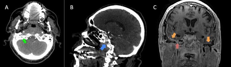
A multi-panel diagnostic imaging study featuring CT and MRI scans of the skull base and temporal bones. Panel A (Axial CT): Shows focal sclerosis of the skull base (green arrow), typical of chronic sequelae from osteomyelitis. Panel B (Sagittal CT): Demonstrates postsurgical changes in the right temporal region (blue arrow) without acute bony destruction. Panel C (Coronal T1-weighted post-contrast MRI): Displays dural thickening and abnormal enhancement (orange arrows) along the bilateral petrous pyramids and internal auditory canals (IAC). A central region of persistent skull base osteomyelitis (red arrow) is identified, extending across the midline between the right and left temporal bones. This series illustrates clinical findings of persistent or recurrent skull base osteomyelitis with associated pachymeningeal involvement, relevant for neuroradiology and infectious disease specialties.
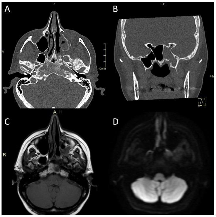
This composite diagnostic image showcases CT and MRI findings in a case of atypical bacterial skull-base osteomyelitis (ASBO). Panel A (axial CT bone algorithm) and Panel B (coronal CT bone algorithm) reveal significant bony alterations at the skull base, characterized by expansion of the left pterygopalatine fossa, opacification of the ipsilateral maxillary and sphenoid sinuses, and mixed erosive and hyperostotic changes. Panel B specifically highlights asymmetric expansion and destruction of the left Vidian canal. Panel C (axial MRI T1-weighted) demonstrates replacement of normal high-signal fatty marrow with low-signal edema in the clivus, while Panel D (axial MRI Diffusion-Weighted Imaging/DWI) shows restricted diffusion (increased signal) involving the left pterygoid base, pterygopalatine fossa, and infratemporal fossa. These multimodal findings illustrate the hallmark features of invasive sinogenic infection and osteomyelitis, emphasizing the use of high-resolution CT for cortical assessment and MRI for soft tissue and marrow involvement.
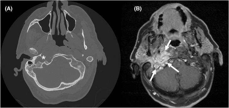
This dual-modality comparison presents axial diagnostic imaging of the head, focusing on the right central skull base. Panel (A) is a non-contrast Computed Tomography (CT) scan in a bone window, clearly demonstrating significant cortical erosion and bony destruction of the right skull base and occipital bone (marked with an asterisk). Panel (B) shows a T1-weighted Dixon Magnetic Resonance Imaging (MRI) sequence at a corresponding anatomical level. The MRI highlights an extensive, hyperintense infiltrating soft tissue mass (indicated by white arrows) involving the right skull base, upper neck, and extending toward the occipital bone and jugular bulb region. The image illustrates the complementary roles of CT for evaluating cortical integrity and MRI for assessing soft tissue infiltration and marrow replacement. Clinically, these findings are characteristic of skull base osteomyelitis (e.g., malignant otitis externa) or aggressive neoplastic processes. Anatomical landmarks visible include the maxillary sinuses, sphenoid bone, and posterior fossa structures.
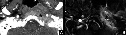
This dual-modality clinical imaging set illustrates skull base osteomyelitis in a 74-year-old male. Image A is an axial contrast-enhanced CT scan demonstrating a destructive mass (marked by an asterisk) at the level of the left skull base. The lesion is characterized by ill-defined, irregular margins and increased density, indicating infiltration between the petrous apex and the temporomandibular joint with associated bone destruction. Image B is a T1-weighted contrast-enhanced MRI with fat saturation at the same anatomical level. The corresponding area (asterisk) shows intense, irregular enhancement, highlighting active inflammation or infection and soft tissue infiltration. The pathological process involves the left jugular foramen, providing a clinical explanation for associated cranial nerve deficits such as hypoglossal nerve palsy and vocal cord paralysis (VCP). This case emphasizes the diagnostic features of aggressive skull base infections, including the loss of normal anatomical fat planes, cortical erosions on CT, and avid enhancement on fat-suppressed MRI sequences.
Skull Base Osteomyelitis (SBO)
Definition and Historical Background
Epidemiology
- Incidence is increasing - from 67 cases (1999-2000) to 421 cases (2012-2013) in UK Hospital Episode Statistics
- Predominantly affects elderly (>60 years) and diabetics (65-100% of cases in various series)
- Male:female ratio 2:1
- Immunosuppressed states are increasingly recognized: HIV/AIDS (often fungal, younger patients, CD4 <50), steroid use, chemotherapy, haematological malignancies (leukaemia, lymphoma), renal transplantation
- HIV patients: younger, higher risk of fungal infection
- Pediatric SBO: rare; associated with IgA deficiency, monocytic leukaemia, neutropenia - Scott-Brown's, Vol. 2
Pathophysiology
- Impaired polymorphonuclear leukocyte function (poor phagocytosis of Pseudomonas)
- Microangiopathy and endarteritis - small vessel obliteration reduces local immune response
- Elevated EAC cerumen pH in diabetics - favours bacterial colonisation
- Breakdown of the cutaneous barrier of the EAC - eczema, seborrhoea, self-induced trauma from pruritus, ear canal irrigation
- CN VII (facial nerve) - most common; close relation to EAC at the stylomastoid foramen
- CN IX, X, XI - jugular foramen involvement
- CN XII (hypoglossal) - further medial spread
- CN VI, V - petrous apex involvement
- CN II - optic nerve, late extension
Bacteriology
| Organism | Notes |
|---|---|
| Pseudomonas aeruginosa | >90% of cases; Gram-negative, obligate aerobe; produces collagenases, elastases, endotoxins, and neurotoxins causing necrotizing vasculitis |
| Staphylococcus aureus/epidermidis | Second-line bacteria |
| Proteus mirabilis | Occasional |
| Klebsiella spp. | Occasional |
| Aspergillus fumigatus | Most common fungal; HIV/AIDS patients (CD4 <50); worse prognosis than Pseudomonas; originates from middle ear/mastoid rather than EAC |
| Other fungi: A. niger, A. flavus, Candida spp., Scedosporium apiospermum | Rare |
Clinical Features
Symptoms
- Severe, deep-seated, nocturnal otalgia - disproportionate to examination findings; resistant to analgesics (prevalence 75-100%). This is the most common presenting symptom.
- Unilateral purulent otorrhoea - second most common
- Conductive hearing loss - from EAC oedema/granulation
- History of ear canal trauma (often after wax irrigation) is common
Signs
- Granulation tissue at the bony-cartilaginous junction (isthmus of EAC) - the cardinal hallmark sign; should never be underestimated
- Oedematous, exquisitely tender ear canal
- Exposed bone may be visible at the floor of the canal
- Tympanic membrane often intact initially; may be obscured by polyp or granuloma
- HIV patients often lack granulation tissue - making diagnosis harder
Cranial Nerve Involvement (in order of progression)
- CN VII (facial nerve palsy) - up to 25% of patients; implies infection encasing the extratemporal portion or involving the stylomastoid foramen
- CN IX, X, XI - jugular foramen involvement (glossopharyngeal, vagus, accessory)
- CN XII - hypoglossal palsy, vocal cord paralysis
- CN VI, V - petrous apex spread
- Multiple cranial neuropathies = worse prognosis
Rare/Late Features
- Septic thrombophlebitis of sigmoid sinus - "picket fence" pattern of fevers
- Meningitis, cerebral abscess - late, poor prognosis
- Parotitis, trismus - masseter myositis and TMJ involvement
- Children: facial nerve palsies occur earlier (more medial fissures of Santorini, undeveloped mastoid)
Diagnosis
Laboratory
- ESR - elevated, non-specific; useful for monitoring treatment response and recurrence
- CRP - elevated; use to monitor treatment response
- WBC and fever may be absent (do not exclude diagnosis)
- Blood glucose - check and optimise
Biopsy
- Mandatory - granulation tissue must be biopsied to rule out malignancy, histiocytosis, tuberculosis, and for culture
- Silver staining on histology identifies fungal pathogens
- Histology shows inflammation without neoplasia
Microbiology
- EAC debris: culture and stain for bacteria, fungi, acid-fast bacilli
- If tympanic membrane intact and topical antibiotics have failed: tympanotomy/tympanocentesis for culture
Imaging
CT Scan
- Shows bony erosion and reduced density at skull base
- Only detects bony demineralisation >30% - limited early sensitivity; demineralisation may take months to appear
- A negative CT does not exclude SBO
- Contrast-CT: evaluates extratemporal soft tissue extent, abscesses, mastoid/TMJ involvement, petrous apex and carotid canal involvement
- Cannot reliably assess treatment response or distinguish neoplasia from inflammation
- Contrast not necessary unless abscess is suspected
MRI
- Superior for soft tissue and dural involvement; detects bone marrow oedema
- Better than CT for evaluating lateral sinus blood flow and intracranial extension
- Cannot reliably assess treatment response - may remain abnormal months after successful treatment
- Combine with MR angiography to evaluate dural sinus patency
Nuclear Medicine (key to monitoring)
| Scan | Role | Notes |
|---|---|---|
| Tc-99m MDP bone scan | Diagnosis - near 100% sensitivity | Detects osteoblastic activity early (before CT); cannot monitor response (stays positive long after cure) |
| Gallium-67 citrate | Monitor treatment response | Normalizes as infection resolves; repeat every 4 weeks until negative; the gold standard for assessing treatment response |
| Indium-111 labelled leucocytes | Detects neutrophil-mediated inflammation | SPECT version improves anatomical localisation |
| FDG-PET | Most accurate single modality | Better than individual scans but not significantly better than Tc-99 + leucocyte combination; often unavailable |
Treatment
1. Aural Toilet
- Regular microsuction/debridement of the EAC - controls granulations and pain
- Controversy exists over concurrent topical antimicrobials (may hinder culture results)
2. Glycaemic Control
- Aggressive optimisation of blood glucose is as important as antibiotics in diabetic patients
- Poor glycaemic control makes SBO harder to treat and maintain in remission
3. Systemic Antibiotics (Mainstay of Treatment)
- Duration: Minimum 6-8 weeks; may extend to months in advanced cases
- Ideally continue until Gallium-67 scan is negative
- First line: Oral ciprofloxacin 750 mg twice daily (monotherapy; excellent antipseudomonal activity, good bone penetration, high oral bioavailability)
- Some experts advocate initial parenteral therapy, switching to oral when ESR/CRP begin to fall - no conclusive evidence either way
- Rifampicin 600 mg twice daily has been suggested as an addition - no direct evidence of added benefit
For Fluoroquinolone-Resistant Pseudomonas:
- Antipseudomonal cephalosporins: ceftazidime, cefepime
- Antipseudomonal penicillins: ticarcillin-clavulanate, piperacillin-tazobactam
- Aminoglycosides (in combination)
- Double coverage against Pseudomonas is started empirically after cultures
For Fungal SBO:
- Amphotericin B - antifungal of choice
- Can transition to oral itraconazole after successful amphotericin course
4. Hyperbaric Oxygen (HBO)
- Proposed mechanism: increases tissue pO2, relieves hypoxia, enhances oxidative killing of microbes
- A Cochrane review found no evidence from RCTs to support its use; remains adjunctive/investigational
5. Surgery
- Role is now very limited compared to the era when radical debridement was routine
- Indications:
- Obtaining specimens for culture and histology
- Local debridement of granulation tissue
- Drainage of associated abscesses
- Removal of dead sequestra
- Facial nerve decompression is NOT indicated - infection involves the extratemporal portion of the nerve, not the intratemporal canal, so decompression fails to address the actual site of involvement
Prognosis / Poor Prognostic Factors
- Cranial neuropathies (especially multiple)
- EAC granulations (paradoxically, their presence correlates with severity)
- Bilateral symptoms
- Positive Aspergillus cultures
- Meningitis, sigmoid sinus thrombosis, cerebral abscess - late complications with poor prognosis
Imaging Gallery
Recent Literature (2023-2025)
| PMID | Title | Year |
|---|---|---|
| 40351881 | Current Evidence in the Management of Central Skull Base Osteomyelitis: A Systematic Review - J Neurol Surg B Skull Base | 2025 |
| 37479637 | Skull Base Osteomyelitis: Historical Perspective, Diagnosis and Management Update - Otolaryngol Clin North Am | 2023 |
| 37235302 | Atypical Skull-Base Osteomyelitis: Comprehensive Review and Multidisciplinary Management Viewpoints - Trop Med Infect Dis | 2023 |
| 36382775 | Skull Base Infections, Their Complications, and Management - Neuroradiol J | 2024 |
Grades of skull base osteomyelitis
"skull base osteomyelitis" AND staging AND classification
PMID: 33640201
PMID: 35832975
Chandler staging classification skull base osteomyelitis malignant otitis externa grades stages
https://drsanu.com/articles/malignant-otits-externa-clinical…
Grades / Staging of Skull Base Osteomyelitis
1. Chandler's Staging System (1977) - Classic / Most Widely Used
| Stage | Features |
|---|---|
| Stage I | Clinical evidence of SBO/MOE with infection of soft tissues beyond the EAC. Technetium-99 bone scan negative. |
| Stage II | Soft tissue infection beyond the EAC. Technetium-99 bone scan positive (confirms bony involvement). |
| Stage III | Stage II features + Cranial nerve palsy - IIIa: Single cranial nerve palsy - IIIb: Multiple cranial nerve palsies |
| Stage IV | Intracranial complications: meningitis, empyema, sigmoid/dural sinus thrombosis, brain abscess. |
- Progression from I → IV represents increasing severity and worsening prognosis
- The Tc-99 bone scan is the pivotal investigation that separates Stage I from Stage II
- Facial nerve (CN VII) is the most commonly affected nerve in Stage III (up to 25-60% of cases)
- Stage IV carries the worst prognosis; intracranial extension historically associated with very high mortality
- Stage IIIb (multiple cranial nerve palsies) and Stage IV are considered advanced disease
2. Chandler's Original Grading (1968) - Historical
3. Lee et al. Prognosis-Based Classification (2008)
- Presence/absence of cranial nerve involvement
- Bacterial vs. fungal aetiology (fungal = worse prognosis)
- Extent of bony involvement on imaging
- Response to initial therapy
4. Takahashi et al. Radiological Staging System (2022) - HRCT/MRI Based
| Stage | Radiological Extent |
|---|---|
| Stage 1 | Lesions limited to the temporal bone |
| Stage 2 | Extension beyond temporal bone to less than half of the skull base |
| Stage 3 | Extension exceeding the midline |
| Stage 4 | Extension involving the whole of the clivus |
5. Özer et al. Clinicoradiological Grading System (2021)
| Clinical Grade (CG) | Features |
|---|---|
| CG1 | Radiological involvement without cranial nerve dysfunction |
| CG2 | Radiological involvement + single cranial nerve palsy |
| CG3 | Radiological involvement + multiple cranial nerve palsies |
- Regression rate (disease control) decreased from CG1 → CG3 (p = 0.029)
- Duration of hospitalization increased with higher CG (p = 0.047)
- 5-year overall survival: 45.8% overall; CG significantly correlated with overall survival (p = 0.017)
- Surgery had no significant effect on regression at discharge (p = 0.41)
- 1-year survival 82.2%, 2-year 70.8%, 5-year 45.8%
Summary Comparison
| System | Year | Basis | Key Feature |
|---|---|---|---|
| Chandler | 1977 | Clinical + Tc-99 bone scan | Most widely used; 4 stages; exam standard |
| Lee et al. | 2008 | Clinical + microbiological + prognosis | Emphasises outcome prediction |
| Takahashi et al. | 2022 | HRCT/MRI anatomical extent | Predicts infection control and mortality |
| Özer et al. CGS | 2021 | MRI scoring + CN dysfunction | Integrated clinicoradiological; survival correlation |
Important Prognostic Notes
- CN VII palsy alone - earlier literature suggested poor prognosis; more recent data (Corey, Soudry, Mani) suggests facial nerve palsy alone does not necessarily worsen prognosis
- Multiple cranial nerve palsies (IIIb) - consistently associated with worse outcomes across all systems
- Positive Aspergillus cultures - worse prognosis than Pseudomonas in all grading contexts
- Intracranial extension (Stage IV / Stage 3-4 Takahashi) - highest mortality; associated with sigmoid sinus thrombosis, meningitis, brain abscess
Meliodosis
"melioidosis"[MeSH Terms] AND management
melioidosis Burkholderia pseudomallei clinical features diagnosis treatment 2024 guidelines
melioidosis Burkholderia pseudomallei clinical skin abscess chest X-ray
https://digitallibrary.health.nt.gov.au/nthealthserver/api/c…
Melioidosis
Definition and Causative Organism
Epidemiology
- Southeast Asia (northeastern Thailand is hyperendemic), Philippines, Indonesia, Malaysia, Singapore
- Northern Australia (Darwin region is hyperendemic - the 30-year Darwin Prospective Melioidosis Study has been landmark)
- South Asia (India), southern China, Hong Kong, Taiwan
- Increasingly recognized in: Africa, Middle East, Central and South America
- Gulf Coast of the USA (B. pseudomallei found in environment; local cases reported in Texas)
- Contaminated imported products (e.g., aromatherapy spray from India) have caused outbreaks in non-endemic areas
- Diabetes mellitus - the single most common risk factor (present in majority of cases)
- Alcohol use disorder
- Chronic kidney disease / renal failure
- Chronic lung disease (COPD, bronchiectasis, cystic fibrosis in endemic areas)
- Heart disease, smoking
- HIV/AIDS and other immunosuppressive states
- Military personnel and travelers to endemic areas
Microbiology / Organism Characteristics
| Feature | Detail |
|---|---|
| Gram stain | Gram-negative bacillus with bipolar ("safety-pin") staining |
| Motility | Motile (peritrichous flagella) |
| Oxidase | Oxidase positive |
| Growth | Grows readily on routine culture media (MacConkey, blood agar); produces wrinkled colonies |
| Selective media | Ashdown's medium (contains gentamicin + crystal violet) - preferred for isolation from non-sterile sites |
| Virulence mechanism | Type III secretion system - inhibits autophagy in host cells; subverts phagolysosomal maturation in macrophages; causes cell lysis via caspase-1-dependent pyroptosis; produces endotoxins |
| Special feature | Can establish latency with reactivation years to decades later (Vietnam veterans relapsed years after return) |
| Bioterrorism | Listed as a potential bioterrorism agent (Category B) |
Routes of Infection
- Percutaneous inoculation - direct contact of abraded/lacerated skin with contaminated soil or water (most common)
- Inhalation of aerosolized bacteria (especially during heavy rain/typhoons; probable during storms)
- Ingestion of contaminated water or food
- Sexual intercourse (rare, documented)
- Laboratory acquisition - biosafety concern
Clinical Spectrum
1. Asymptomatic / Subclinical Infection
- Demonstrated by seroprevalence studies in endemic areas
- Most common in healthy individuals
2. Acute Localized Infection
- Skin ulcers, cellulitis, subcutaneous abscesses
- Genitourinary infection (prostatic abscesses - particularly common in men in endemic areas; important clue)
- Lymphadenitis
- Septic arthritis, osteomyelitis
- Parotid abscesses (characteristic in children in Thailand)
3. Pulmonary Melioidosis (Most Common Form - ~50% of cases)
- Fever, productive cough, dyspnea
- Hemoptysis and pleuritic chest pain may be prominent
- Can present as rapid respiratory failure with signs of sepsis
- CXR: consolidation, often upper lobe involvement (~50%), multilobe in ~1/3
- Can progress to bacteraemia (63% of primary pneumonia cases) and septic shock (33%)
- Symptoms >2 months
- Fever, weight loss, chronic productive cough ± hemoptysis
- CXR: upper lobe cavitary disease mimicking tuberculosis
- Diffuse miliary nodules that can expand and cavitate
4. Septicaemic Melioidosis
- Most severe form
- Fever, prostration, signs of sepsis/septic shock
- Metastatic abscesses form in multiple organs: liver, spleen, kidneys, lungs, brain
- "Miliary" pattern of abscesses on imaging
- Over half of septicaemic patients have positive blood cultures
- Mortality 20-50% overall; up to 50-90% in untreated septicaemia (reduced to ~20% with aggressive ICU care)
5. Neurological Melioidosis
- Encephalomyelitis - described in the neurology literature (Adams and Victor)
- Brainstem encephalitis (particularly in northern Australia)
- Flaccid paralysis, fever, cranial nerve involvement
- Rare but severe manifestation
6. Chronic / Disseminated Melioidosis
- Characterized by abscesses and granulomas at multiple sites
- Can mimic tuberculosis, disseminated fungal infections, sarcoidosis
- Cutaneous manifestations: cellulitis, subcutaneous abscesses, granulomatous lesions, ecthyma gangrenosum, purpura, pustules, Sweet syndrome, urticaria, necrotizing fasciitis
Diagnosis
Gold Standard: Culture
- Culture of B. pseudomallei from any clinical specimen confirms diagnosis
- Specimens: blood, sputum, urine, pus/abscess aspirate, wound swab
- >50% of patients have positive blood cultures in septicaemia
- Grows readily on routine media; Ashdown's selective medium used for non-sterile sites
- Sensitivity of culture: as low as 60% overall
Microbiology Clues
- Gram stain: Gram-negative bacillus, bipolar ("safety-pin") staining
- Wrinkled, cream-coloured colonies; metallic odour
Molecular / PCR
- Multiplex PCR-based assays: more sensitive and species-specific than culture - increasingly used
- Useful when antibiotics already started
Serology
- Complement-fixing and agglutinating antibodies appear within 4-6 weeks of infection
- IHA (indirect haemagglutination assay) - used widely in endemic areas
- Not adequate alone in endemic regions (high background seropositivity); useful in non-endemic areas
- A 4-fold rise in titre is significant
Imaging
- CT chest: consolidation, upper lobe cavitation, miliary nodules
- CT abdomen/pelvis: hepatic, splenic, renal, or prostatic abscesses
- CT/MRI brain: if neurological involvement suspected
Key Points for Diagnosis
- Requires high index of suspicion - mimics many other conditions (TB, pneumonia, sepsis)
- Ask about travel to or residence in endemic areas
- Culture laboratories must be alerted (biosafety risk - BSL-3 pathogen)
Treatment
Phase 1: Intensive Phase (IV Antibiotics)
| Drug | Dose | Notes |
|---|---|---|
| Ceftazidime (first line, ward) | 2 g IV every 6 hours (50 mg/kg up to 2 g in children) | Standard intensive therapy; can use 24-hour continuous infusion (6g/24h) for HITH/outpatient |
| Meropenem (first line, ICU) | 1 g IV every 8 hours (25 mg/kg up to 1 g in children) | Preferred for ICU patients, septic shock, neurological melioidosis, persistent bacteraemia, CNS disease |
| Meropenem 2g over 3 hours | For septic shock + augmented renal clearance (CrCl >130 mL/min), double dose | Extended infusion to optimise PK/PD targets |
| Imipenem | 1 g IV every 6 hours | Alternative carbapenem |
| Focus / Presentation | Minimum IV Duration |
|---|---|
| Skin abscess | 2 weeks |
| Bacteraemia (no focus) | 2 weeks |
| Unilobar pneumonia, no lymphadenopathy, no ICU, negative blood cultures | 2 weeks |
| Multilobar pneumonia OR positive blood cultures | 3 weeks |
| Pneumonia + lymphadenopathy or ICU admission | 3+ weeks |
| Deep-seated infections: neurological, osteomyelitis, septic arthritis, prostatic abscess | 4-8 weeks or longer |
- Clinical deterioration on ceftazidime
- Persistent bacteraemia
- CNS involvement develops
- ICU admission required
Phase 2: Eradication Phase (Oral Antibiotics)
| Drug | Dose | Duration |
|---|---|---|
| TMP-SMX (trimethoprim + sulfamethoxazole) - first line | Adult 40-60 kg: 240+1200 mg; >60 kg: 320+1600 mg orally (children: 6+30 mg/kg up to 240+1200 mg) | Minimum 3 months for most presentations |
| Amoxicillin-clavulanate | 875/125 mg twice daily | Alternative if TMP-SMX not tolerated; less preferred |
| Doxycycline | Used in some regimens | Less favoured; higher relapse rate |
- Most presentations: 3 months
- Deep-seated/disseminated disease: may extend to 6-12 months
- Goal: prevent relapse (relapse rate ~10% without adequate eradication)
Drugs B. pseudomallei is intrinsically resistant to:
- First/second-generation penicillins (ampicillin, amoxicillin alone)
- First/second-generation cephalosporins
- Fluoroquinolones (ciprofloxacin, levofloxacin) - NOT effective empirically
- Macrolides
- Aminoglycosides
- Polymyxins
- Chloramphenicol (active but toxicity limits use in developed countries)
Complications and Prognosis
| Complication | Notes |
|---|---|
| Septic shock | High mortality without aggressive management |
| Multi-organ failure | Liver, kidneys, lung |
| Metastatic abscess formation | Liver, spleen, prostate, brain, bone |
| Neurological melioidosis | Brainstem encephalitis, flaccid paralysis |
| Relapse | ~10% rate; can occur after decades of latency |
- Overall: 10-40% depending on presentation and setting
- Septicaemic form: 20-50% (up to 90% untreated)
- Indonesia (2024): mortality rate ~41%
- With aggressive ICU management: reduced to ~20%
- CNS involvement: poorest prognosis
- Septicaemic presentation
- CNS involvement
- Deep-seated abscesses
- Delayed diagnosis
- Immunosuppression / poorly controlled diabetes
- Absence of treatment in intensive phase with appropriate antibiotics
Special Considerations
Latency and Reactivation
- B. pseudomallei can remain latent in the body for decades
- US Vietnam War veterans developed melioidosis years after return ("Vietnamese time bomb")
- Reactivation can be triggered by immunosuppression, illness, or trauma
Bioterrorism
- B. pseudomallei (and B. mallei causing glanders) are listed as potential bioterrorism agents
- Biosafety Level 3 (BSL-3) pathogen - laboratory workers need protection
- Vaccine candidates in Phase 1 clinical trials (no licensed vaccine yet as of 2025-2026)
Paediatric Melioidosis
- Parotid gland abscesses are a characteristic presentation in children in Thailand
- Generally better prognosis than adults
The Darwin 30-Year Prospective Study
- The landmark natural history study (Currie et al., Lancet Infect Dis 2021) - 30 years of prospective data from Darwin, Australia, providing the most robust treatment and outcome data available
Recent Evidence (2022-2026)
| PMID | Study | Year |
|---|---|---|
| 37307278 | Systematic review + network meta-analysis: Drug treatment for severe melioidosis and eradication therapy (PLoS Negl Trop Dis) | 2023 |
| 41571490 | Treatment of Melioidosis - review (Infect Dis Clin North Am) | 2026 |
| 35942848 | Progress in epidemiology, diagnosis, treatment and vaccination - Currie BJ (Curr Opin Infect Dis) | 2022 |