The database didn't return veterinary-specific text results, so I'll provide a comprehensive answer entirely from veterinary ophthalmology knowledge (Slatter's Fundamentals of Veterinary Ophthalmology, Gelatt's Veterinary Ophthalmology, Kirk's Current Veterinary Therapy, and standard ACVO references).
Persistent Pupillary Membrane (PPM) in Veterinary Medicine
1. What Is PPM?
Persistent Pupillary Membrane (PPM) is a congenital ocular anomaly in which remnants of the embryonic pupillary membrane (a fetal vascular structure) fail to regress normally after birth. These strands appear as iris-to-iris, iris-to-lens, or iris-to-cornea tissue bridges crossing the pupil.
Embryology Recap:
During fetal development, the lens and anterior chamber are covered by a vascular membrane called the tunica vasculosa lentis (TVL), which includes the pupillary membrane anteriorly. This membrane supplies the developing lens with nutrients. In most species, this membrane completely regresses before or shortly after birth. When regression is incomplete, the remnants = PPM.
2. Species Affected
| Species | Prevalence | Notes |
|---|
| Dogs | Very common | Basenji most studied; also common in Chow Chow, Mastiff, Pembroke Welsh Corgi |
| Cats | Less common than dogs | Usually mild; often incidental finding |
| Horses | Occasionally reported | Can cause significant vision impairment |
| Cattle, small ruminants | Rare | |
3. Classification of PPM
PPM is classified by where the strands attach:
Type 1 – Iris-to-Iris (Iris-Iris)
- Strands run from one part of the iris to another part of the iris
- Most common and mildest form
- Usually do NOT affect vision
- No lens or corneal involvement
- Appear as thin, thread-like bands crossing the pupil
Type 2 – Iris-to-Lens (Iris-Lenticular)
- Strands run from the iris to the anterior lens capsule
- Can cause focal anterior lens opacity (cataract) at attachment point
- The area where PPM attaches to the lens may develop into a "rosette" or "pie-crust" cataract
- Potentially vision-threatening if large or centrally located
- Clinically significant
Type 3 – Iris-to-Cornea (Iris-Corneal)
- Strands run from the iris to the posterior corneal surface
- Can cause focal corneal opacity (leukoma) at the attachment site
- May impair vision if in the visual axis
- Most clinically significant type
Type 4 – Sheets / Plaques
- Large, sheet-like membranes rather than individual strands
- Can significantly obstruct the visual axis
- Rare but severe
4. Clinical Signs & Presentation
Visual Appearance
- Fine, brownish or pigmented filaments or strands crossing the pupil
- May look like a spider web across the pupil
- Originate from the iris collarette (not the pupillary margin — this differentiates them from synechiae)
- May be single or multiple strands
- Present in one or both eyes (bilateral is common, especially in dogs)
Symptoms Depending on Type
| Type | Symptoms |
|---|
| Iris-Iris | Usually asymptomatic; found incidentally |
| Iris-Lens | Possible vision impairment, squinting (blepharospasm), photosensitivity if cataract develops |
| Iris-Cornea | Corneal cloudiness/white spot, vision impairment, possible secondary complications |
| Sheet PPM | Obvious visual obstruction, possible amblyopia if present from birth |
5. Diagnosis
A. Ophthalmic Examination
- Pupil dilation (mydriasis) using:
- Tropicamide 1% (drug of choice) — dilates pupil to reveal full extent of PPM strands
- Allows complete visualization of strand insertion points
- Slit-lamp biomicroscopy — most accurate tool to trace strand origin and insertion
- Retroillumination technique — backlight through the lens, PPM strands appear as dark silhouettes against the tapetal reflex
B. Differentiation from Other Conditions
| Feature | PPM | Posterior Synechia |
|---|
| Origin | Iris collarette | Iris pupillary margin |
| Cause | Congenital | Acquired (uveitis) |
| Attachment | Iris/lens/cornea | Lens only |
| Age of onset | Present from birth | Develops after inflammatory episode |
| Treatment urgency | Rarely urgent | Urgent if causing glaucoma |
C. Electroretinogram (ERG)
- Used if vision impairment is suspected to evaluate retinal function behind any opacity
6. Genetics & Breeding Implications
- In Basenjis, PPM is autosomal dominant with incomplete penetrance — extensively studied by OFA (Orthopedic Foundation for Animals) and CERF (Canine Eye Registry Foundation, now part of OFA-CAER)
- Affected dogs should NOT be used for breeding in lines where PPM severity is high
- Iris-to-iris forms in Basenjis are graded on a severity scale (Grade 1–5):
- Grade 1–2: Minor, non-vision-threatening
- Grade 3–5: Significant, breeding discouraged
7. Treatment
A. No Treatment Required (Most Cases)
- Iris-to-iris PPM without vision impact = monitor only
- Many animals live normal lives with no intervention
- Re-examine annually (especially young animals, as strands sometimes regress in the first few months of life)
B. Medical Management
Used when there is secondary corneal or lenticular involvement:
| Drug | Purpose | Dose (Approximate) |
|---|
| Tropicamide 1% ophthalmic drops | Maintain pupil dilation to prevent synechiae formation, reduce light sensitivity | 1 drop BID-TID |
| Cyclosporine A 0.2% ophthalmic ointment | If keratitis/corneal opacity secondary to iris-cornea contact | Applied BID |
| Topical corticosteroids (e.g., prednisolone acetate 1%) | Reduce secondary inflammation | Only if no corneal ulceration; BID-QID |
| Artificial tear lubricants | Supportive for corneal health | BID-QID as needed |
C. Surgical Management
Indicated when PPM causes significant vision impairment or non-regressing corneal/lens opacity:
- Surgical membrane dissection / membranectomy
- PPM strands are carefully cut and removed under general anesthesia
- Performed with fine iris scissors or vitrectomy instruments
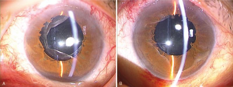
- Post-op image (from database) shows the result after bilateral PPM resection — clear visual axis achieved:
Post-op slit-lamp image showing successful bilateral PPM resection with clear, unobstructed pupillary zones. Note minor iris margin irregularities at former attachment points — this is expected and normal post-surgically.
-
Phacoemulsification (cataract surgery)
- If iris-to-lens PPM has caused a significant cataract affecting vision
- Combined with intraocular lens (IOL) implantation
- Performed by a veterinary ophthalmologist (DACVO)
-
Nd:YAG laser treatment
- Laser photodisruption of PPM strands — used in human medicine, experimental in veterinary patients
- Limited availability; requires specialized equipment
8. Prognosis
| Type | Prognosis |
|---|
| Iris-Iris | Excellent — virtually no vision impact, no treatment needed |
| Iris-Lens (minor) | Good — small focal cataracts, stable over time in most cases |
| Iris-Lens (major) | Guarded — may progress; monitor for cataract maturation |
| Iris-Cornea | Fair to Good — depends on location; corneal leukoma may be permanent |
| Sheet PPM | Guarded — may need surgery; risk of amblyopia if vision axis obstructed early in life |
9. Examples to Illustrate
Example 1 — Basenji Dog, 8 Weeks Old
Presentation: Breeder brings a Basenji puppy for eye exam before sale. On retroillumination, 2 fine pigmented strands are visible crossing the pupil from the 2 o'clock to 8 o'clock position on the iris collarette. No corneal or lens involvement.
Diagnosis: PPM Type 1, Iris-Iris, Grade 2
Action: No treatment. Annual CAER exam. Inform breeder — puppy is visually functional but should be flagged in breeding records. Strands may partially regress in the next 4–6 weeks.
Example 2 — Domestic Shorthair Cat, 4 Months Old
Presentation: Owner notices a "white foggy spot" on the cat's eye. On slit-lamp exam after tropicamide dilation, a single PPM strand is found running from the iris collarette to the anterior lens capsule, with a focal 2mm anterior polar cataract at attachment.
Diagnosis: PPM Type 2, Iris-Lens, with focal anterior polar cataract
Action: Monitor cataract size every 3–6 months. Topical tropicamide BID to maintain dilation and prevent further adhesion. Refer to veterinary ophthalmologist if cataract expands. Prognosis good if stable.
10. What to Tell the Owner (Owner Communication Guide)
"Your pet has been diagnosed with Persistent Pupillary Membrane, or PPM. Let me explain what this means for your animal."
What is PPM?
"During development before birth, the eye has a temporary 'covering' over the pupil that supplies blood to the growing lens. Normally, this covering disappears before or shortly after birth. In your pet, small strands of this covering remained behind — these are called Persistent Pupillary Membranes."
Is it serious?
"In most cases, no. The most common form — where the strands connect only to the iris itself — does not affect vision at all and requires no treatment. It is simply something we monitor once a year."
"However, if the strands are attached to the clear outer surface of the eye (cornea) or to the lens, they can sometimes cause cloudiness. This is less common but may need treatment or referral to a specialist."
Will my pet go blind?
"For the vast majority of animals with PPM, the answer is no. Most live completely normal, full-quality lives with no vision problems. Only the rare, severe forms involving large membranes or significant cataracts could potentially affect vision, and even those can often be managed or surgically treated."
Is this genetic? Can my dog/cat pass it on?
"PPM has a hereditary component in certain breeds, especially Basenjis. If you plan to breed your pet, I would recommend having a formal eye certification done through OFA-CAER to document the findings and inform breeding decisions."
What do I watch for at home?
"Watch for any cloudiness or white spots developing in the eye, squinting, excessive tearing, or if your pet seems to be bumping into things. If you notice any of these, contact us promptly."
Follow-up?
"I recommend an eye check every 6–12 months to make sure things remain stable. No special care is needed at home for now."
Summary Table
| Feature | Details |
|---|
| Definition | Failure of fetal pupillary membrane to regress |
| Species most affected | Dogs > Cats > Horses |
| Types | Iris-Iris / Iris-Lens / Iris-Cornea / Sheet |
| Most common type | Iris-Iris (mildest, best prognosis) |
| Diagnosis tool | Tropicamide dilation + slit-lamp / retroillumination |
| Key DDx | Posterior synechia (originates from pupillary margin, acquired) |
| Treatment | Monitor (iris-iris), topical meds, surgery (severe cases) |
| Hereditary? | Yes — dominant trait in Basenjis; OFA-CAER certification recommended |
| Prognosis | Excellent for iris-iris; guarded for sheet/severe iris-cornea types |
References: Gelatt KN — Veterinary Ophthalmology (5th ed.); Slatter D — Fundamentals of Veterinary Ophthalmology (4th ed.); OFA-CAER Breed-Specific Eye Disease Guidelines; ACVO Genetics Committee Reports on PPM in Basenjis.