
I am studying bsc operation theatre and anaesthesia technology 2nd I need notes you give notes based on dr mgr medical University
Which 2nd year BSc OTAT subject do you need notes for? · Do you have a specific topic/unit within that subject, or do you want a general overview of the whole subject?
Explain in detail about sterilization 10 Mark's
Sterilization - 10 Mark Answer
Definition
Classification of Sterilization Methods
A. Physical Methods
- Flaming: direct exposure to flame (e.g., wire loops, mouths of culture tubes) - instant but limited to metal items.
- Incineration: burning to ashes - used for disposal of contaminated dressings, pathological waste, and syringes.
- Hot Air Oven: 160°C for 1-2 hours; used for glassware, oils, powders, and metal instruments that can withstand high dry heat. Dry heat requires longer exposure than moist heat because dry protein is more heat-resistant.
- Below 100°C: Pasteurization (used for milk/fluids, not a sterilization method as spores survive).
- At 100°C: Boiling for 10-30 minutes - kills vegetative bacteria and most viruses but not all spores.
- Above 100°C - Autoclaving: the gold standard for OT sterilization (details below).
- Ultraviolet (UV) rays: used for air and surface disinfection in OT, laminar flow units; poor penetration power.
- Ionizing radiation (gamma rays): used industrially to sterilize pre-packaged disposable items like syringes, catheters, and sutures.
B. Chemical Methods
- Ethylene oxide (ETO) gas: sterilizes heat-sensitive items (plastic tubing, catheters, electronic anaesthesia equipment); requires 4 hours exposure plus 12 hours aeration to remove toxic residue - flammable and carcinogenic, so used only when alternatives are unsuitable.
- Glutaraldehyde (2%): used for endoscopes, laryngoscopes, and anaesthesia breathing circuits; achieves high-level disinfection/sterilization with 6-10 hours immersion.
- Hydrogen peroxide plasma: vaporized H2O2 with radiofrequency energy generates free radicals; efficient, no toxic residue, has largely replaced ETO for heat-sensitive instruments.
- Formaldehyde vapor: used for fumigation of OT rooms.
- Peracetic acid: oxidizing agent, breaks down to nontoxic acetic acid and oxygen.
C. Filtration
The Autoclave (Key Method for OT)
- Mechanical indicators: pressure gauge, temperature chart recorder.
- Chemical indicators: autoclave tape/Bowie-Dick test strips that change color when correct temperature is reached.
- Biological indicators: spore strips (Geobacillus stearothermophilus) placed in test packs - considered the most reliable proof of sterility since they directly test the ability to kill the most resistant organism.
Sterilization vs Disinfection vs Antisepsis (differentiation, often asked together)
| Term | Meaning | Example |
|---|---|---|
| Sterilization | Complete kill of all microbes including spores | Autoclaving instruments |
| Disinfection | Kills most pathogens, not necessarily spores; applied to inanimate objects | Sodium hypochlorite for floors |
| Antisepsis | Chemical agents applied to living tissue to inhibit growth | Povidone-iodine skin prep |
Importance in Anaesthesia and OT Technology
- Prevents surgical site infections and cross-transmission between patients via reusable equipment (laryngoscope blades, breathing circuits, endotracheal tube stylets).
- Ensures safety of invasive procedures (central lines, spinal/epidural needles, surgical instruments).
- Selection of method depends on the material: metal instruments -> autoclave; heat-sensitive plastics/optics -> ETO or H2O2 plasma or glutaraldehyde; disposable single-use items -> gamma irradiation.
Generate a study guide PDF on sterilization for your exams
- Definitions & Basic Concepts - sterilization vs disinfection vs antisepsis, spectrum of activity, D-value/kill curves
- Physical Methods - Dry Heat - flaming, incineration, hot air oven (temp/time, uses, limitations)
- Physical Methods - Moist Heat - boiling, pasteurization, and autoclaving (principle, cycles, pressure-temp-time relationships)
- Radiation Sterilization - UV rays (OT air disinfection) and ionizing/gamma radiation (industrial, disposables)
- Chemical Sterilization - ethylene oxide, glutaraldehyde, hydrogen peroxide plasma, formaldehyde, peracetic acid (mechanism, uses, hazards)
- Filtration - HEPA filters, membrane filtration for heat-labile liquids and OT air
- Autoclave Deep-Dive - construction, working principle, standard cycles, types (gravity displacement vs pre-vacuum), troubleshooting
- Sterility Assurance & Quality Control - chemical indicators, biological indicators (spore strips), Bowie-Dick test, autoclave validation
- Applications in OT/Anaesthesia Practice - instrument-specific protocols (linen, gloves, scopes, anaesthesia circuits, sharps), infection control policies
Which part of the Sterilization study guide should I develop in full detail first for your PDF?
Definition
Classification of Sterilization Methods
A. Physical Methods
- Flaming: direct exposure to flame (e.g., wire loops, mouths of culture tubes) - instant but limited to metal items.
- Incineration: burning to ashes - used for disposal of contaminated dressings, pathological waste, and syringes.
- Hot Air Oven: 160°C for 1-2 hours; used for glassware, oils, powders, and metal instruments that can withstand high dry heat. Dry heat requires longer exposure than moist heat because dry protein is more heat-resistant.
- Below 100°C: Pasteurization (used for milk/fluids, not a sterilization method as spores survive).
- At 100°C: Boiling for 10-30 minutes - kills vegetative bacteria and most viruses but not all spores.
- Above 100°C - Autoclaving: the gold standard for OT sterilization (details below).
- Ultraviolet (UV) rays: used for air and surface disinfection in OT, laminar flow units; poor penetration power.
- Ionizing radiation (gamma rays): used industrially to sterilize pre-packaged disposable items like syringes, catheters, and sutures.
B. Chemical Methods
- Ethylene oxide (ETO) gas: sterilizes heat-sensitive items (plastic tubing, catheters, electronic anaesthesia equipment); requires 4 hours exposure plus 12 hours aeration to remove toxic residue - flammable and carcinogenic, so used only when alternatives are unsuitable.
- Glutaraldehyde (2%): used for endoscopes, laryngoscopes, and anaesthesia breathing circuits; achieves high-level disinfection/sterilization with 6-10 hours immersion.
- Hydrogen peroxide plasma: vaporized H2O2 with radiofrequency energy generates free radicals; efficient, no toxic residue, has largely replaced ETO for heat-sensitive instruments.
- Formaldehyde vapor: used for fumigation of OT rooms.
- Peracetic acid: oxidizing agent, breaks down to nontoxic acetic acid and oxygen.
C. Filtration
The Autoclave (Key Method for OT)
- Mechanical indicators: pressure gauge, temperature chart recorder.
- Chemical indicators: autoclave tape/Bowie-Dick test strips that change color when correct temperature is reached.
- Biological indicators: spore strips (Geobacillus stearothermophilus) placed in test packs - considered the most reliable proof of sterility since they directly test the ability to kill the most resistant organism.
Sterilization vs Disinfection vs Antisepsis (differentiation, often asked together)
| Term | Meaning | Example |
|---|---|---|
| Sterilization | Complete kill of all microbes including spores | Autoclaving instruments |
| Disinfection | Kills most pathogens, not necessarily spores; applied to inanimate objects | Sodium hypochlorite for floors |
| Antisepsis | Chemical agents applied to living tissue to inhibit growth | Povidone-iodine skin prep |
Importance in Anaesthesia and OT Technology
- Prevents surgical site infections and cross-transmission between patients via reusable equipment (laryngoscope blades, breathing circuits, endotracheal tube stylets).
- Ensures safety of invasive procedures (central lines, spinal/epidural needles, surgical instruments).
- Selection of method depends on the material: metal instruments -> autoclave; heat-sensitive plastics/optics -> ETO or H2O2 plasma or glutaraldehyde; disposable single-use items -> gamma irradiation.
- Definitions & Basic Concepts - sterilization vs disinfection vs antisepsis, spectrum of activity, D-value/kill curves
- Physical Methods - Dry Heat - flaming, incineration, hot air oven (temp/time, uses, limitations)
- Physical Methods - Moist Heat - boiling, pasteurization, and autoclaving (principle, cycles, pressure-temp-time relationships)
- Radiation Sterilization - UV rays (OT air disinfection) and ionizing/gamma radiation (industrial, disposables)
- Chemical Sterilization - ethylene oxide, glutaraldehyde, hydrogen peroxide plasma, formaldehyde, peracetic acid (mechanism, uses, hazards)
- Filtration - HEPA filters, membrane filtration for heat-labile liquids and OT air
- Autoclave Deep-Dive - construction, working principle, standard cycles, types (gravity displacement vs pre-vacuum), troubleshooting
- Sterility Assurance & Quality Control - chemical indicators, biological indicators (spore strips), Bowie-Dick test, autoclave validation
- Applications in OT/Anaesthesia Practice - instrument-specific protocols (linen, gloves, scopes, anaesthesia circuits, sharps), infection control policies
Define local anesthesia . Explain the mechanism of action, uses and various preparation of lignocaine 10 marks
Local Anesthesia and Lignocaine - 10 Mark Answer
Definition of Local Anesthesia
Mechanism of Action
- Structure: Local anesthetics like lignocaine are weak bases, existing in equilibrium between a charged (protonated, NH+) form and an uncharged (neutral) form, depending on tissue pH and the drug's pKa.
- Penetration: The uncharged (lipid-soluble) form crosses the lipid nerve membrane (axolemma) and enters the axoplasm.
- Channel block: Once inside, the drug re-equilibrates, and the charged (NH+) form binds to a specific receptor site on the inner surface of the voltage-gated sodium channel (from the intracellular side).
- Effect: This binding stabilizes the channel in its inactivated state, preventing the influx of Na+ ions during depolarization. Without sodium influx, the nerve cannot reach threshold potential, so the action potential cannot be generated or propagated.
- Result: Conduction block occurs first in small, unmyelinated fibers (pain, temperature - C and A-delta fibers) before larger myelinated fibers (touch, motor, proprioception), which explains the characteristic differential sensory block seen clinically.
- Lignocaine also shows use-dependent (phasic) block - it binds more effectively to channels that are frequently opening (rapidly firing nerves), which is why it is more active on damaged or repetitively firing pain fibers.
Uses of Lignocaine (Lidocaine)
- Topical anesthesia: mucous membranes (throat spray before laryngoscopy/endoscopy, EMLA cream for skin before venipuncture in children)
- Infiltration anesthesia: minor surgical procedures, suturing wounds
- Nerve block anesthesia: dental procedures, peripheral nerve blocks for limb surgery
- Spinal anesthesia: offers quick onset and short duration - useful for short procedures
- Epidural anesthesia: can be topped up postoperatively or used as continuous infusion for labor/postoperative analgesia
- Intravenous regional anesthesia (Bier's block)
- Antiarrhythmic agent (Class Ib): IV lignocaine blocks sodium channels preferentially in ischemic myocardial tissue, used historically for ventricular arrhythmias (especially post-MI ventricular tachycardia)
- Attenuates the pressor/cough response to laryngoscopy and intubation when given IV before induction
Various Preparations of Lignocaine
| Preparation | Concentration | Use |
|---|---|---|
| Plain injection solution | 0.5%, 1%, 2% | Infiltration, nerve blocks, IV regional block |
| With adrenaline (epinephrine) | 1-2% with 1:200,000 adrenaline | Prolongs duration, reduces bleeding and systemic absorption (never used in digits/nose/ear/penis - end-artery areas) |
| Topical/viscous gel | 2%, 4% | Oral mucosa, urethral instrumentation |
| Spray | 4%, 10% | Laryngeal/pharyngeal anesthesia before intubation, bronchoscopy |
| Jelly | 2% | Lubricant-anesthetic for catheterization, endoscopy |
| EMLA cream (eutectic mixture) | 2.5% lignocaine + 2.5% prilocaine | Skin anesthesia before venipuncture, especially in children |
| Ointment | 5% | Hemorrhoids, minor dermal lesions |
| Hyperbaric solution | 5% in dextrose | Spinal anesthesia |
| IV preparation (preservative-free) | 1-2% | Antiarrhythmic use, attenuating intubation response |
Classification of bronchodialaters and add a note on theophylline
Classification of Bronchodilators
I. Sympathomimetics (Beta-2 Adrenergic Agonists)
- Salbutamol (Albuterol)
- Terbutaline
- Fenoterol
- Salmeterol
- Formoterol
- Vilanterol (ultra-long acting, used once daily)
II. Anticholinergics (Muscarinic Antagonists)
- Ipratropium bromide
- Tiotropium
- Glycopyrronium
- Umeclidinium
III. Methylxanthines
- Theophylline
- Aminophylline (theophylline ethylenediamine salt, water-soluble, used IV)
- Doxophylline
IV. Other/Adjunct Agents (often grouped with bronchodilator therapy)
- Leukotriene receptor antagonists: Montelukast, Zafirlukast (block bronchoconstriction from leukotrienes)
- Mast cell stabilizers: Sodium cromoglycate (prophylactic, not a direct bronchodilator)
- Anti-IgE / anti-IL5 monoclonal antibodies: Omalizumab, Mepolizumab, Benralizumab, Dupilumab (add-on biologics for severe persistent asthma, not classic bronchodilators but part of the therapeutic ladder)
Note on Theophylline
- Non-selective phosphodiesterase (PDE) inhibition - increases intracellular cAMP and cGMP, producing smooth muscle relaxation.
- Adenosine receptor antagonism - blocks adenosine-mediated bronchoconstriction.
- Possesses additional anti-inflammatory and immunomodulatory activity, though the exact mechanism for this effect remains unclear.
- Chronic asthma (add-on maintenance therapy when symptoms are not controlled with inhaled agents)
- COPD (mild bronchodilation, may improve diaphragmatic contractility and respiratory drive)
- Available as oral tablets (immediate and sustained-release) and as aminophylline for intravenous use in severe acute bronchospasm.
- Metabolized in the liver via CYP1A2, giving it a narrow therapeutic index (10-20 mcg/mL) and numerous drug interactions - levels are increased by cimetidine, ciprofloxacin, and erythromycin (enzyme inhibitors), and decreased by enzyme inducers like phenytoin and rifampicin.
- Requires serum concentration monitoring during chronic use.
- Common: nausea, vomiting, tremor, sinus tachycardia, insomnia, headache
- Severe overdose (serum level > 100 mg/L): seizures (often resistant to standard anticonvulsants), hypotension, hypokalemia, hyperglycemia, and life-threatening cardiac arrhythmias (atrial tachycardia, PVCs, ventricular tachycardia)
- Management of toxicity: activated charcoal with whole bowel irrigation, beta-blockers (e.g., propranolol, esmolol) for beta-mediated hypotension/tachycardia, and hemodialysis for very high levels or intractable seizures
Classification of bronchodialaters and add a note on theophylline 10 marks
Classification of Bronchodilators - 10 Mark Answer
I. Sympathomimetics (Beta-2 Adrenergic Agonists)
- Salbutamol (Albuterol)
- Terbutaline
- Fenoterol
- Salmeterol
- Formoterol
- Vilanterol (ultra-long acting, once daily)
II. Anticholinergics (Muscarinic Antagonists)
- Ipratropium bromide
- Tiotropium
- Glycopyrronium
- Umeclidinium
III. Methylxanthines
- Theophylline
- Aminophylline (theophylline ethylenediamine salt, water-soluble, used IV)
- Doxophylline
IV. Other/Adjunct Agents (often grouped with bronchodilator therapy)
- Leukotriene receptor antagonists: Montelukast, Zafirlukast (block bronchoconstriction from leukotrienes)
- Mast cell stabilizers: Sodium cromoglycate (prophylactic, not a direct bronchodilator)
- Anti-IgE / anti-IL5 monoclonal antibodies: Omalizumab, Mepolizumab, Benralizumab, Dupilumab (add-on biologics for severe persistent asthma)
Note on Theophylline
- Non-selective phosphodiesterase (PDE) inhibition - increases intracellular cAMP and cGMP, producing bronchial smooth muscle relaxation.
- Adenosine receptor antagonism - blocks adenosine-mediated bronchoconstriction.
- Possesses additional anti-inflammatory and immunomodulatory activity, though the exact mechanism for this effect is unclear.
- Chronic asthma (add-on maintenance therapy when symptoms are not controlled with inhaled agents)
- COPD (mild bronchodilation, may improve diaphragmatic contractility and respiratory drive)
- Available as oral tablets (immediate and sustained-release) and as aminophylline for intravenous use in severe acute bronchospasm.
- Metabolized in the liver via CYP1A2, giving it a narrow therapeutic index (10-20 mcg/mL) and numerous drug interactions - levels increased by cimetidine, ciprofloxacin, erythromycin (enzyme inhibitors); decreased by phenytoin, rifampicin (enzyme inducers).
- Requires serum concentration monitoring during chronic use.
- Common: nausea, vomiting, tremor, sinus tachycardia, insomnia, headache
- Severe overdose (serum level > 100 mg/L): seizures (often resistant to standard anticonvulsants), hypotension, hypokalemia, hyperglycemia, and life-threatening cardiac arrhythmias (atrial tachycardia, PVCs, ventricular tachycardia)
- Management of toxicity: activated charcoal with whole bowel irrigation, beta-blockers (e.g., propranolol, esmolol) for beta-mediated hypotension/tachycardia, and hemodialysis for very high levels or intractable seizures
Prevention of hospital acquired infection 5 marks
Prevention of Hospital Acquired Infection - 5 Mark Answer
Preventive Measures
Classify muscle relaxant 5 marks
Classification of Muscle Relaxants - 5 Mark Answer
I. Depolarizing Muscle Relaxants
- Succinylcholine (Suxamethonium) - only clinically used drug in this class; rapid onset (30-60 sec), ultra-short duration (5-10 min); used for rapid sequence intubation.
- Side effects: hyperkalemia, malignant hyperthermia trigger, bradycardia, myalgia, prolonged apnea in pseudocholinesterase deficiency.
II. Non-Depolarizing Muscle Relaxants
- Aminosteroid compounds: Vecuronium, Rocuronium, Pancuronium
- Benzylisoquinolinium compounds: Atracurium, Cisatracurium, Mivacurium, Tubocurarine (d-tubocurarine, the prototype)
- Short-acting: Mivacurium
- Intermediate-acting: Vecuronium, Rocuronium, Atracurium, Cisatracurium
- Long-acting: Pancuronium, d-Tubocurarine, Pipecuronium
Other Classification (Broader "Muscle Relaxant" Category)
- Baclofen, Diazepam (benzodiazepines), Tizanidine, Methocarbamol, Cyclobenzaprine, Chlorzoxazone
Paracetamol 5 marks
Paracetamol (Acetaminophen) - 5 Mark Answer
Definition/Class
Mechanism of Action
- Weak inhibition of COX-1 and COX-2 in peripheral tissues, with much greater activity centrally (in the CNS) than peripherally - this explains its antipyretic/analgesic effect without significant peripheral anti-inflammatory action.
- Central inhibition of prostaglandin synthesis in the hypothalamus contributes to its antipyretic effect.
- Possible modulation of the endogenous cannabinoid/vanilloid receptor system in the brain, and interaction with descending serotonergic pain pathways - proposed but not fully confirmed mechanisms.
- Unlike aspirin/NSAIDs, it has minimal effect on platelet function or gastric mucosa, since peripheral COX inhibition is weak (inactivated in the presence of peroxides found at inflamed tissue sites).
Uses
- Mild to moderate pain (headache, myalgia, dental pain, postoperative pain)
- Fever/pyrexia of any cause
- Preferred analgesic/antipyretic in patients where NSAIDs are contraindicated (peptic ulcer, asthma, bleeding disorders, children with viral fever - avoids Reye syndrome risk associated with aspirin)
- Used as part of multimodal/balanced analgesia in the perioperative period, providing an opioid-sparing effect and reducing postoperative nausea/vomiting
- Available in oral, rectal, and intravenous (IV) preparations - IV form allows rapid onset (max effect ~1 hour) and is useful when oral/rectal route is unreliable
Dosage
- Adults: 500-1000 mg orally every 4-6 hours (max 4 g/day)
- Onset of action: 30-40 minutes (oral); shorter with IV
Adverse Effects / Toxicity
- Generally safe at therapeutic doses with an excellent safety profile and few drug interactions
- Hepatotoxicity is the major concern in overdose (acute ingestion of 10-15 g or more can cause fatal hepatic necrosis) - due to depletion of glutathione stores, allowing accumulation of the toxic metabolite NAPQI (N-acetyl-p-benzoquinone imine)
- Antidote for overdose: N-acetylcysteine (NAC), which replenishes glutathione
- Caution in chronic alcoholics and malnourished patients (reduced glutathione reserve, increased toxicity risk)
NSAID 5 marks
NSAIDs (Non-Steroidal Anti-Inflammatory Drugs) - 5 Mark Answer
Definition
Mechanism of Action
- NSAIDs inhibit cyclooxygenase (COX-1 and COX-2) enzymes, blocking conversion of arachidonic acid to prostaglandins and thromboxanes.
- COX-1 is constitutive, present in most tissues, and maintains gastric mucosal protection, renal blood flow, and platelet aggregation (via thromboxane A2).
- COX-2 is inducible, upregulated at sites of inflammation, and mediates pain, fever, and inflammation.
- Reduced prostaglandin synthesis produces the anti-inflammatory, analgesic, and antipyretic effects, but inhibition of COX-1 also accounts for most adverse effects (GI ulceration, bleeding, renal impairment).
Classification
-
Non-selective COX inhibitors (inhibit both COX-1 and COX-2)
- Salicylates: Aspirin
- Propionic acid derivatives: Ibuprofen, Naproxen, Ketoprofen
- Acetic acid derivatives: Diclofenac, Indomethacin, Ketorolac
- Oxicams: Piroxicam, Tenoxicam
- Fenamates: Mefenamic acid
- Pyrazolone derivatives: Phenylbutazone
-
Preferential COX-2 inhibitors
- Nimesulide, Meloxicam, Etodolac
-
Selective COX-2 inhibitors (Coxibs)
- Celecoxib, Etoricoxib, Parecoxib (used to reduce GI toxicity, but with increased cardiovascular risk)
- Short-acting: Ibuprofen, Diclofenac
- Long-acting: Piroxicam, Naproxen
Uses
- Musculoskeletal pain: osteoarthritis, rheumatoid arthritis, ankylosing spondylitis
- Postoperative and dental pain (multimodal analgesia, opioid-sparing)
- Fever
- Dysmenorrhea, renal colic, migraine
- Low-dose aspirin: antiplatelet use in cardiovascular prophylaxis
- Closure of patent ductus arteriosus (indomethacin)
Adverse Effects
- Gastrointestinal: gastritis, peptic ulcer, GI bleeding (due to COX-1 inhibition reducing protective mucosal prostaglandins)
- Renal: decreased renal blood flow, sodium/water retention, acute kidney injury (especially in dehydration/renal impairment)
- Cardiovascular: increased risk of thrombotic events with COX-2 selective agents
- Hematological: inhibition of platelet aggregation (especially aspirin - irreversible)
- Hypersensitivity: bronchospasm/aspirin-induced asthma
- Avoid in peptic ulcer disease, bleeding disorders, severe renal/hepatic impairment, and near term in pregnancy (risk of premature closure of ductus arteriosus)
Gram stains 5 marks
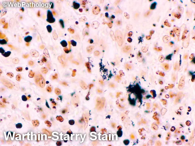
Gram Stain - 5 Mark Answer
Definition
Principle
- Gram-positive bacteria have a thick peptidoglycan layer that traps the crystal violet-iodine complex within the cell wall even after decolorization, so they retain the purple/violet color.
- Gram-negative bacteria have a thin peptidoglycan layer and an outer lipid membrane that is disrupted by the alcohol/acetone decolorizer, allowing the crystal violet-iodine complex to wash out; they then take up the counterstain and appear pink/red.
Procedure (4 Steps)
| Step | Reagent | Action |
|---|---|---|
| 1. Primary stain | Crystal violet | Stains all bacteria purple |
| 2. Mordant | Gram's iodine | Forms crystal violet-iodine complex, fixes dye in cell wall |
| 3. Decolorizer | Alcohol or acetone | Washes out complex from thin-walled (Gram-negative) cells only |
| 4. Counterstain | Carbol fuchsin or safranin | Stains decolorized (Gram-negative) cells pink/red |
Results/Interpretation
- Gram-positive bacteria: appear purple/blue (e.g., Staphylococcus, Streptococcus, Bacillus, Clostridium)
- Gram-negative bacteria: appear pink/red (e.g., E. coli, Klebsiella, Pseudomonas, Neisseria)
- Certain organisms stain poorly or unpredictably (e.g., Mycobacteria - resist penetration by crystal violet due to a waxy, lipid-rich cell wall, requiring acid-fast staining instead; fungi like Aspergillus may also appear Gram-negative due to failure to retain crystal violet)
Clinical Significance / Uses
- Rapid presumptive identification of bacteria directly from clinical specimens (pus, sputum, CSF, urine, wound swabs) within minutes, guiding empirical antibiotic therapy before culture results are available
- Assessment of specimen quality (e.g., sputum - presence of pus cells vs epithelial cells)
- Determines bacterial morphology (cocci, bacilli, arrangement - clusters, chains, pairs) alongside Gram reaction
- Used in the operation theatre/anaesthesia setting for rapid diagnosis of surgical site infections, catheter-related infections, and to guide initial antibiotic choice in sepsis
Advantages and Limitations
- Advantages: quick, inexpensive, technically simple, widely available
- Limitations: cannot identify the exact species, does not work well for organisms lacking a typical cell wall (e.g., Mycoplasma), and cell walls of dead/degenerated bacteria or over-decolorized smears can give false results
Anti arrhythmic drugs 5 marks
Anti-Arrhythmic Drugs - 5 Mark Answer
Definition
Classification (Vaughan Williams / Singh-Vaughan Williams Classification)
Class I - Sodium Channel Blockers
- Class IA (moderate Na+ block, prolongs action potential/repolarization): Quinidine, Procainamide, Disopyramide
- Class IB (weak Na+ block, shortens action potential): Lidocaine, Mexiletine, Phenytoin - useful for ventricular arrhythmias
- Class IC (strong Na+ block, minimal effect on repolarization): Flecainide, Propafenone
Class II - Beta-Adrenergic Blockers
- Propranolol, Metoprolol, Esmolol, Atenolol
Class III - Potassium Channel Blockers
- Amiodarone, Sotalol, Dofetilide, Ibutilide, Bretylium
Class IV - Calcium Channel Blockers
- Verapamil, Diltiazem
Class 0 / Miscellaneous (Not fitting the 4 main classes)
- Adenosine - activates K+ channels via A1 receptors, transiently blocks AV node conduction; drug of choice for acute termination of paroxysmal supraventricular tachycardia (PSVT)
- Digoxin - increases vagal tone, slows AV conduction; used in atrial fibrillation for rate control
- Magnesium sulfate - used in torsades de pointes
- Ivabradine - inhibits the If ("funny") current in the SA node
Clinical Notes
- The Sicilian Gambit is a newer, more complex classification based on channels, receptors, and clinical arrhythmogenic mechanisms, offering a more individualized approach than Vaughan Williams.
- Amiodarone is unique as it has properties of all four classes (broad-spectrum activity) and is widely used in both supraventricular and ventricular arrhythmias.
- Relevant to OT/anaesthesia practice: lidocaine (Class IB) is used both as a local anesthetic and IV for ventricular arrhythmias; esmolol (Class II) is used for rapid intraoperative rate/rhythm control due to its very short half-life; adenosine is used in emergency management of SVT during the perioperative period.
Salmonella typhi 5 marks
Salmonella typhi - 5 Mark Answer
Introduction
Morphology and Characteristics
- Gram-negative bacillus, non-spore forming, non-capsulated
- Motile with peritrichate flagella (H antigen)
- Facultative anaerobe, does not ferment lactose (distinguishes it on MacConkey/DCA/XLD media)
- Grows well on selective media: Deoxycholate Citrate Agar (DCA), Wilson and Blair medium (produces black colonies with metallic sheen due to H2S production), XLD agar
Antigenic Structure
- O antigen (somatic, cell wall lipopolysaccharide)
- H antigen (flagellar)
- Vi antigen (capsular, virulence antigen) - present in fresh isolates, associated with virulence and used in Vi vaccine
Mode of Transmission
- Fecal-oral route via contaminated water and food
- Human is the only natural host/reservoir
- Carrier state: chronic carriers (e.g., gallbladder carriers) shed the organism in feces/urine and act as important reservoirs for spread ("Typhoid Mary" phenomenon)
Pathogenesis
- Organism invades through Peyer's patches in the terminal ileum, multiplies in mesenteric lymph nodes, enters bloodstream causing primary bacteremia, then localizes in the reticuloendothelial system (liver, spleen, bone marrow), followed by secondary bacteremia with clinical symptoms.
- Intestinal involvement causes ulceration over Peyer's patches, which can lead to complications like intestinal perforation and hemorrhage.
Clinical Features
- Step-ladder pattern of fever, relative bradycardia, hepatosplenomegaly, rose spots on the trunk, abdominal pain, and constipation (early) or diarrhea (later stage)
- Complications: intestinal hemorrhage, perforation, encephalopathy, osteomyelitis
Laboratory Diagnosis
- Blood culture: gold standard, positive during the first week of fever (bacteremic phase)
- Stool and urine culture: positive later in the disease course
- Widal test: serological test detecting agglutinating antibodies against O and H antigens of S. Typhi; a rising titer over paired sera (2 weeks apart) is more significant than a single titer; still commonly used in the Indian subcontinent despite limited specificity/sensitivity
- Bone marrow culture: most sensitive, useful even after antibiotics started
Treatment and Prevention
- Antibiotics: Ceftriaxone, Azithromycin, Fluoroquinolones (depending on resistance patterns)
- Prevention: safe drinking water, sanitation, hand hygiene, and typhoid vaccination (Vi polysaccharide vaccine, typhoid conjugate vaccine, or oral live attenuated Ty21a vaccine)
- Relevance to OT/anaesthesia: chronic carriers among hospital staff (especially food handlers) are a source of nosocomial spread; proper stool screening and hygiene practices are part of infection control.
Laboratory diagnosis of HIV infection. 5 marks
Laboratory Diagnosis of HIV Infection - 5 Mark Answer
Overview
1. Screening Tests
- Most widely used initial screening test - highly sensitive and relatively inexpensive
- Detects antibodies against HIV-1 and HIV-2, and in 4th generation assays, also the p24 antigen (allows earlier detection, narrowing the "window period")
- A positive ELISA must be confirmed with a second test before a diagnosis is made, since false positives can occur
- Point-of-care immunochromatographic tests giving results within 15-30 minutes
- Useful in emergency settings, labor rooms, and resource-limited areas; based on similar antigen-antibody principle as ELISA
2. Confirmatory Tests
- Traditional gold-standard confirmatory test; detects antibodies to specific HIV-1 viral proteins (gp41, gp120, p24, etc.) separated by electrophoresis
- Highly specific (>99.99% when combined with ELISA), but can give indeterminate results in early infection or with HIV-2
- Largely replaced in many current protocols by newer differentiation assays and molecular tests due to cost, turnaround time, and indeterminate results
- Newer standard confirmatory approach after a positive 4th generation ELISA, distinguishing HIV-1 from HIV-2 antibodies
3. Molecular/Nucleic Acid Tests (NAT)
- PCR (Polymerase Chain Reaction) - detects viral RNA or DNA directly
- Used for: early diagnosis during the window period (before antibodies develop), diagnosis in infants born to HIV-positive mothers (since maternal antibodies persist in the infant's blood for months, making antibody tests unreliable), and blood bank screening to reduce transfusion-transmitted risk
4. Monitoring/Staging Tests (not diagnostic but essential for management)
- CD4+ T-cell count - assesses degree of immunosuppression, guides initiation of antiretroviral therapy and opportunistic infection prophylaxis
- Viral load (HIV RNA quantification) - monitors response to antiretroviral therapy and disease progression
Window Period
Relevance to OT/Anaesthesia Practice
- Pre-operative HIV screening (with consent) is part of standard protocol in many settings for infection control and universal precaution planning
- Needle-stick injury protocols rely on rapid testing of source patient and post-exposure prophylaxis (PEP) initiation based on results
Heparin 3 marks
Heparin - 3 Mark Answer
Definition and Type
Mechanism of Action
Uses
- Prevention and treatment of deep vein thrombosis (DVT) and pulmonary embolism
- Cardiopulmonary bypass surgery and extracorporeal circuits (dialysis) - prevents clotting in the circuit
- Acute coronary syndrome, unstable angina
- During vascular/cardiac surgery to prevent intraoperative thrombosis
- Prophylaxis in immobilized/postoperative patients
Adverse Effects
- Bleeding/hemorrhage
- Heparin-Induced Thrombocytopenia (HIT) - immune-mediated, paradoxically prothrombotic
- Osteoporosis with prolonged use
Monitoring and Reversal
- UFH monitored via aPTT (activated partial thromboplastin time); LMWH generally does not require routine monitoring
- Reversed by Protamine sulfate (specific antidote)
Incineration 3 marks
Incineration - 3 Mark Answer
Definition
Principle and Process
- Waste is burnt in a controlled chamber; suitable waste requires a minimum heating value of about 2,000 kcal/kg for single-chamber incinerators and above 3,500 kcal/kg for pyrolytic double-chamber incinerators.
- Typical operating temperatures range from 900-1000°C, high enough to destroy pathogens completely and reduce waste to a small quantity of ash and bone fragments.
- No chemical pretreatment is required before incineration, except for microbiological, laboratory, and highly infectious waste.
- The resulting incineration ash must still be disposed of safely (e.g., in designated pits), as it is considered biomedical waste.
Uses in Hospital/OT Setting
- Disposal of human tissues, organs, body parts, and fetus (below viability period) - collected in yellow non-chlorinated plastic bags and sent for incineration or plasma pyrolysis
- Disposal of contaminated dressings, cotton, gauze, and sharps contaminated with blood/body fluids
- Disposal of pathological and anatomical waste generated during surgery
Advantages
- Complete destruction of pathogens and reduction of waste volume/weight
- Suitable for hazardous, infectious, and anatomical waste unsuitable for landfill
Disadvantages
- Requires proper equipment and fuel; can be a source of air pollution (release of dioxins, particulate matter) if not properly maintained
- Not economical for waste with low heating value or high moisture content
Succinylcholine 3 marks
Succinylcholine - 3 Mark Answer
Definition/Class
Mechanism of Action
Pharmacokinetics
- Onset: very rapid (30-60 seconds)
- Duration: ultra-short (5-10 minutes) due to rapid hydrolysis by plasma pseudocholinesterase
- Prolonged action seen in patients with pseudocholinesterase deficiency (genetic or acquired, e.g., liver disease, pregnancy)
Uses
- Drug of choice for rapid sequence induction/intubation (RSI) due to fast onset and short duration
- Short surgical procedures requiring brief muscle relaxation (e.g., ECT, endoscopy)
- Management of laryngospasm
Adverse Effects
- Hyperkalemia (dangerous in burns, crush injury, neuromuscular disease, renal failure)
- Malignant hyperthermia - triggers this life-threatening hypermetabolic reaction in genetically susceptible individuals (RYR1/CACNA1S mutations); treated with IV dantrolene
- Bradycardia (especially with repeated doses, more common in children)
- Postoperative myalgia
- Masseter muscle spasm/jaw rigidity
- Prolonged apnea in pseudocholinesterase deficiency
Contraindications
- History of malignant hyperthermia (personal or family)
- Hyperkalemia risk states (burns, crush injury, denervation, neuromuscular disease)
- Pseudocholinesterase deficiency
Adverse reactions of insulin 3 marks
Adverse Reactions of Insulin - 3 Mark Answer
1. Hypoglycemia
2. Weight Gain
3. Lipodystrophy
- Lipohypertrophy: thickened, fatty swelling at repeated injection sites (from repeated injection at the same site)
- Lipoatrophy: localized loss of fat at injection site
- Minimized by rotating injection sites
4. Local Injection Site Reactions
5. Allergic/Immunologic Reactions
- Rare systemic hypersensitivity reactions (urticaria, angioedema, anaphylaxis) - more common historically with animal-derived insulin, rare with human/recombinant insulin
- Insulin antibody formation can occasionally cause insulin resistance
6. Other Effects
- Hypokalemia (insulin drives potassium into cells, used therapeutically in hyperkalemia management but a risk in overdose)
- Edema (insulin promotes renal sodium retention) in some patients starting therapy
Mantoux test 3 marks
Mantoux Test - 3 Mark Answer
Definition
Principle
Procedure
- 1 TU (tuberculin unit) of PPD-RT23 (standard strength used in India) is injected intradermally in 0.1 mL, on the flexor surface of the left forearm, midway between elbow and wrist.
- Injected using a tuberculin syringe with the needle bevel facing upward - correct placement produces a pale wheal 6-10 mm in diameter.
- Reading: done after 48-96 hours, ideally at 72 hours. Only the induration (not erythema) is measured, using a transparent ruler, as the transverse diameter in millimeters.
Interpretation
| Induration | Result |
|---|---|
| < 6 mm | Negative |
| 6-9 mm | Doubtful (may be due to atypical mycobacteria or M. tuberculosis) |
| ≥ 10 mm | Positive |
| ≥ 20 mm | Strong reactor - higher risk of developing active TB |
Clinical/Public Health Significance
- Positive test indicates past or present TB infection, not necessarily active disease
- Used for TB prevalence surveys, contact tracing, and screening (e.g., before starting immunosuppressive therapy)
- False negatives can occur in immunosuppressed patients (anergy), malnutrition, and overwhelming TB infection
- BCG vaccination can cause false-positive reactions
Streptococci 3 marks
Streptococci - 3 Mark Answer
General Characteristics
Classification
- Beta (β) hemolysis: complete lysis of RBCs, clear zone around colonies - most pyogenic (disease-causing) streptococci, e.g., S. pyogenes
- Alpha (α) hemolysis: partial/incomplete hemolysis, greenish discoloration around colonies - e.g., S. pneumoniae, viridans streptococci
- Gamma (γ) hemolysis: no hemolysis (non-hemolytic) - e.g., enterococci
- Group A: Streptococcus pyogenes
- Group B: Streptococcus agalactiae
- Group D: Enterococci
Medically Important Species
- Streptococcus pyogenes (Group A) - causes pharyngitis, scarlet fever, rheumatic fever, post-streptococcal glomerulonephritis, cellulitis, necrotizing fasciitis
- Streptococcus agalactiae (Group B) - neonatal sepsis and meningitis
- Streptococcus pneumoniae - alpha-hemolytic, causes pneumonia, meningitis, otitis media (important in surgical/anaesthesia settings for aspiration pneumonia risk)
- Viridans group streptococci - part of oral flora, important cause of infective endocarditis following dental/surgical procedures
Clinical Relevance (OT/Anaesthesia)
Anti emetics 3 marks
Antiemetics - 3 Mark Answer
Definition
Classification (by Mechanism/Receptor Target)
- Ondansetron, Granisetron, Palonosetron
- Block 5-HT3 receptors centrally in the CTZ/vomiting center and peripherally on vagal afferents in the gut
- First-line for postoperative nausea/vomiting (PONV) and chemotherapy-induced nausea/vomiting (CINV)
- Phenothiazines: Prochlorperazine, Chlorpromazine
- Butyrophenones: Droperidol, Haloperidol
- Benzamides: Metoclopramide, Domperidone (also has prokinetic action via D2 blockade in the gut)
- Hyoscine (Scopolamine) - useful for motion sickness and PONV, available as transdermal patch
- Promethazine, Cyclizine, Diphenhydramine, Dimenhydrinate - useful for motion sickness and vestibular causes
- Aprepitant (oral), Fosaprepitant (IV) - block substance P/NK-1 receptors, particularly effective for delayed CINV
- Dexamethasone - used as an adjuvant, mechanism not fully clear, effective in PONV and CINV prophylaxis
- Dronabinol, Nabilone - used in chemotherapy-induced nausea refractory to other agents
- Lorazepam - adjunct, useful for anticipatory nausea
Clinical Relevance (OT/Anaesthesia)
Control of wound infections
Control of Wound Infection - Exam Answer
Definition
A. Preoperative Measures
- Preoperative bathing/skin preparation - antiseptic wash (chlorhexidine) the night before/morning of surgery
- Hair removal - avoid shaving; if necessary, use clipping just before surgery (shaving causes micro-abrasions that increase infection risk)
- Treatment of remote infections - identify and treat any existing infection (UTI, skin infection) before elective surgery
- Screening and decolonization of MRSA carriers where indicated
- Antibiotic prophylaxis - single dose of appropriate antibiotic given within 60 minutes before skin incision for indicated procedures
- Control of blood glucose and nutritional optimization in diabetic/malnourished patients
- Smoking cessation advice preoperatively (impairs wound healing)
B. Intraoperative Measures
- Hand hygiene/surgical scrub - since Semmelweis first demonstrated its impact on mortality, hand hygiene remains foundational
- Aseptic technique - sterile gowns, gloves, drapes; strict maintenance of the sterile field
- Sterilization of instruments - proper autoclaving/sterilization of all surgical instruments
- OT environment control - laminar airflow, HEPA filtration, restricted traffic/minimizing OT personnel movement, positive pressure ventilation
- Skin antisepsis at the surgical site with antiseptic solution (povidone-iodine or chlorhexidine)
- Meticulous surgical technique - minimizing tissue trauma, maintaining hemostasis, avoiding dead space and hematoma formation, adequate tissue oxygenation
- Maintenance of normothermia - hypothermia impairs immune function and increases SSI risk
- Wound classification-based care - clean, clean-contaminated, contaminated, dirty wounds managed differently (e.g., delayed primary closure for contaminated wounds)
C. Postoperative Measures
- Aseptic wound dressing technique - sterile dressing changes, appropriate dressing choice based on wound exudate
- Early detection/surveillance - monitoring for signs of infection (erythema, purulent discharge, fever) and prompt culture/antibiotic adjustment
- Drain care - proper aseptic management and timely removal of surgical drains
- Nutrition and glycemic control postoperatively to support wound healing
- Hand hygiene by staff before and after every wound dressing/contact
- Isolation of patients with resistant organism infections (e.g., MRSA) to prevent cross-transmission
D. Hospital/Administrative Measures
- Infection control committee surveillance of SSI rates
- Staff training and adherence to CDC/WHO guidelines for prevention of surgical site infection
- Rational antibiotic policy to prevent resistance
Control of wound infections 3 marks
Control of Wound Infection - 3 Mark Answer
Preoperative Measures
- Antiseptic skin preparation (chlorhexidine/povidone-iodine wash) before surgery
- Avoid shaving; use clipping just before surgery if hair removal needed
- Treat any existing remote infections before elective surgery
- Antibiotic prophylaxis - single dose within 60 minutes before skin incision
Intraoperative Measures
- Strict hand hygiene and surgical scrub
- Aseptic technique - sterile gowns, gloves, drapes, and sterilized instruments
- Skin antisepsis at the surgical site
- OT environment control - laminar airflow, HEPA filtration, restricted traffic
- Meticulous surgical technique - minimize tissue trauma, maintain hemostasis, avoid dead space/hematoma
- Maintain patient normothermia during surgery
Postoperative Measures
- Aseptic dressing technique for wound care
- Early detection of infection signs (redness, discharge, fever) with prompt treatment
- Proper aseptic drain care and timely removal
- Good nutrition and glycemic control to support healing
- Isolation of patients with resistant organisms to prevent cross-transmission
Pasteurization 3 marks
Pasteurization - 3 Mark Answer
Definition
Methods
- Milk heated to 62.5-65.6°C (145°F) for 30 minutes, then rapidly cooled to 4°C
- Milk heated to 71.6°C (161°F) for 15-20 seconds, then rapidly cooled - more commonly used commercially today
- 72°C for 5 seconds (short-term pasteurization)
Purpose/Uses
- Renders milk safe by destroying pathogenic organisms such as Mycobacterium tuberculosis, Brucella, Salmonella, Coxiella burnetii, and coliforms, without spoiling its taste or nutritive value
- Used for human breast milk in milk banks to inactivate pathogens like CMV, HIV, HBV
- Applied to fruit juices to reduce foodborne illness risk (E. coli O157, Salmonella)
Important Points
- After pasteurization, milk must be rapidly cooled to 4°C to prevent multiplication of any surviving/recontaminating organisms
- Test of efficacy: absence of coliform organisms in pasteurized milk confirms adequate pasteurization; their presence indicates improper pasteurization or post-pasteurization contamination
- Since spores survive, pasteurization is classified as disinfection, not sterilization
Aspirin 3 marks
Aspirin - 3 Mark Answer
Definition/Class
Mechanism of Action
- Antiplatelet effect: At low doses (75-150 mg/day), aspirin selectively inhibits platelet COX-1, blocking thromboxane A2 (a potent vasoconstrictor and platelet aggregator) synthesis. Since platelets are anucleate and cannot synthesize new enzyme, this effect lasts for the life of the platelet (8-10 days).
- Anti-inflammatory/analgesic/antipyretic effect: At higher doses (300-900 mg or more), it inhibits COX in other tissues, reducing prostaglandin-mediated pain, fever, and inflammation.
Uses
- Low-dose: antiplatelet prophylaxis in coronary artery disease, myocardial infarction, stroke prevention
- Higher dose: mild-moderate pain, fever, rheumatic/inflammatory conditions (rheumatoid arthritis, rheumatic fever)
- Kawasaki disease (pediatrics)
Adverse Effects
- Gastric irritation, peptic ulceration, GI bleeding (COX-1 inhibition reduces protective mucosal prostaglandins)
- Increased bleeding tendency (antiplatelet effect)
- Reye syndrome - avoided in children with viral febrile illness
- Hypersensitivity/bronchospasm in aspirin-sensitive asthmatics
- Salicylism/overdose - tinnitus, hyperventilation, metabolic acidosis
Relevance to OT/Anaesthesia
Combine all 3 marks in single pdf
mkdir -p /tmp/workspace/otat-notes && ls -la /tmp/workspace/
~/otat-notes/generate_pdf.py
# -*- coding: utf-8 -*-
from reportlab.lib.pagesizes import A4
from reportlab.lib.units import cm
from reportlab.lib.styles import getSampleStyleSheet, ParagraphStyle
from reportlab.lib.enums import TA_CENTER
from reportlab.platypus import (SimpleDocTemplate, Paragraph, Spacer, ListFlowable,
                                 ListItem, PageBreak, Table, TableStyle, HRFlowable)
from reportlab.lib import colors

doc = SimpleDocTemplate(
    "/tmp/workspace/otat-notes/3_Marks_Notes_OTAT.pdf",
    pagesize=A4,
    topMargin=1.6*cm, bottomMargin=1.6*cm, leftMargin=1.8*cm, rightMargin=1.8*cm,
    title="3 Mark Notes - BSc OTAT", author="Orris"
)

styles = getSampleStyleSheet()

title_style = ParagraphStyle('TitleX', parent=styles['Title'], fontSize=20, spaceAfter=4, textColor=colors.HexColor('#1a3d5c'))
subtitle_style = ParagraphStyle('SubtitleX', parent=styles['Normal'], fontSize=11, alignment=TA_CENTER, textColor=colors.HexColor('#555555'), spaceAfter=18)
h1 = ParagraphStyle('H1', parent=styles['Heading1'], fontSize=15, textColor=colors.white, backColor=colors.HexColor('#1a3d5c'),
                     spaceBefore=6, spaceAfter=10, leftIndent=6, borderPadding=(6,6,6,6))
h2 = ParagraphStyle('H2', parent=styles['Heading2'], fontSize=12, textColor=colors.HexColor('#1a3d5c'), spaceBefore=10, spaceAfter=4)
body = ParagraphStyle('BodyX', parent=styles['BodyText'], fontSize=10.3, leading=14.5, spaceAfter=5)
bullet = ParagraphStyle('BulletX', parent=body, leftIndent=14, bulletIndent=4, spaceAfter=3)
source_style = ParagraphStyle('SourceX', parent=styles['Normal'], fontSize=8.5, textColor=colors.HexColor('#666666'), spaceBefore=6, spaceAfter=2, leftIndent=0)
toc_style = ParagraphStyle('TocX', parent=styles['Normal'], fontSize=11, spaceAfter=6, leftIndent=10)

def P(text):
    return Paragraph(text, body)

def B(items):
    return ListFlowable([ListItem(Paragraph(i, bullet)) for i in items], bulletType='bullet', start='circle', leftIndent=14)

def SRC(text):
    return Paragraph(f"<i>Sources: {text}</i>", source_style)

story = []

story.append(Paragraph("3 Mark Notes", title_style))
story.append(Paragraph("BSc Operation Theatre and Anaesthesia Technology - 2nd Year (Dr. MGR Medical University)", subtitle_style))
story.append(HRFlowable(width="100%", thickness=1, color=colors.HexColor('#1a3d5c')))
story.append(Spacer(1, 12))

topics = ["Heparin", "Incineration", "Succinylcholine", "Adverse Reactions of Insulin",
          "Mantoux Test", "Streptococci", "Anti-emetics", "Control of Wound Infection",
          "Pasteurization", "Aspirin"]
story.append(Paragraph("Contents", h2))
toc_items = [Paragraph(f"{i+1}. {t}", toc_style) for i, t in enumerate(topics)]
story.append(ListFlowable(toc_items, bulletType='1'))
story.append(PageBreak())

# ---------------- 1. HEPARIN ----------------
story.append(Paragraph("1. Heparin", h1))
story.append(Paragraph("<b>Definition and Type</b>", h2))
story.append(P("Heparin is a naturally occurring anticoagulant, a sulfated glycosaminoglycan, available as Unfractionated Heparin (UFH) and Low Molecular Weight Heparin (LMWH) (e.g., enoxaparin, dalteparin)."))
story.append(Paragraph("<b>Mechanism of Action</b>", h2))
story.append(P("Heparin acts by activating antithrombin (previously antithrombin III), greatly accelerating (up to 1000-fold) the rate at which antithrombin inactivates clotting enzymes, mainly thrombin (Factor IIa) and Factor Xa. Heparin acts as a catalytic cofactor without being consumed; once the antithrombin-protease complex forms, heparin is released intact to bind further antithrombin molecules. UFH inhibits both thrombin and Factor Xa, while LMWH preferentially inhibits Factor Xa."))
story.append(Paragraph("<b>Uses</b>", h2))
story.append(B([
    "Prevention and treatment of deep vein thrombosis (DVT) and pulmonary embolism",
    "Cardiopulmonary bypass surgery and extracorporeal circuits (dialysis)",
    "Acute coronary syndrome, unstable angina",
    "During vascular/cardiac surgery to prevent intraoperative thrombosis",
    "Prophylaxis in immobilized/postoperative patients",
]))
story.append(Paragraph("<b>Adverse Effects</b>", h2))
story.append(B(["Bleeding/hemorrhage", "Heparin-Induced Thrombocytopenia (HIT)", "Osteoporosis with prolonged use"]))
story.append(Paragraph("<b>Monitoring and Reversal</b>", h2))
story.append(P("UFH is monitored via aPTT (activated partial thromboplastin time); LMWH generally does not require routine monitoring. Reversed by Protamine sulfate (specific antidote)."))
story.append(SRC("Katzung's Basic and Clinical Pharmacology, 16th Ed.; Braunwald's Heart Disease, 2 Vol Set; Harrison's Principles of Internal Medicine, 22nd Ed."))
story.append(PageBreak())

# ---------------- 2. INCINERATION ----------------
story.append(Paragraph("2. Incineration", h1))
story.append(Paragraph("<b>Definition</b>", h2))
story.append(P("Incineration is a method of waste disposal in which combustible waste is burnt at high temperature to reduce it to ash, gases, and heat. It is the method of choice for disposal of hazardous, infectious, and biomedical waste, especially where suitable land for burial/landfill is not available."))
story.append(Paragraph("<b>Principle and Process</b>", h2))
story.append(B([
    "Suitable waste requires a minimum heating value of about 2,000 kcal/kg for single-chamber incinerators, and above 3,500 kcal/kg for pyrolytic double-chamber incinerators",
    "Typical operating temperatures range from 900-1000 degrees C, high enough to destroy pathogens completely and reduce waste to ash and bone fragments",
    "No chemical pretreatment is required before incineration, except for microbiological, laboratory, and highly infectious waste",
    "The resulting incineration ash must still be disposed of safely, as it is considered biomedical waste",
]))
story.append(Paragraph("<b>Uses in Hospital/OT Setting</b>", h2))
story.append(B([
    "Disposal of human tissues, organs, body parts, and fetus (below viability period)",
    "Disposal of contaminated dressings, cotton, gauze, and sharps contaminated with blood/body fluids",
    "Disposal of pathological and anatomical waste generated during surgery",
]))
story.append(Paragraph("<b>Advantages and Disadvantages</b>", h2))
story.append(P("Advantages: complete destruction of pathogens and reduction of waste volume/weight, suitable for hazardous and anatomical waste unsuitable for landfill. Disadvantages: requires proper equipment and fuel, can cause air pollution (dioxins, particulate matter) if not properly maintained; not economical for low heating value or high moisture waste."))
story.append(SRC("Park's Textbook of Preventive and Social Medicine; Sherris and Ryan's Medical Microbiology, 8th Ed."))
story.append(PageBreak())

# ---------------- 3. SUCCINYLCHOLINE ----------------
story.append(Paragraph("3. Succinylcholine", h1))
story.append(Paragraph("<b>Definition/Class</b>", h2))
story.append(P("Succinylcholine (Suxamethonium) is the only clinically used depolarizing neuromuscular blocking agent, consisting of two acetylcholine molecules joined together."))
story.append(Paragraph("<b>Mechanism of Action</b>", h2))
story.append(P("Acts as an agonist at nicotinic acetylcholine receptors at the neuromuscular junction, causing sustained depolarization of the motor end plate. Initial depolarization produces visible muscle fasciculations, followed by flaccid paralysis because the receptor remains desensitized; succinylcholine is only slowly degraded by plasma pseudocholinesterase."))
story.append(Paragraph("<b>Pharmacokinetics</b>", h2))
story.append(B([
    "Onset: very rapid (30-60 seconds)",
    "Duration: ultra-short (5-10 minutes) due to rapid hydrolysis by plasma pseudocholinesterase",
    "Prolonged action seen in patients with pseudocholinesterase deficiency",
]))
story.append(Paragraph("<b>Uses</b>", h2))
story.append(B(["Drug of choice for rapid sequence induction/intubation (RSI)", "Short surgical procedures requiring brief muscle relaxation", "Management of laryngospasm"]))
story.append(Paragraph("<b>Adverse Effects</b>", h2))
story.append(B(["Hyperkalemia", "Malignant hyperthermia (treated with IV dantrolene)", "Bradycardia", "Postoperative myalgia", "Masseter muscle spasm/jaw rigidity", "Prolonged apnea in pseudocholinesterase deficiency"]))
story.append(Paragraph("<b>Contraindications</b>", h2))
story.append(B(["History of malignant hyperthermia", "Hyperkalemia risk states (burns, crush injury, denervation)", "Pseudocholinesterase deficiency"]))
story.append(SRC("Morgan and Mikhail's Clinical Anesthesiology, 7th Ed.; Barash, Cullen, and Stoelting's Clinical Anesthesia, 9e; Miller's Anesthesia, 10e."))
story.append(PageBreak())

# ---------------- 4. INSULIN ADVERSE REACTIONS ----------------
story.append(Paragraph("4. Adverse Reactions of Insulin", h1))
story.append(Paragraph("<b>1. Hypoglycemia</b>", h2))
story.append(P("The most common and serious adverse effect. Occurs due to excess dose relative to food intake/activity, especially with short-acting insulin. Presents with sweating, tremors, palpitations, confusion, and if severe, seizures or coma."))
story.append(Paragraph("<b>2. Weight Gain</b>", h2))
story.append(P("Occurs due to the anabolic effect of insulin (promotes glucose uptake, glycogen and fat synthesis)."))
story.append(Paragraph("<b>3. Lipodystrophy</b>", h2))
story.append(B(["Lipohypertrophy: thickened, fatty swelling at repeated injection sites", "Lipoatrophy: localized loss of fat at injection site", "Minimized by rotating injection sites"]))
story.append(Paragraph("<b>4. Local Injection Site Reactions</b>", h2))
story.append(P("Redness, swelling, itching, or mild allergic reaction at the site of subcutaneous injection."))
story.append(Paragraph("<b>5. Allergic/Immunologic Reactions</b>", h2))
story.append(P("Rare systemic hypersensitivity reactions (urticaria, angioedema, anaphylaxis); more common historically with animal-derived insulin, rare with human/recombinant insulin. Insulin antibody formation can occasionally cause insulin resistance."))
story.append(Paragraph("<b>6. Other Effects</b>", h2))
story.append(B(["Hypokalemia (insulin drives potassium into cells)", "Edema (insulin promotes renal sodium retention) in some patients starting therapy"]))
story.append(P("<b>Relevance to OT/Anaesthesia:</b> Perioperative insulin therapy in diabetic patients requires close blood glucose monitoring to avoid intraoperative hypoglycemia, which can be masked under general anesthesia."))
story.append(SRC("Lippincott Illustrated Reviews - Pharmacology; Symptom to Diagnosis: An Evidence-Based Guide, 4th Ed."))
story.append(PageBreak())

# ---------------- 5. MANTOUX TEST ----------------
story.append(Paragraph("5. Mantoux Test", h1))
story.append(Paragraph("<b>Definition</b>", h2))
story.append(P("The Mantoux test (tuberculin skin test) is a delayed-type hypersensitivity (Type IV) skin test used to detect past or present infection with Mycobacterium tuberculosis. Discovered by Von Pirquet (1907)."))
story.append(Paragraph("<b>Principle</b>", h2))
story.append(P("Tuberculin, in the form of Purified Protein Derivative (PPD), is injected intradermally. In a sensitized person, sensitized T-lymphocytes react with the antigen, producing local erythema and induration over 48-96 hours."))
story.append(Paragraph("<b>Procedure</b>", h2))
story.append(B([
    "1 TU of PPD-RT23 (standard strength used in India) injected intradermally in 0.1 mL on the flexor surface of the left forearm, midway between elbow and wrist",
    "Injected with the needle bevel facing upward, producing a pale wheal 6-10 mm in diameter",
    "Reading done after 48-96 hours, ideally at 72 hours; only induration (not erythema) is measured in mm",
]))
story.append(Paragraph("<b>Interpretation</b>", h2))
table_data = [["Induration", "Result"],
              ["< 6 mm", "Negative"],
              ["6-9 mm", "Doubtful"],
              [">= 10 mm", "Positive"],
              [">= 20 mm", "Strong reactor - higher TB risk"]]
t = Table(table_data, colWidths=[6*cm, 9*cm])
t.setStyle(TableStyle([
    ('BACKGROUND', (0,0), (-1,0), colors.HexColor('#1a3d5c')),
    ('TEXTCOLOR', (0,0), (-1,0), colors.white),
    ('FONTSIZE', (0,0), (-1,-1), 9.5),
    ('GRID', (0,0), (-1,-1), 0.5, colors.grey),
    ('ROWBACKGROUNDS', (0,1), (-1,-1), [colors.white, colors.HexColor('#eef3f7')]),
    ('VALIGN', (0,0), (-1,-1), 'MIDDLE'),
    ('TOPPADDING', (0,0), (-1,-1), 5), ('BOTTOMPADDING', (0,0), (-1,-1), 5),
]))
story.append(t)
story.append(Spacer(1, 8))
story.append(Paragraph("<b>Clinical/Public Health Significance</b>", h2))
story.append(B([
    "Positive test indicates past or present TB infection, not necessarily active disease",
    "Used for TB prevalence surveys, contact tracing, and screening",
    "False negatives can occur in immunosuppressed patients, malnutrition, and overwhelming TB infection",
    "BCG vaccination can cause false-positive reactions",
]))
story.append(SRC("Park's Textbook of Preventive and Social Medicine; Robbins and Kumar Basic Pathology; Jawetz, Melnick and Adelberg's Medical Microbiology, 28th Ed."))
story.append(PageBreak())

# ---------------- 6. STREPTOCOCCI ----------------
story.append(Paragraph("6. Streptococci", h1))
story.append(Paragraph("<b>General Characteristics</b>", h2))
story.append(P("Gram-positive cocci arranged in chains or pairs, catalase-negative (distinguishing them from Staphylococcus), non-motile, facultative anaerobes. They ferment carbohydrates producing lactic acid."))
story.append(Paragraph("<b>Classification</b>", h2))
story.append(P("<b>A. Based on Hemolysis on Blood Agar:</b>"))
story.append(B([
    "Beta (b) hemolysis: complete lysis of RBCs, clear zone - most pyogenic streptococci, e.g. S. pyogenes",
    "Alpha (a) hemolysis: partial/incomplete hemolysis, greenish discoloration - e.g. S. pneumoniae, viridans streptococci",
    "Gamma (g) hemolysis: no hemolysis (non-hemolytic) - e.g. enterococci",
]))
story.append(P("<b>B. Lancefield Classification</b> (serological, based on cell wall carbohydrate antigen, developed by Rebecca Lancefield): divides beta-hemolytic streptococci into serogroups A through H and K through V."))
story.append(B(["Group A: Streptococcus pyogenes", "Group B: Streptococcus agalactiae", "Group D: Enterococci"]))
story.append(Paragraph("<b>Medically Important Species</b>", h2))
story.append(B([
    "Streptococcus pyogenes (Group A) - pharyngitis, scarlet fever, rheumatic fever, glomerulonephritis, cellulitis, necrotizing fasciitis",
    "Streptococcus agalactiae (Group B) - neonatal sepsis and meningitis",
    "Streptococcus pneumoniae - alpha-hemolytic, causes pneumonia, meningitis, otitis media",
    "Viridans group streptococci - oral flora, important cause of infective endocarditis",
]))
story.append(P("<b>Clinical Relevance (OT/Anaesthesia):</b> Viridans streptococci are a key reason for antibiotic prophylaxis before dental and certain surgical procedures in patients with valvular heart disease, to prevent bacteremia-induced infective endocarditis."))
story.append(SRC("Sherris and Ryan's Medical Microbiology, 8th Ed.; Jawetz, Melnick and Adelberg's Medical Microbiology, 28th Ed.; Medical Microbiology 9e; Henry's Clinical Diagnosis and Management by Laboratory Methods."))
story.append(PageBreak())

# ---------------- 7. ANTIEMETICS ----------------
story.append(Paragraph("7. Anti-emetics", h1))
story.append(Paragraph("<b>Definition</b>", h2))
story.append(P("Drugs used to prevent or treat nausea and vomiting, acting at the chemoreceptor trigger zone (CTZ), vomiting center, and peripheral vagal afferents."))
story.append(Paragraph("<b>Classification (by Mechanism/Receptor Target)</b>", h2))
story.append(B([
    "5-HT3 Receptor Antagonists: Ondansetron, Granisetron, Palonosetron - first-line for PONV and CINV",
    "Dopamine (D2) Receptor Antagonists: Prochlorperazine, Chlorpromazine, Droperidol, Haloperidol, Metoclopramide, Domperidone",
    "Anticholinergics: Hyoscine (Scopolamine) - motion sickness and PONV, transdermal patch",
    "Antihistamines (H1 Antagonists): Promethazine, Cyclizine, Diphenhydramine, Dimenhydrinate",
    "NK-1 Receptor Antagonists: Aprepitant (oral), Fosaprepitant (IV) - effective for delayed CINV",
    "Corticosteroids: Dexamethasone - adjuvant for PONV and CINV prophylaxis",
    "Cannabinoids: Dronabinol, Nabilone - refractory chemotherapy-induced nausea",
    "Benzodiazepines: Lorazepam - adjunct for anticipatory nausea",
]))
story.append(P("<b>Clinical Relevance (OT/Anaesthesia):</b> A multimodal approach combining agents from at least two different classes (e.g., 5-HT3 antagonist + dexamethasone +/- droperidol) is standard practice for prevention of postoperative nausea and vomiting (PONV)."))
story.append(SRC("Katzung's Basic and Clinical Pharmacology, 16th Ed.; Berek and Novak's Gynecology; Ganong's Review of Medical Physiology, 26th Ed."))
story.append(PageBreak())

# ---------------- 8. CONTROL OF WOUND INFECTION ----------------
story.append(Paragraph("8. Control of Wound Infection", h1))
story.append(Paragraph("<b>Preoperative Measures</b>", h2))
story.append(B([
    "Antiseptic skin preparation (chlorhexidine/povidone-iodine wash) before surgery",
    "Avoid shaving; use clipping just before surgery if hair removal needed",
    "Treat any existing remote infections before elective surgery",
    "Antibiotic prophylaxis - single dose within 60 minutes before skin incision",
]))
story.append(Paragraph("<b>Intraoperative Measures</b>", h2))
story.append(B([
    "Strict hand hygiene and surgical scrub",
    "Aseptic technique - sterile gowns, gloves, drapes, and sterilized instruments",
    "Skin antisepsis at the surgical site",
    "OT environment control - laminar airflow, HEPA filtration, restricted traffic",
    "Meticulous surgical technique - minimize tissue trauma, maintain hemostasis, avoid dead space/hematoma",
    "Maintain patient normothermia during surgery",
]))
story.append(Paragraph("<b>Postoperative Measures</b>", h2))
story.append(B([
    "Aseptic dressing technique for wound care",
    "Early detection of infection signs (redness, discharge, fever) with prompt treatment",
    "Proper aseptic drain care and timely removal",
    "Good nutrition and glycemic control to support healing",
    "Isolation of patients with resistant organisms to prevent cross-transmission",
]))
story.append(SRC("Sabiston Textbook of Surgery (Sterility and Environmental Issues, Antibiotic Prophylaxis); Dermatology 2-Volume Set, 5th Ed. (Management of Wound Infection)."))
story.append(PageBreak())

# ---------------- 9. PASTEURIZATION ----------------
story.append(Paragraph("9. Pasteurization", h1))
story.append(Paragraph("<b>Definition</b>", h2))
story.append(P("Pasteurization is a method of moist heat disinfection (not sterilization) that uses temperatures below 100 degrees C to kill pathogenic vegetative bacteria in liquids, most commonly milk, without significantly altering nutritional/organoleptic quality. It does not destroy bacterial spores."))
story.append(Paragraph("<b>Methods</b>", h2))
story.append(B([
    "Holder (Batch) Method: milk heated to 62.5-65.6 degrees C (145 F) for 30 minutes, then rapidly cooled to 4 degrees C",
    "Flash (HTST) Method: milk heated to 71.6 degrees C (161 F) for 15-20 seconds, then rapidly cooled - commonly used commercially",
    "Short-term/Ultra methods (e.g., human breast milk in milk banks): 72 degrees C for 5 seconds",
]))
story.append(Paragraph("<b>Purpose/Uses</b>", h2))
story.append(B([
    "Renders milk safe by destroying pathogens (M. tuberculosis, Brucella, Salmonella, Coxiella burnetii, coliforms) without spoiling taste/nutritive value",
    "Used for human breast milk in milk banks to inactivate CMV, HIV, HBV",
    "Applied to fruit juices to reduce foodborne illness risk",
]))
story.append(Paragraph("<b>Important Points</b>", h2))
story.append(B([
    "After pasteurization, milk must be rapidly cooled to 4 degrees C to prevent multiplication of surviving/recontaminating organisms",
    "Test of efficacy: absence of coliform organisms in pasteurized milk confirms adequate pasteurization",
    "Since spores survive, pasteurization is classified as disinfection, not sterilization",
]))
story.append(SRC("Park's Textbook of Preventive and Social Medicine; Sherris and Ryan's Medical Microbiology, 8th Ed."))
story.append(PageBreak())

# ---------------- 10. ASPIRIN ----------------
story.append(Paragraph("10. Aspirin", h1))
story.append(Paragraph("<b>Definition/Class</b>", h2))
story.append(P("Aspirin (acetylsalicylic acid) is a salicylate NSAID with analgesic, antipyretic, anti-inflammatory, and antiplatelet properties. It is unique among NSAIDs as an irreversible inhibitor of cyclooxygenase."))
story.append(Paragraph("<b>Mechanism of Action</b>", h2))
story.append(P("Aspirin irreversibly acetylates a serine residue on the cyclooxygenase (COX-1 and COX-2) enzyme, permanently inactivating it, blocking conversion of arachidonic acid to prostaglandins and thromboxane A2."))
story.append(B([
    "Antiplatelet effect: at low doses (75-150 mg/day), selectively inhibits platelet COX-1, blocking thromboxane A2 synthesis; effect lasts the life of the platelet (8-10 days) since platelets cannot synthesize new enzyme",
    "Anti-inflammatory/analgesic/antipyretic effect: at higher doses (300-900 mg or more), inhibits COX in other tissues, reducing prostaglandin-mediated pain, fever, and inflammation",
]))
story.append(Paragraph("<b>Uses</b>", h2))
story.append(B([
    "Low-dose: antiplatelet prophylaxis in coronary artery disease, myocardial infarction, stroke prevention",
    "Higher dose: mild-moderate pain, fever, rheumatic/inflammatory conditions",
    "Kawasaki disease (pediatrics)",
]))
story.append(Paragraph("<b>Adverse Effects</b>", h2))
story.append(B([
    "Gastric irritation, peptic ulceration, GI bleeding",
    "Increased bleeding tendency",
    "Reye syndrome - avoided in children with viral febrile illness",
    "Hypersensitivity/bronchospasm in aspirin-sensitive asthmatics",
    "Salicylism/overdose - tinnitus, hyperventilation, metabolic acidosis",
]))
story.append(P("<b>Relevance to OT/Anaesthesia:</b> Patients on aspirin therapy require careful assessment before surgery due to increased bleeding risk; decisions to continue or withhold aspirin perioperatively are individualized based on cardiovascular risk versus bleeding risk."))
story.append(SRC("Katzung's Basic and Clinical Pharmacology, 16th Ed.; Harrison's Principles of Internal Medicine, 22nd Ed.; Lippincott Illustrated Reviews - Pharmacology."))

doc.build(story)
print("PDF generated successfully")
cd /tmp/workspace/otat-notes && python3 generate_pdf.py
ls -la /tmp/workspace/otat-notes/
3 Marks Notes OTAT
PDF Document · PDF