
Pictureize the elbow crutch and axillary crutch and also label them.
axillary crutch labeled parts diagram
elbow crutch forearm crutch labeled diagram
crutch
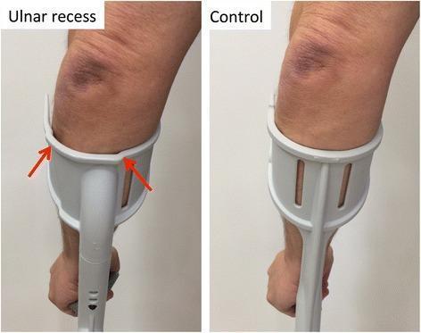
This clinical comparison photograph displays two types of forearm crutch cuffs being used on a human subject. The left image shows an 'ulnar recess' anatomic cuff, while the right image shows a 'control' (conventional) cuff. In the ulnar recess design, red arrows highlight two specific indentations along the superior aspect of the plastic cuff. These indentations create a visual gap between the cuff material and the forearm, specifically targeting the area over the ulnar bone and proximal forearm to redistribute weight-bearing pressure. In contrast, the conventional control cuff demonstrates a uniform, continuous curve that maintains direct contact with the limb. Both forearms exhibit visible skin discoloration and bruising near the elbow joint, likely related to chronic crutch use. This image illustrates an orthopedic intervention designed to mitigate ulnar nerve compression, periosteal pain, and skin irritation in patients requiring long-term mobility aids by optimizing the crutch-forearm interface.
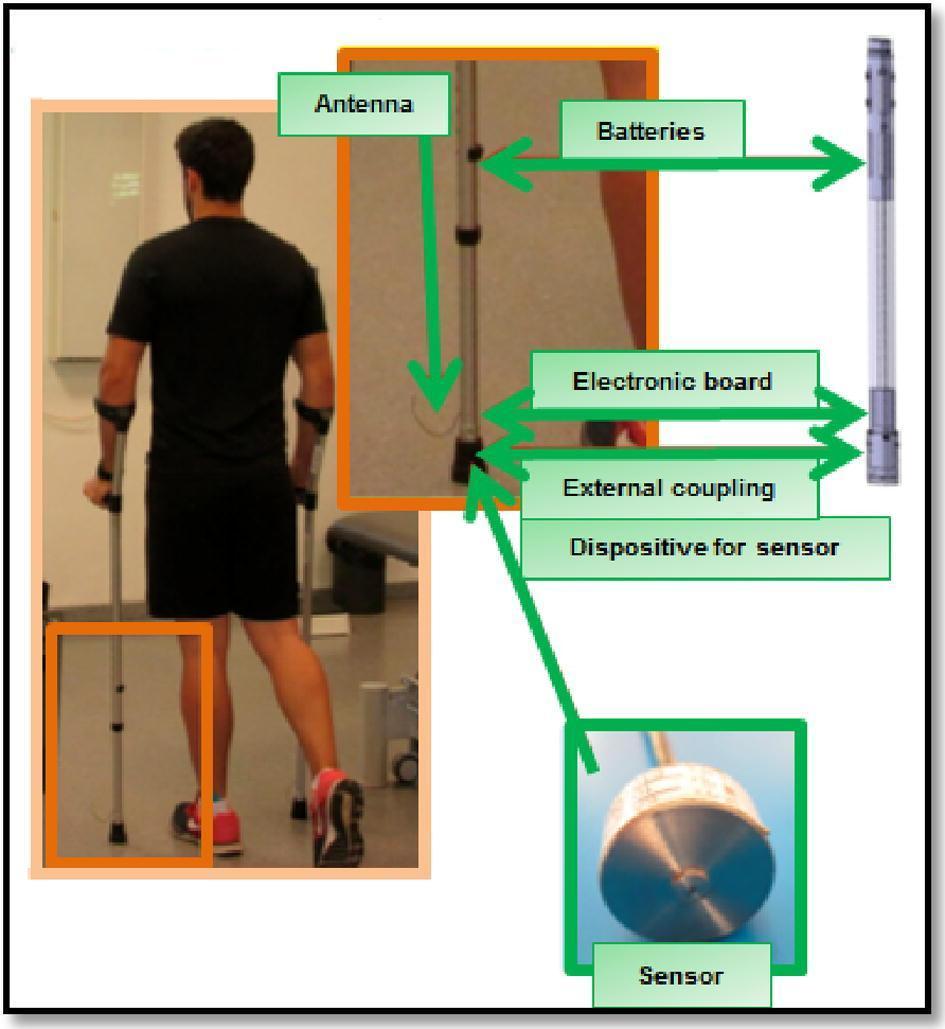
This composite educational illustration demonstrates the GCH 2.0 load measurement system integrated into a standard forearm crutch for gait analysis. The image consists of a clinical photograph showing a male subject using bilateral forearm crutches in a laboratory setting, a schematic of the internal hardware, and detailed callouts of the electronic components. The distal part of the crutch contains a miniaturized force sensor positioned at the base to measure ground reaction forces. Moving superiorly along the crutch shaft, the assembly includes an external coupling, an electronic board for signal processing, power batteries, and an antenna for wireless data transmission to a receiver. This system is designed for rehabilitation and orthopedic research to monitor load-bearing compliance during unilateral assisted gait (UAG). Educational focuses include biomechanics, physical therapy monitoring, and the use of assistive technology to evaluate weight-bearing percentages (e.g., 25% or 50% of body weight) during recovery from lower limb injury or surgery.
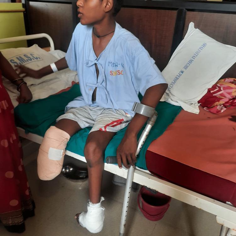
A clinical photograph of a pediatric or adolescent patient seated on a hospital bed, demonstrating post-operative management of the lower extremities. The right leg displays a below-knee amputation (transtibial) with a residual limb (stump) that is wrapped in a beige compression bandage secured with white tape, typical for post-surgical shaping and edema control. The left foot and ankle are encased in a white medical bandage or light cast, consistent with a post-operative state for congenital talipes equinovarus (CTEV) correction via wedge tarsectomy. The patient is shown holding a metal forearm crutch (Loftstrand crutch) in the left hand, indicating active participation in mobility or gait training. The patient is wearing a light blue hospital gown and grey shorts. This image illustrates post-surgical orthopedic rehabilitation, prosthetic preparation, and the use of assistive devices in a multidisciplinary clinical setting.
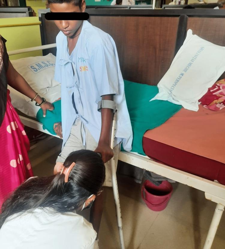
Clinical photograph depicting a pediatric or adolescent patient undergoing postoperative gait training in a hospital setting. The patient is shown standing next to a hospital bed, wearing a light blue medical gown and gray shorts. To facilitate ambulation, the patient is utilizing a forearm crutch (Lofstrand crutch) on the left side. A physical therapist is positioned at the patient's feet, manually assisting with foot placement and supporting the lower extremity during the gait cycle. The clinical context involves rehabilitation following a right below-knee amputation and a left-foot wedge tarsectomy for congenital talipes equinovarus (CTEV). The image demonstrates the multidisciplinary approach to post-surgical recovery, focusing on independent mobility, weight-bearing stabilization, and the use of assistive devices. The hospital environment includes labeled pillows and standard medical furniture, emphasizing the transition from acute surgical care to active physical therapy and prosthetic preparation.
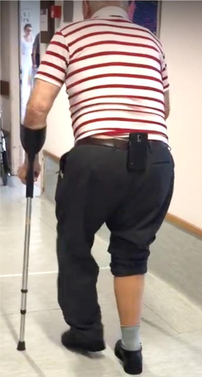
A clinical photograph depicting a participant in a rehabilitation research setting, viewed from behind. The individual is performing a functional mobility assessment, likely a Six-Minute Walk Test (6MWT). The participant utilizes a forearm crutch (Lofstrand crutch) in the left hand for gait stabilization. An Android smartphone is mounted vertically to the center of the participant's belt at the lower back (L5/sacral region), serving as a wearable inertial measurement unit (IMU) to collect tri-axial accelerometer and gyroscope data. The participant's lower extremities show asymmetry: the left leg is fully clothed in dark trousers, while the right trouser leg is absent, revealing a grey sock and black shoe on the right foot. This setup is characteristic of gait analysis studies focused on automated step-detection, spatiotemporal gait parameters, and the integration of mobile health (mHealth) technology in physical therapy and geriatric rehabilitation.
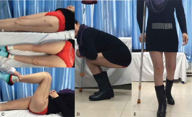
This composite of clinical photographs documents the 6-month post-operative recovery of a patient following a complex orthopedic procedure, likely tumor resection and revision knee arthroplasty of the left femur. Panel A shows the left lower limb in extension, highlighting a long (approx. 25 cm), well-healed longitudinal surgical scar on the anterior aspect of the thigh and knee. Panel B provides a bilateral comparison of the legs, showing relatively equal limb length. Panel C demonstrates passive knee flexion, confirming a functional range of motion and the stability of the surgical site. Panel D illustrates the patient’s functional mobility, showing the ability to perform an independent squat, albeit with support from an axillary crutch. Panel E shows the patient ambulating with the assistance of a single crutch and wearing a supportive boot on the left leg. This educational series highlights post-operative outcomes in orthopedic oncology and reconstructive surgery, emphasizing the restoration of range of motion and gait following significant limb salvage procedures.
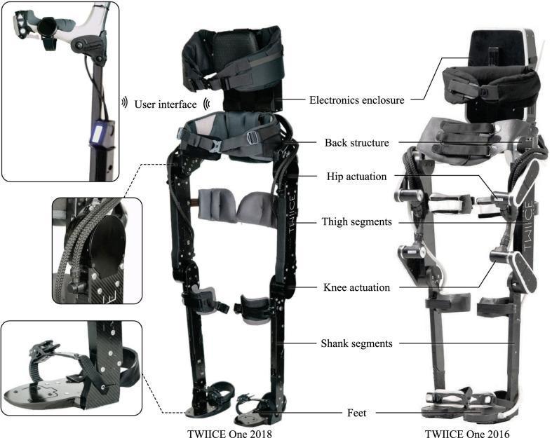
This technical infographic presents an overview of the TWIICE One lower-limb exoskeleton, an assistive robotic device designed for individuals with motor-complete spinal cord injury. The central image compares the 2018 model (left) with the 2016 version (right). The device is a powered hip-knee-ankle-foot orthosis featuring four actuated joints to facilitate gait in the sagittal plane. Structural components include a height-adjustable thoracic belt, waist belt, carbon-fiber reinforced thigh and shank segments, and specialized footplates with straps. Key highlighted features include the electronics enclosure mounted on the back structure, hip and knee actuation motors, and a complex user interface. Inset panels detail the interface components: a forearm crutch handle equipped with push-buttons and a trigger, a smartwatch for high-level commands, and an integrated LCD display on the crutch for visual mode feedback. Detailed views also show the custom motor housings and the ergonomic foot design. The visualization illustrates the engineering advancements in medical rehabilitative technology aimed at restoring mobility and gait stability.
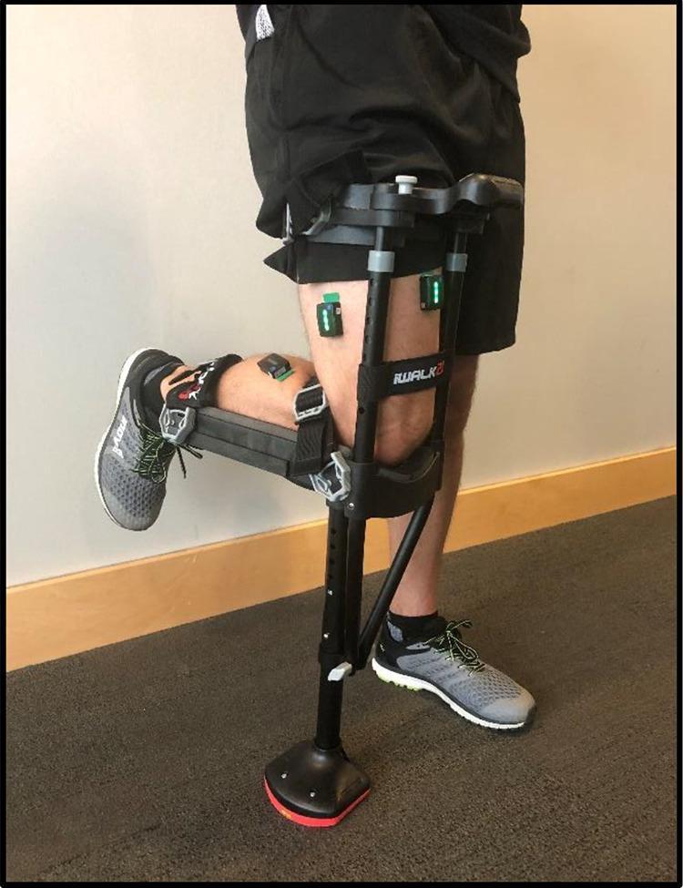
A clinical photograph illustrating a hands-free single crutch (HFSC), specifically the iWALKFree model, fitted to a user's dominant lower extremity. The device is designed for non-weight-bearing restrictions, showing the lower leg bent at a 90-degree angle at the knee and resting horizontally on a padded knee plate. The HFSC is secured with adjustable straps around the upper thigh and lower leg. Also visible are three Trigno Avanti wireless Bluetooth electromyography (EMG) sensors attached to the skin using adhesive pads. These sensors are strategically placed over the bellies of the rectus femoris, vastus lateralis, and lateral gastrocnemius muscles to monitor muscle activity and engagement during gait. The image demonstrates the clinical application of ambulatory aids combined with wearable sensor technology used to assess muscle atrophy prevention and gait analysis in orthopedic rehabilitation.
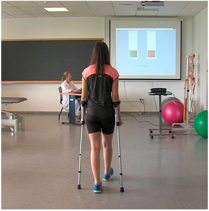
This clinical photograph illustrates a physical therapy and gait rehabilitation setting focusing on assistive device training. A female patient is seen from behind, ambulating with a pair of adjustable forearm crutches. These crutches are equipped with integrated force sensors designed to monitor partial weight-bearing (PWB) compliance. In the background, a large projection screen provides real-time visual biofeedback through two vertical bar charts; the left bar is green and the right is orange, representing the load distribution for each crutch. A healthcare professional monitors the session from a workstation. The setting includes common rehabilitation equipment such as an anatomical skeleton model and therapy balls. This setup demonstrates a closed-loop feedback mechanism in gait training, where sensor-based data is used to help patients self-correct their loading technique and adhere to prescribed weight-bearing limits during the recovery of lower limb pathologies.
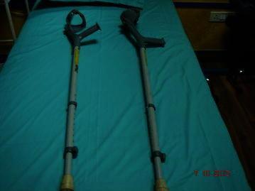
This clinical photograph displays a pair of adjustable elbow crutches, also known as Loftstrand or forearm crutches, resting on a flat teal surface. Each mobility aid consists of a contoured, semi-circular forearm cuff and an ergonomic hand grip at the top, typically made of high-density plastic or rubber. The main support structure is a metallic telescopic shaft featuring a series of adjustment holes and spring-loaded locking pins to customize the height for the user's specific anthropometrics. At the distal end, each crutch is fitted with a wide, non-slip rubber tip (ferrule) to provide traction and stability during ambulation. These devices are commonly utilized in physical therapy and orthopedic rehabilitation for patients with paraplegia, spinal cord injuries, or lower limb impairments who possess sufficient upper body strength and coordination to transition from more stable aids like walkers to less restrictive mobility tools.
A clear medical illustration of an axillary crutch (underarm crutch) with labeled parts pointing to: axillary pad (underarm pad), hand grip, upper shaft with height adjustment holes and pin, lower adjustable shaft, and rubber tip (ferrule) at the base. Clean white background, diagram style with label lines and text.
A clear medical illustration of an elbow crutch (forearm/Lofstrand crutch) with labeled parts pointing to: forearm cuff, hand grip, adjustable metal shaft with height adjustment holes and locking pin, and rubber tip (ferrule) at the base. Clean white background, diagram style with label lines and text.
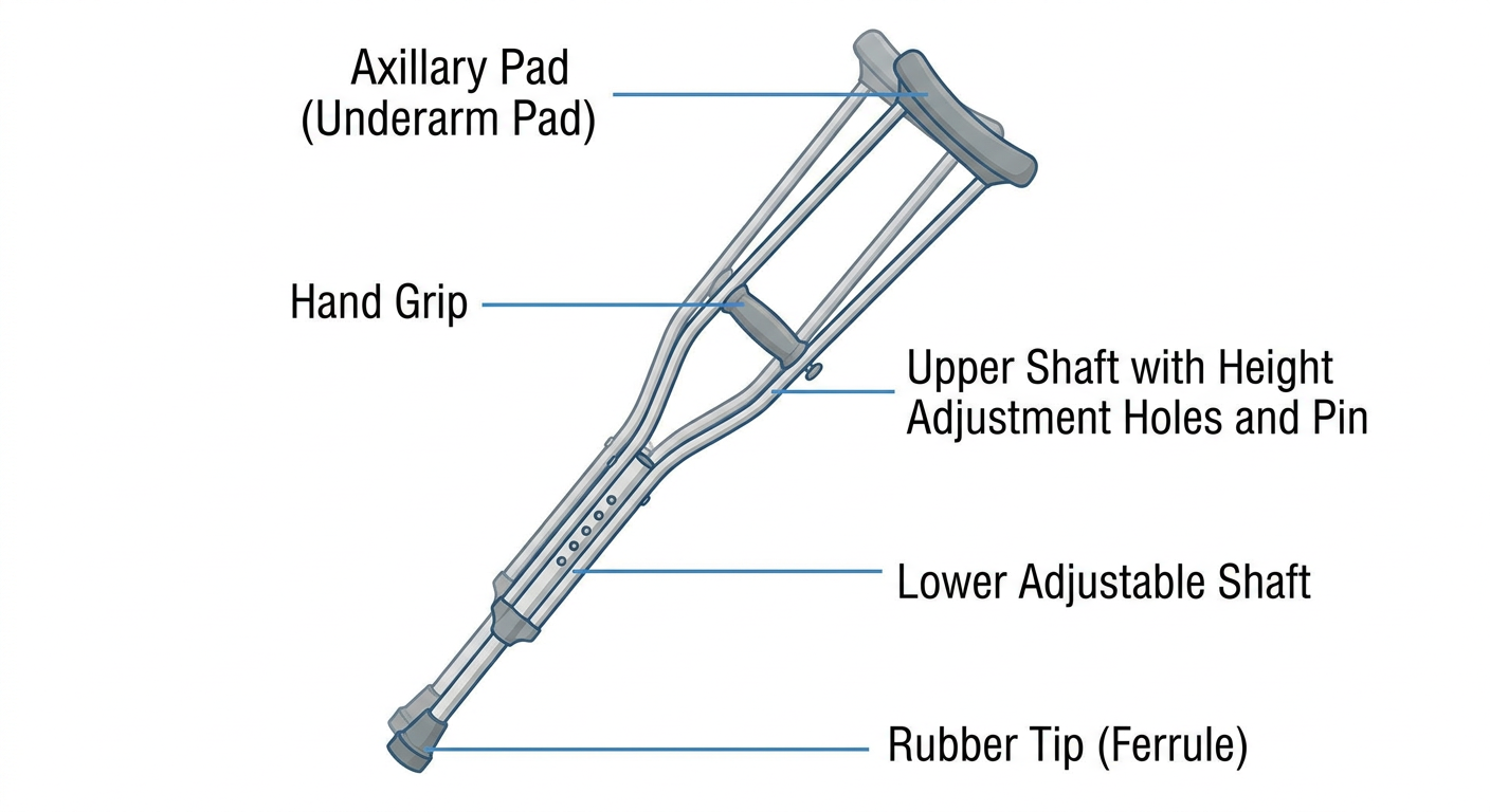
- Axillary pad - top padded piece that rests against the ribcage below the armpit (should NOT bear weight directly on the axilla, to avoid nerve compression)
- Hand grip - horizontal bar gripped by the hand, positioned so the elbow is slightly flexed (~15-30 degrees)
- Upper and lower shafts - adjustable telescoping bars with holes and a locking pin to set overall height and hand-grip height independently
- Rubber tip (ferrule) - non-slip base that contacts the ground
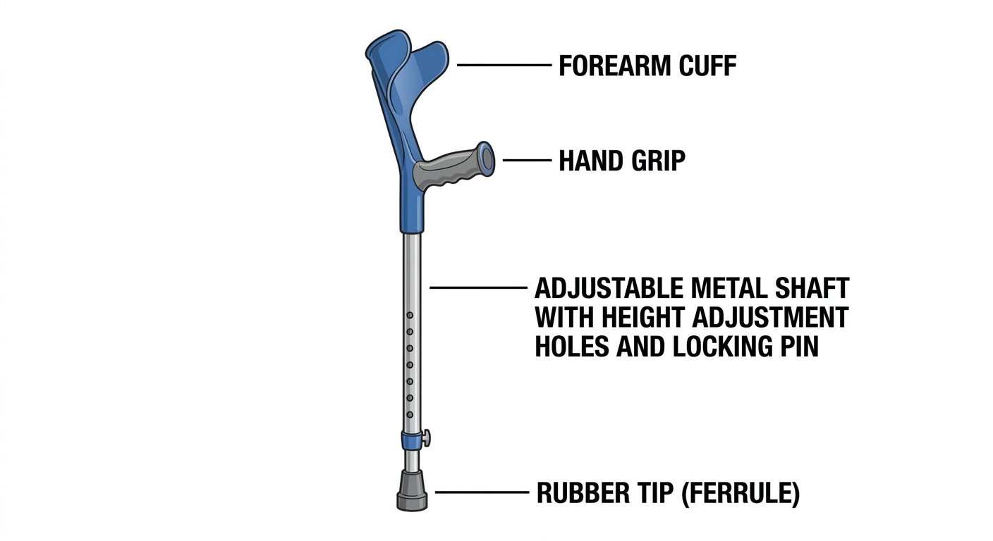
- Forearm cuff - semi-circular or open cuff that encircles the forearm just below the elbow, providing lateral support without an underarm pad
- Hand grip - gripped bar for weight-bearing through the hand
- Adjustable metal shaft - telescoping shaft with adjustment holes/pin for height
- Rubber tip (ferrule) - non-slip base