I am dermatology resident, I want complete long case of leprosy. Start from detailed history and significance of asking questions p. Also provide detailed clinical examination. Include also how to do all the test in sequence and its significance. Also includes nerve examinations in detail,where to palpate,and its course. Include Differential diagnosis. And management as per Nepal guidelines. Lastly detailed viva questions and quiz with answer side by

Reading File
Finding Sources
Searching the Web

Nepal national leprosy guidelines 2023 2024 MDT treatment protocol

Searching PubMed

"leprosy"[MeSH Terms] AND "diagnosis"[MeSH Terms]

Reading File
Reading File
Reading File
Reading Web Page

https://shisiradhikari.com/storage/uploads/National%20Lepros…

Reading File
Reading File
Finding Sources
Reading File
Reading File
Reading File
Searching the Web

Nepal leprosy treatment protocol MDT 2024 WHO classification paucibacillary multibacillary dose

Loading Image
Loading Image
I now have all the material needed to write the comprehensive long case. Let me compile it.

LEPROSY (Hansen's Disease) - Complete Long Case for Dermatology Residency

Sources: Andrews' Diseases of the Skin (Clinical Dermatology), Park's Textbook of Preventive & Social Medicine, Red Book 2021, Nepal National Leprosy Strategy 2021-2025, WHO Guidelines, PMC11346855

PART 1: DETAILED HISTORY TAKING

Opening Statement

"I am going to ask you some questions about your illness. Please answer freely - everything you say is confidential."

A. PRESENTING COMPLAINTS

Chief Complaint Format: Note the primary complaint with duration (in the patient's own words):
  • Skin patch/lesion with or without numbness
  • Thickened nerves
  • Weakness of hands or feet
  • Deformity of fingers/toes
  • Nodules on face/ears/body
  • Painless wounds or ulcers on hands/feet

B. HISTORY OF PRESENTING ILLNESS

1. Skin Lesions - Ask in detail:

QuestionClinical Significance
When did the patch first appear?Duration correlates with type: TT often older; LL insidious onset
How many patches are there?Key for WHO classification: 1-5 = PB; >5 = MB
Where did it start?TT favors face, limbs, trunk; LL diffuse/symmetric
Has it changed in number, size, or color?Progression from indeterminate to TT or LL; borderline is unstable
Is the surface of the patch dry/scaly/hairless?Anhidrosis and hair loss = autonomic nerve damage (TT/BT)
Is there change in sensation over the patch?Cardinal feature: anesthesia/hypoesthesia in skin lesion = diagnostic
What is the earliest sensation affected?Temperature is affected first, then light touch, then pain
Does the patch itch?Leprosy lesions typically do NOT itch - important negative
Has the lesion/patch become inflamed, red, swollen recently?Suggests Type 1 Reaction (reversal reaction) in borderline types
Has new nodular lesion appeared on old flat patch?Suggests upgrading in reversal reaction
Has there been ulceration of the patch?Can occur during Type 1 reaction

2. Neural Symptoms - Ask systematically:

QuestionSignificance
Do you have numbness anywhere?Neuropathy: earliest symptom in 90% of patients
Do you feel difficulty distinguishing hot from cold?Temperature discrimination lost first - dissociation of sensation is suspicious
Do you have tingling or burning sensation?Painful neuropathy; also early neuritis
Have you burned yourself without feeling it?Loss of protective sensation - risk of trophic ulcers
Do you have weakness of hand grip?Ulnar nerve involvement - claw hand
Can you spread your fingers?Ulnar nerve - interossei weakness
Do you have foot drop?Common peroneal nerve palsy
Do you have wrist drop?Radial nerve involvement
Do you have difficulty closing the eye completely?Facial nerve (zygomatic branch) - lagophthalmos - risk of corneal ulcer
Do you have pain in the nerve itself?Neuritis; tender enlarged nerve; common in reactions
Has weakness developed suddenly or gradually?Sudden = reaction-associated; gradual = chronic neuropathy

3. Reactional Episodes:

QuestionSignificance
Has there been sudden redness, swelling, tenderness of existing lesions?Type 1 (Reversal) Reaction - borderline spectrum
Have new painful raised red nodules appeared anywhere?Type 2 (ENL) - BL/LL spectrum
Accompanying fever, joint pain, eye redness?Systemic features of ENL (Type 2)
Painful testes?Orchitis in ENL/LL
History of iritis or red eye?Ocular involvement: iridocyclitis in ENL
Any nerve pain during reaction?Acute neuritis - needs urgent steroid treatment

4. Systemic Review:

QuestionSignificance
Nasal stuffiness, epistaxis, loss of smellNasal mucosa involvement in LL; saddle nose if advanced
Hoarseness of voiceLaryngeal involvement in LL
Loss of eyebrows/eyelashes (Madarosis)Classical LL finding - outer 1/3 eyebrows first
Hair loss from bodyLL - gradual body hair loss
Gynaecomastia, infertility, impotenceTesticular infiltration/atrophy in LL
Joint painsENL reaction; also Lucio phenomenon
Difficulty walking, recurrent foot ulcersPlantar anesthesia + neuropathy

C. PAST HISTORY

QuestionSignificance
Previous skin patches treated elsewhere?Relapse vs. new case; assess treatment history
Previous MDT treatment - how long and what drugs?Determines if currently on treatment; assess compliance
Any reactions during previous treatment?Risk of future reactions; drug adjustment needed
Previous nerve damage or disability?Grade the disability baseline
History of tuberculosisTB shares immune pathways; BCG vaccination status matters
Diabetes mellitusMimics lepromatous neuropathy; co-existing neuropathy complicates assessment

D. DRUG HISTORY

  • Current medications: MDT blister packs (rifampicin, dapsone, clofazimine)?
  • Thalidomide (if given for ENL)
  • Corticosteroids (if on reaction management)
  • Dapsone hypersensitivity syndrome history
  • Allergies to sulfonamides (cross-reactivity with dapsone)

E. FAMILY HISTORY

QuestionSignificance
Anyone in family/household with similar skin patches?Household contacts have 8-10x higher risk; transmission is droplet-based
Family history of leprosyGenetic susceptibility (HLA-DR2, HLA-DR3 in TT; HLA-DQ1 in LL)
Contact screening done?Nepal guidelines mandate household and close contact screening

F. PERSONAL & SOCIAL HISTORY

QuestionSignificance
OccupationAgricultural workers, those with soil/water contact in endemic areas at risk; stigma in occupational settings
Living conditions - crowding, ventilationOvercrowding facilitates droplet transmission
Area of residence - endemic or non-endemic region?Province 2 of Nepal is highest burden; Terai districts endemic
Nutritional statusMalnutrition worsens immune response; delays healing
Alcohol useAffects compliance; worsens peripheral neuropathy
Immigration history, travel to endemic regionsImportant in non-endemic countries
Psychological impact: stigma, depression, social isolationMajor cause of morbidity; essential to address
Employment status - lost job due to disease?Socioeconomic impact; disability allowance eligibility

G. REVIEW OF SYSTEMS (Specific to Leprosy)

  • Eyes: redness, photophobia, watering, decreased vision (corneal ulcer from lagophthalmos)
  • Nose: stuffiness, epistaxis, collapse of nasal bridge
  • Throat: hoarseness
  • Musculoskeletal: joint pains, bone pain, pathological fractures (absorption of phalanges in LL)
  • Genitourinary: testicular pain/swelling, sexual dysfunction (LL)
  • Psychological: depression, anxiety, suicidal ideation (especially due to social stigma)

PART 2: CLINICAL EXAMINATION

A. GENERAL EXAMINATION

Perform in sequence:
  1. General appearance - well/ill, nutritional status, signs of disability
  2. Vital signs - temperature (fever in ENL), pulse, BP
  3. BMI / nutritional status (malnutrition impairs immunity)
  4. Lymphadenopathy - axillary, inguinal (in MB/LL; also in ENL)
  5. Gynecomastia (testicular infiltration in LL)
  6. Eye examination (see below in specific examination)

B. SPECIFIC SKIN EXAMINATION

Expose the entire body in good natural light with patient undressed. Examine systematically from head to toe.

Step 1: Count and describe each lesion

For each lesion, document:
FeatureTuberculoid (TT/BT)Borderline (BB)Lepromatous (BL/LL)
Number1-5 (PB)Variable>5 (MB), may be innumerable
SizeLargeVariableSmall to large, diffuse
ColorHypopigmented or erythematousVariablePale macules, diffuse infiltration
EdgeWell-defined, raised, sharpPunched-out inner edge (BT), blurred outerIll-defined, blends into skin
SurfaceDry, scaly, hairlessLess dryShiny, waxy; hair initially present
DistributionAsymmetricMay be asymmetric or symmetricSymmetric, bilateral
Central healingCommon ("saucer right-side up")PartialAbsent
SensationAbsent (anesthetic)Variable, reducedMinimal loss early; late stocking-glove
SweatingAbsent (anhidrosis)ReducedAbsent late
Satellite lesionsAbsent in TT; small satellites in BTPresentMultiple
Classic sites to examine:
  • Face (cheeks, forehead, chin)
  • Upper limbs (arms, forearms - but NOT axillae)
  • Trunk (front and back)
  • Buttocks
  • Lower limbs (thighs, legs - but NOT groin, perineum, scalp)
  • Ears (helix, earlobe) - early nodules in LL
  • Eyebrows - madarosis (outer 1/3 loss first in LL)
Special feature - "Immune sites" (areas spared in leprosy): Scalp, axillae, groins, perineum, and inner aspects of elbows and knees are warm areas that are spared because M. leprae prefers cooler temperatures.
Tuberculoid leprosy - large annular erythematous plaque on lower leg
Tuberculoid leprosy: large, circular erythematous plaque with raised borders and central clearing (Andrews' Diseases of the Skin)
Lepromatous leprosy - multiple nodules (lepromas) on face
Lepromatous leprosy: diffuse nodular infiltration (lepromas) of the face with involvement of the ear lobe (Andrews' Diseases of the Skin)

Step 2: Sensory Testing Over Lesions

Test in the following sequence within the lesion and compare with adjacent normal skin:
  1. Touch - Use a wisp of cotton wool; touch lightly
  2. Temperature - Use test tubes of hot (~40°C) and cold water; ask patient to differentiate
  3. Pain/Pinprick - Use a sterile pin; ask if sharp or blunt
Method: Ask patient to close eyes. Touch normal skin first so they understand what to feel. Then test within the lesion. Ask "can you feel this?" and "is it sharp or blunt?" and "hot or cold?"
Significance:
  • Temperature is affected first (small fiber neuropathy)
  • Light touch next
  • Pain last
  • Sensory loss in a skin lesion is a cardinal sign of leprosy

Step 3: Sweating Test Over Lesions

Pilocarpin Test (Minor's Starch-Iodine Test):
  • Apply iodine solution to the lesion and normal skin
  • Cover with starch powder
  • Inject pilocarpine SC or ask patient to exercise
  • Normal skin turns dark blue-purple (sweating positive)
  • Leprosy lesion remains pale (anhidrosis in lesion area)
Significance: Autonomic nerve damage - postganglionic sympathetic fibers to sweat glands are involved early, even before sensory loss is obvious.

C. NERVE EXAMINATION - DETAILED

Overview - Nerves Commonly Affected in Leprosy

M. leprae preferentially affects superficially located peripheral nerves in cooler body regions. The key nerves to examine are:
NerveLocation of PalpationTest for Function
Great auricular nervePosterior triangle of neckSensory to angle of jaw, earlobe
Ulnar nerveCubital tunnel (medial epicondyle)Claw hand, interossei, hypothenar
Median nerveCarpal tunnel, medial to FCR at wristThumb opposition, thenar wasting
Radial cutaneous nerveRadial side of forearm, lateral to radial arterySensory - first web space
Common peroneal (lateral popliteal) nerveFibular head, neck of fibulaFoot drop, dorsiflexion
Posterior tibial nerveBehind medial malleolusPlantar sensation, intrinsic foot muscles
Supra-orbital nerveSupra-orbital notch, superomedial orbitForehead sensation
Facial nerveAt stylomastoid foramen / parotidEye closure, smile, facial muscles

NERVE PALPATION TECHNIQUE

General Principles:
  • Use the pads of index and middle fingers
  • Roll the nerve gently over the underlying bony/firm structure
  • Compare both sides
  • Assess: size (thickened?), consistency (firm, soft, nodular, beaded), tenderness, warmth (neuritis)
  • Do NOT compress hard - this causes unnecessary pain

1. GREAT AURICULAR NERVE (GAN)

Course: Arises from C2-C3 nerve roots. It winds around the posterior border of sternocleidomastoid (SCM) muscle, runs up along the surface of the SCM toward the parotid gland, and supplies sensation to the angle of the mandible, parotid area, and earlobe.
Palpation site:
  • Ask the patient to turn the head to the opposite side (this stretches the SCM and makes the nerve more prominent)
  • Palpate the posterior surface of the SCM at the junction of its upper and middle third
  • The nerve runs obliquely across the muscle surface from posterior to anterior-superior
Normal: May be barely palpable or impalpable Abnormal (leprosy): Visibly enlarged, palpable as a firm cord, can be nodular and beaded
Significance: Most commonly enlarged nerve visible to the naked eye in leprosy; enlargement of GAN is almost pathognomonic of leprosy if accompanied by skin lesion
Test for function: Sensory - test skin over the angle of jaw and earlobe for light touch and temperature

2. ULNAR NERVE

Course: Arises from medial cord of brachial plexus (C8, T1). Runs medially down the arm, passes behind the medial epicondyle in the ulnar groove (cubital tunnel), enters the forearm between the two heads of flexor carpi ulnaris (FCU), runs along the medial side of the forearm with the ulnar artery, passes through Guyon's canal at the wrist, and divides into superficial (sensory) and deep (motor) branches in the hand.
Palpation site:
  • Primary site: At the medial epicondyle - patient's elbow flexed 90°, palpate in the groove between medial epicondyle and olecranon. Roll the nerve gently.
  • Secondary site: Mid-upper arm, at the medial intermuscular septum
Abnormal: Thickened, tender, firm or nodular cord; sometimes visibly enlarged
Motor testing - Ulnar nerve:
  • Hand of Benediction sign / Claw hand: Clawing of 4th and 5th fingers (ring and little fingers) due to weakness of 3rd and 4th lumbricals (also intrinsic minus hand). Ask patient to straighten all fingers - if they cannot fully extend the ring and little finger IP joints, ulnar claw is present.
  • Froment's sign: Ask patient to hold a piece of paper between thumb and index finger. Weakness of adductor pollicis (ulnar) causes thumb to flex at IP joint (FPL compensates). Positive = flexion of thumb IP joint.
  • Card test: Ask patient to hold card between adjacent fingers (abduction/adduction) - weakness of interossei
  • Interossei wasting: Look for guttering (hollowing) between metacarpal heads on the dorsum of hand
  • Hypothenar wasting: Flat or wasted hypothenar eminence (abductor digiti minimi)
  • Grip strength: Reduced due to intrinsic muscle weakness
Sensory testing - Ulnar nerve:
  • Medial 1.5 fingers (little and half of ring finger) - dorsal and palmar aspects
  • Medial border of hand and palm

3. MEDIAN NERVE

Course: Formed by lateral and medial cords of brachial plexus (C5-T1). Runs in the medial bicipital groove alongside the brachial artery, enters the cubital fossa medial to the biceps tendon, passes between the heads of pronator teres, runs in the forearm between FDS and FDP, passes through the carpal tunnel under the flexor retinaculum, and divides into recurrent motor branch (thenar) and digital sensory branches.
Palpation site:
  • At the wrist: palpate medial to the tendon of flexor carpi radialis (FCR) and lateral to palmaris longus at the wrist crease
  • Also palpable in the forearm between FDS and FDP
Motor testing - Median nerve:
  • Pen cap test / Thumb-index pinch test: Ask to make a circle with thumb and index finger. Weakness of FPL and FDP index (anterior interosseous branch) causes rectangular shape rather than circular (OK sign test).
  • Thenar wasting: Flat thenar eminence - loss of abductor pollicis brevis
  • Opposition test: Ask to oppose thumb to little finger - lost in median nerve palsy
  • Bottle sign: Patient cannot fully oppose thumb and index finger around a bottle - indicative of thenar wasting
Sensory testing - Median nerve:
  • Radial 3.5 fingers (thumb, index, middle, radial half of ring)
  • Thenar eminence

4. RADIAL NERVE (Cutaneous Branch)

Course: The superficial radial nerve (purely sensory) leaves the main radial nerve about 8 cm above the wrist, passes under the brachioradialis, and emerges at the lateral aspect of the forearm to supply the dorsum of the radial 3.5 fingers and dorsal thenar eminence.
Palpation site: Radial aspect of the wrist, lateral to the radial artery, under the brachioradialis tendon. Roll gently over the radius.
Motor testing (radial nerve proper, less commonly affected in leprosy):
  • Wrist extension, finger extension
  • Wrist drop if radial nerve trunk involved
Sensory testing: Dorsum of the first web space (most reliable for superficial radial nerve)

5. COMMON PERONEAL NERVE (Lateral Popliteal Nerve)

Course: Branch of sciatic nerve. Winds around the fibular head at the neck of fibula (very superficial here), enters the lateral compartment of leg, divides into deep peroneal (motor - foot dorsiflexors, toe extensors; sensory - first web space of foot) and superficial peroneal (motor - evertors; sensory - dorsum of foot).
Palpation site:
  • Neck of fibula: The nerve wraps around the fibular neck from the posterior aspect of the knee to the anterior aspect. Place fingers just below and behind the fibular head and roll gently.
  • The nerve is most superficial here and this is the most common site of leprosy involvement.
Motor testing:
  • Foot drop: Ask patient to walk - high-stepping gait
  • Dorsiflexion: Ask patient to pull toes up toward the shin against resistance
  • Eversion: Ask patient to evert the foot against resistance
  • Toe extension: Ask to extend toes
Sensory testing:
  • Dorsum of foot (superficial peroneal)
  • First web space (deep peroneal)

6. POSTERIOR TIBIAL NERVE

Course: Accompanies posterior tibial artery behind the medial malleolus, passes through the tarsal tunnel (under flexor retinaculum) and divides into medial and lateral plantar nerves.
Palpation site:
  • Behind the medial malleolus between the medial malleolus and the Achilles tendon
  • The nerve lies posterior to the artery (remember: NAV - Nerve, Artery, Vein - posterior to anterior behind medial malleolus)
Motor testing:
  • Intrinsic foot muscles - toe flexion, abduction/adduction of toes
  • Plantar flexion (mainly tibial nerve, but also S1)
Sensory testing:
  • Plantar surface of foot (medial and lateral plantar nerves)
  • Medial and lateral toes
  • Heel (calcaneal branches)
Significance: Posterior tibial nerve damage leads to sensory loss on the plantar surface - this is responsible for painless plantar ulcers (leading cause of disability in leprosy).

7. FACIAL NERVE

Course: Exits the skull at the stylomastoid foramen, enters the parotid gland, and divides into five branches: temporal, zygomatic, buccal, marginal mandibular, and cervical.
Palpation site: Not routinely palpable; examined mainly by function testing. May be thickened at the stylomastoid foramen.
Motor testing - KEY IN LEPROSY:
  • Ask patient to close eyes tightly - look for incomplete closure (lagophthalmos)
  • Look for Bell's phenomenon (eye rolls up when trying to close - protective reflex)
  • Measure the gap of lagophthalmos in mm
  • Ask to raise eyebrows (temporal branch), smile (buccal/zygomatic), show teeth (buccal/marginal mandibular)
  • Corneal reflex - reduced/absent if trigeminal also involved
Significance: Facial nerve + trigeminal nerve (corneal sensation) involvement = risk of exposure keratitis and corneal blindness. The zygomatic branch is most commonly affected in leprosy.

8. SUPRA-ORBITAL NERVE (Branch of Trigeminal V1)

Course: Exits the orbit through the supra-orbital notch/foramen, located at the superior orbital rim at the mid-pupillary line.
Palpation site: Supra-orbital notch - palpate along the superior orbital rim for tenderness and thickening.
Sensory testing: Skin of forehead and anterior scalp.
Significance: In LL, supra-orbital nerve can be thickened; loss of forehead sensation is significant.

D. EYE EXAMINATION

Systematic and urgent in leprosy due to risk of blindness:
  1. Visual acuity - Snellen chart
  2. Cornea - look for corneal anesthesia (touch with wisp of cotton wool), pannus, ulceration, scarring
  3. Iris/pupil - signs of iritis (perilimbal injection, constricted pupil, posterior synechiae, irregular pupil), beaded anterior chamber (pearls = lepromas)
  4. Eyelids - lagophthalmos, madarosis, ectropion, entropion
  5. Conjunctiva - tarsal nodules
  6. Sclera - scleritis (in ENL)
  7. Pupillary reactions - sluggish pupil if autonomic involvement

E. EAR EXAMINATION

  • Inspect the helix and earlobe - early lepromas in LL appear here
  • Thickening/nodularity of earlobes
  • Palpate for GAN thickening

F. NOSE EXAMINATION

  • Collapse of nasal bridge (saddle nose) - destruction of nasal septum cartilage in LL
  • Nasal mucosa: epistaxis, septal perforation
  • Atrophied nostrils

G. HAND EXAMINATION

Sequential examination:
  1. Look (dorsum first):
    • Claw hand (4th and 5th finger) - ulnar nerve
    • Ape thumb deformity - median nerve (thenar wasting, thumb falls back in same plane as fingers)
    • Combined claw (all fingers) - both ulnar and median
    • Wrist drop - radial nerve
    • Trophic changes: dry skin, fissures, ulcers (especially on digits)
    • Shortening/absorption of fingers (LL, advanced)
    • Contractures
    • Interosseous guttering on dorsum of hand
  2. Look (palmar surface):
    • Thenar wasting (median)
    • Hypothenar wasting (ulnar)
    • Palmar fissures/trophic changes
  3. Feel: Warmth, tenderness, swelling
  4. Move: As described in nerve examination section

H. FOOT EXAMINATION

  1. Inspection:
    • Foot drop - dropped posture, high stepping gait
    • Trophic ulcers: usually on pressure points (ball of foot, heel, metatarsal heads)
    • Fissures, callus
    • Absorption/shortening of toes
    • Claw toes (intrinsic muscle weakness)
  2. Sensation: Test dorsum and plantar surface; monofilament if available
  3. Pulses: Peripheral vascular disease mimics and coexists in some patients

I. MUSCULOSKELETAL EXAMINATION

  • Charcot joints: painless deformity of joints (rare, due to repeated trauma without sensation)
  • Wasting of intrinsic hand and foot muscles
  • Contractures of digits
  • Shortening and auto-amputation of digits (LL - advanced)

PART 3: INVESTIGATIONS

Perform in the following sequence with significance:

1. SLIT-SKIN SMEAR (SSS) - MOST IMPORTANT INVESTIGATION

How to perform:
  1. Select sites: both earlobes, chin, forehead, and the edge of the most active skin lesion (total 4-6 sites)
  2. Clean the site with spirit and let dry
  3. Make a small fold of skin between thumb and forefinger - this blanches the area and reduces bleeding
  4. Use a fresh disposable lancet or scalpel (#15 blade)
  5. Make a small incision 5mm long and 2-3mm deep in the dermis (NOT too deep - dermis only)
  6. Scrape the walls of the incision firmly to obtain tissue fluid and cells
  7. Spread the material evenly on a glass slide (circular smear, 5-7mm diameter)
  8. Allow to air dry, fix with methanol
  9. Stain with Ziehl-Neelsen (modified - using 5% H2SO4 not HCl) or Fite-Faraco stain
  10. Examine under oil immersion (100x)
What to look for:
  • Bacillary Index (BI): Logarithmic scale 0-6+ (Ridley's scale)
    • 0 = no bacilli in 100 fields
    • 1+ = 1-10 bacilli in 100 fields
    • 2+ = 1-10 bacilli in 10 fields
    • 3+ = 1-10 bacilli in 1 field
    • 4+ = 10-100 bacilli per field
    • 5+ = 100-1000 bacilli per field
    • 6+ = >1000 bacilli / clumping per field
  • Morphological Index (MI): Percentage of solid-staining (viable) bacilli. Normal bacteria stain solid (viable, infective). Fragmented/granular = dead bacilli (good response to treatment).
  • Globi: Round clumps of bacilli - seen in LL (Virchow's lepra cells)
Significance:
  • Positive SSS = definitive diagnosis; confirms MB leprosy
  • Negative SSS does not exclude leprosy (PB cases are negative)
  • Used for classification into PB/MB
  • MI used to monitor treatment response (solid forms decrease with effective therapy)
  • BI used to monitor for relapse (should fall by 1 unit/year on MDT)

2. SKIN BIOPSY AND HISTOPATHOLOGY

Technique:
  1. Select the active border of the most representative lesion
  2. Include normal skin and transition zone at the edge
  3. Full-thickness punch biopsy (4-6mm), ensure it reaches subcutaneous fat
  4. Fix in 10% formalin
  5. Request Fite-Faraco stain (optimal for M. leprae - uses peanut oil/xylene in decolorization step to preserve AFB staining, unlike standard Ziehl-Neelsen which may decolorize leprae organisms)
  6. Also request H&E stain and S-100 stain (to highlight nerve involvement)
Histopathological Features:
TypeHistology
IndeterminateNon-specific lymphocytic infiltrate around neurovascular bundles; minimal bacilli; no granuloma
Tuberculoid (TT)Epithelioid granulomas with Langhan's giant cells; no grenz zone (touches epidermis); perineural concentric fibrosis; few/no AFB
Borderline Tuberculoid (BT)Similar to TT but with grenz zone; some vacuolation of epithelioid cells; occasional bacilli
Borderline (BB)Poorly organized granulomas; macrophages with foamy cytoplasm; no giant cells; moderate bacilli
Borderline Lepromatous (BL)Foamy histiocytes predominant; lymphocytes dispersed in granuloma; abundant organisms; perineural lymphocytic infiltration
Lepromatous (LL)Virchow's lepra cells (foam cells, lipid-laden, bacilli-laden); abundant AFB in globi; clear grenz zone; no giant cells; sparse lymphocytes (suppressed CMI)
Histoid LeprosySpindle cells (histiocytes) in storiform pattern; solid staining bacilli very abundant; distinct from surrounding skin
The grenz zone: A clear strip of uninvolved papillary dermis between the inflammatory infiltrate and the epidermis. Absent in TT (infiltrate extends to epidermis), present in BT and LL.
Significance: Allows Ridley-Jopling (RJ) classification; determines prognosis; guides treatment intensity.

3. LEPROMIN (MITSUDA) TEST

Preparation of Lepromin:
  • Integral lepromin (Dharmendra antigen): heat-killed M. leprae
  • Mitsuda lepromin: crude preparation from infected human/armadillo tissue
Method:
  1. Inject 0.1 mL of standard lepromin intradermally on the flexor aspect of forearm
  2. Read results at two time points:
    • Fernandez reaction (early): Read at 24-48 hours - indicates delayed hypersensitivity (like tuberculin)
    • Mitsuda reaction (late): Read at 28 days - the main clinically used reading
Interpretation of Mitsuda reaction:
ResultIndurationInterpretation
Negative<3mm or no papuleNo CMI; suggests lepromatous spectrum
Positive (+)3-5mm firm noduleModerate CMI
Strongly positive (++)5-10mm noduleGood CMI; TT spectrum
Strongly positive (+++)>10mm with ulcerationVery strong CMI; TT response
Significance:
  • Correlates with CMI status and position on Ridley-Jopling spectrum
  • TT: Strongly positive (++/+++)
  • BT: Positive (+/++)
  • BB: Weakly positive or negative
  • BL: Negative
  • LL: Strongly negative
  • NOT a diagnostic test (positive in BCG vaccinated, tuberculosis contacts)
  • Useful as a prognostic indicator
  • Not routinely done in Nepal; used in research/classification settings

4. SENSORY TESTING - FORMAL METHODS

Semmes-Weinstein Monofilament Test:
  • Nylon filaments of graded stiffness
  • Apply perpendicular to skin until filament bends; patient reports sensation
  • Map areas of sensory loss
Hot-Cold Test: Formally using two test tubes (one filled with water at 40-45°C, other at 5-10°C); test random areas of skin and nerve territories with eyes closed.
Ballpoint pen test / Two-point discrimination: For fine touch assessment
Significance: Documents the extent of neuropathy; baseline before treatment to monitor; identifies at-risk areas for trophic ulcers; grades disability.

5. VOLUNTARY MUSCLE TESTING (VMT)

How to grade (MRC grading):
GradeDescription
0No contraction
1Visible contraction, no movement
2Movement with gravity eliminated
3Movement against gravity
4Movement against resistance (reduced strength)
5Normal strength
Muscles to test systematically:
  • Ulnar nerve: First dorsal interosseous, abductor digiti minimi, hypothenar muscles
  • Median nerve: Abductor pollicis brevis (APB) - ask patient to point thumb straight up perpendicular to palm and resist downward pressure
  • Radial nerve: Wrist extensors, finger extensors
  • Common peroneal: Tibialis anterior (dorsiflexion), extensor hallucis longus, peronei (eversion)
  • Posterior tibial: Intrinsic foot muscles
  • Facial nerve: Orbicularis oculi, orbicularis oris, frontalis
Significance: Grading impairment; WHO disability grading requires VMT; monitoring during reactions; outcome of steroid treatment.

6. WHO DISABILITY GRADING

GradeHandsEyesFeet
0No problemNo problemNo problem
1Diminished sensation, no deformityReduced vision (VA 6/60 or better), no lagophthalmosDiminished sensation, no deformity
2Visible deformity (claw, wrist drop, absorption)Severe visual impairment/blindness OR lagophthalmos/corneal diseaseVisible deformity (drop foot, absorption, ulcer)
Significance: Standard measure for surveillance in Nepal National Program; determines disability allowance; used to assess program performance.

7. NERVE CONDUCTION STUDIES (NCS) / ELECTROMYOGRAPHY

  • Objective assessment of large fiber neuropathy
  • Confirms axonal vs. demyelinating pattern
  • Useful in pure neural leprosy (no skin lesions)
  • May show abnormalities before clinical deficits appear
  • Not routinely available at district level in Nepal

8. HIGH-RESOLUTION ULTRASONOGRAPHY (HRUS) OF NERVES

  • A 2026 systematic review [PMID: 41773898] confirms HRUS is accurate for detecting peripheral nerve enlargement in leprosy
  • Shows increased cross-sectional area of affected nerves
  • Ulnar nerve at medial epicondyle: enlargement starts at the ulnar sulcus, reaches maximum 4 cm above medial epicondyle
  • Non-invasive, real-time assessment; useful in pure neural leprosy
  • Increasingly available at teaching hospitals in Nepal

9. PCR FOR M. LEPRAE

  • Detects M. leprae DNA in tissue, blood, skin
  • Useful in pure neural leprosy or pauci-bacillary cases where SSS and biopsy are negative
  • Sensitivity ~50% in PB cases; higher in MB
  • Not widely available in Nepal; used at reference centers

10. SEROLOGY

  • Anti-PGL-1 (phenolic glycolipid 1) antibody: Positive in 90% of LL, only 50% of TT
  • Fluorescent Leprosy Antibody (FLA-ABS) test
  • Poor sensitivity in PB leprosy
  • Not routinely used for diagnosis; mainly for research/contact screening

11. ADDITIONAL INVESTIGATIONS

InvestigationPurpose
CBCBaseline before MDT; monitor for dapsone-induced hemolytic anemia (especially G6PD deficiency)
G6PD levelsBefore starting dapsone; G6PD deficiency risk of severe hemolysis
LFTsRifampicin hepatotoxicity monitoring
RFTsBaseline; clofazimine accumulation in renal failure
Blood glucoseRule out diabetic neuropathy as differential/co-morbidity
CXRRule out tuberculosis (co-infection; also before rifampicin)
Slit-lamp examinationENL uveitis; iritis
X-ray hands/feetBone absorption (LL), neuropathic arthropathy

PART 4: DIFFERENTIAL DIAGNOSIS

Primary Differentials for Hypopigmented/Anesthetic Patch:

ConditionDistinguishing Features
VitiligoComplete depigmentation (chalk-white, not just hypopigmented); NORMAL sensation; no nerve thickening; skin texture normal; Wood's lamp: bright fluorescence
Tinea Versicolor (Pityriasis versicolor)Hypopigmented/hyperpigmented macules; fine powdery scale; NORMAL sensation; KOH shows spaghetti and meatball appearance (Malassezia); responds to antifungals
Pityriasis AlbaIll-defined hypopigmented patches; usually children; face; associated with atopic tendency; normal sensation; spontaneously resolves
Nevus depigmentosusCongenital hypopigmented patch; normal sensation; stable; no nerve thickening
Hypopigmented mycosis fungoidesHypopigmented patches; scale; normal or reduced sensation; biopsy: lymphocytic epidermotropism (Pautrier microabscesses); CD4 T-cells
Lichen sclerosusWhite sclerotic patches; genital involvement; normal sensation

Primary Differentials for Thickened Nerves:

ConditionDistinguishing Features
Charcot-Marie-Tooth DiseaseHereditary; bilateral symmetric; no skin lesions; pes cavus; no AFB
Amyloidosis (primary)May cause nerve enlargement; systemic features; biopsy: amyloid deposits
NeurofibromatosisMultiple neurofibromas along nerve course; Café-au-lait patches; Lisch nodules in iris; normal sensation in patches
Peroneal muscular atrophyNo skin lesions; family history

For Nodular Lesions in LL:

ConditionDistinguishing Features
Leishmaniasis (post-kala azar dermal)Nepal endemic; hypopigmented macules and nodules; absence of nerve involvement; slit smear: Leishmania amastigotes
RhinoscleromaGranulomatous nasal disease; Klebsiella rhinoscleromatis; Mikulicz cells on biopsy
SarcoidosisNon-caseating granulomas; systemic features; elevated ACE; bilateral hilar adenopathy
XanthomasYellow color; lipid abnormalities; no sensory loss

For Erythematous Lesions (Borderline):

ConditionDistinguishing Features
Granuloma annulareAnnular erythematous papules/plaques; NO sensory loss; no nerve thickening; biopsy: palisading granuloma
PsoriasisSilvery scales; Auspitz sign; nail changes; normal sensation
Tinea corporisErythematous scaly annular lesion; KOH positive; pruritic; normal sensation; antifungal responsive
Lupus vulgarisTB of skin; apple jelly nodules; no anesthesia; Mycobacterium tuberculosis
Discoid lupus erythematosusFollicular plugging; ANA positive; no nerve changes
Annular sarcoidosisNon-tender annular plaques; systemic sarcoidosis features; biopsy differentiates

For ENL (Type 2 Reaction):

ConditionDistinguishing Features
Erythema nodosumPainful nodules anterior shin; NO previous leprosy; biopsy: septal panniculitis; associated with systemic diseases
Sweet syndromeAcute febrile neutrophilic dermatosis; no leprosy history; biopsy: dense neutrophilic infiltrate
VasculitisOther systemic features; palpable purpura; ANA/ANCA positive

PART 5: MANAGEMENT AS PER NEPAL GUIDELINES

Nepal follows WHO/NLEP guidelines aligned with the National Leprosy Strategy 2021-2025, with the goal of achieving "Leprosy Free Nepal."

A. CLASSIFICATION FOR TREATMENT

WHO Operational Classification (used in Nepal):
TypeDefinitionTreatment Category
Paucibacillary (PB)1-5 skin lesions; SSS negative; no more than 1 nerve trunk involvedPB-MDT
Multibacillary (MB)>5 skin lesions; OR SSS positive; OR more than 1 nerve trunk involvedMB-MDT
Pure Neuritic LeprosyNerve involvement only, no skin lesionsTreat as MB (SSS of nerve territory)
Note: From 2023-2025, WHO and India's NLEP updated to Uniform MDT (3 drugs for both PB and MB) - Nepal's National Program is moving toward alignment. The current Nepal guideline uses the above classification.

B. MDT REGIMEN - NEPAL/WHO STANDARD (Updated 2024)

PB-MDT (1-5 lesions) - Duration: 6 months

DrugAdult DoseChild (10-14 years)Child (<10 years or <40kg)
Rifampicin600 mg once monthly (supervised)450 mg once monthly10 mg/kg once monthly
Dapsone100 mg daily50 mg daily2 mg/kg daily
Clofazimine300 mg once monthly + 50 mg daily150 mg once monthly + 50 mg alternate days100 mg once monthly + 50 mg twice weekly
Note: Clofazimine is now included for PB cases too (updated WHO 2023 uniform MDT)

MB-MDT (>5 lesions) - Duration: 12 months

DrugAdult DoseChild (10-14 years)Child (<10 years or <40kg)
Rifampicin600 mg once monthly (supervised)450 mg once monthly10 mg/kg once monthly
Clofazimine300 mg once monthly + 50 mg daily150 mg once monthly + 50 mg alternate days6 mg/kg/month + 1mg/kg/day
Dapsone100 mg daily50 mg daily2 mg/kg daily
MDT is provided FREE at all public health facilities in Nepal.

C. IMPORTANT COUNSELING POINTS BEFORE MDT

  1. Rifampicin: Turns urine, sweat, tears orange-red - normal; soft contact lenses staining; hepatotoxicity (monitor LFTs)
  2. Clofazimine: Causes skin discoloration (brownish-black, especially in darker skin); takes months to years to resolve after stopping; also causes GI discomfort, ichthyosis
  3. Dapsone: May cause hemolytic anemia - test G6PD before starting; methemoglobinemia; peripheral neuropathy (uncommon); dapsone hypersensitivity syndrome (rare, first 6 weeks)
  4. Explain that the disease is curable and MDT kills bacteria within days - patient is non-infectious very quickly after starting treatment
  5. Address stigma - leprosy is NOT highly contagious; routine household contact is generally safe

D. MANAGEMENT OF LEPROSY REACTIONS

Type 1 Reaction (Reversal Reaction)

  • Indication for treatment: Nerve function impairment OR severe skin inflammation
  • First-line treatment: Prednisolone 40-60 mg/day (1 mg/kg/day), taper over 6 months minimum
  • If no nerve involvement: NSAIDs may be sufficient for skin inflammation alone
  • Continue MDT throughout reactions - do not stop MDT
  • Monitor nerve function regularly during and after steroid treatment
  • Long-term steroid: add methotrexate 7.5-15 mg weekly as steroid-sparing agent

Type 2 Reaction (Erythema Nodosum Leprosum - ENL)

  • Mild: NSAIDs, chloroquine
  • Moderate to severe: Prednisolone 40-60 mg/day; taper as symptoms resolve
  • Severe/recurrent ENL: Thalidomide 100-400 mg/day (NOT in women of childbearing age - teratogenic)
  • Continue MDT throughout ENL treatment
  • Treat associated features: fever (antipyretics), orchitis (scrotal support), iritis (topical steroids + atropine, ophthalmology referral)
  • Clofazimine has anti-inflammatory properties - increase to 300 mg/day during ENL (may reduce steroid dependence)

E. MANAGEMENT OF DISABILITIES

Eye Care (Grade 1/2):

  • Lagophthalmos: Artificial tears; taping the eyelid at night; protective glasses; if severe - lateral tarsorrhaphy
  • Iritis/iridocyclitis: Topical steroids + atropine (ophthalmology referral urgent)
  • Regular ophthalmologic review
  • Patient education: visual inspection of eyes daily; report redness or pain immediately

Hand Care:

  • Soaking in warm water + moisturizing oils daily (clofazimine causes dry skin)
  • Protective footwear and gloves
  • Wound care for trophic ulcers (debridement, dressing)
  • Physiotherapy: passive and active exercises to prevent contracture
  • Splinting for claw hand
  • Reconstructive surgery if stable disability: tendon transfers for claw hand, foot drop correction

Foot Care:

  • Custom footwear (molded shoes with soft insoles)
  • Daily inspection of feet; patient education (they cannot feel injuries)
  • Plantar ulcer management: complete offloading; regular debridement; wound care

F. POST-EXPOSURE PROPHYLAXIS (SDR-PEP)

  • Single Dose Rifampicin (SDR-PEP): Given to household and close contacts of newly diagnosed leprosy patients
  • Adult dose: Rifampicin 600 mg single dose
  • Reduces risk of developing leprosy by approximately 57% in contacts
  • Exclusion criteria: Already on rifampicin-containing treatment (e.g., active TB), known rifampicin allergy, children <2 years
  • Nepal National Program mandates contact screening and SDR-PEP as per 2021-2025 strategy

G. CONTACT TRACING AND SCREENING (Nepal Guidelines)

  1. Identify and examine all household contacts (those living in the same house)
  2. Identify and examine close social contacts (regular close contact for >6 months)
  3. Screen for skin patches, nerve thickening, and sensory loss
  4. Offer SDR-PEP to eligible contacts
  5. Register contacts in the Nikusth online platform (Nepal's digital leprosy surveillance system)
  6. Re-examine contacts annually for 5 years

H. BCG VACCINATION

  • BCG provides 28-60% protection against leprosy
  • Already part of Nepal's routine immunization program
  • Additional BCG dose used in some high-burden provinces as adjunct prophylaxis

I. DISABILITY PREVENTION AND REHABILITATION

Following Nepal's national strategy pillars:
  • Regular disability assessment at each follow-up visit
  • Teaching self-care (skin moisturization, daily inspection, protective footwear)
  • Physiotherapy referral
  • Psychological support and counseling
  • Vocational rehabilitation
  • Leprosy welfare allowance (provided by Government of Nepal to registered disabled patients)
  • Anti-stigma education for community and family

J. FOLLOW-UP SCHEDULE

TimingWhat to do
MonthlySupervised rifampicin dose; examine for reactions; check for new lesions or nerve symptoms; dispensing remaining MDT
At treatment completionFull clinical examination; SSS (MB cases); disability grading; issue release from treatment card
Post-treatment (MB)Annual review for 5 years (relapse/reaction monitoring); BI falls 1+ unit per year
Post-treatment (PB)2-year follow-up

PART 6: VIVA QUESTIONS AND QUIZ (WITH ANSWERS)


SECTION A: VIVA QUESTIONS


Q1. What are the WHO cardinal signs of leprosy?
A: There are three cardinal signs:
  1. Hypopigmented or erythematous skin patch with definite sensory loss
  2. Thickened/enlarged peripheral nerve with sensory loss and/or motor weakness
  3. Positive slit-skin smear (SSS) for acid-fast bacilli
The presence of any one of these is sufficient for diagnosis. The presence of two or more makes the diagnosis certain.

Q2. Which sensation is lost first in leprosy and why?
A: Temperature sensation is lost first, followed by light touch, and then pain. This is because M. leprae has a predilection for cooler areas and selectively damages small myelinated (A-delta) and unmyelinated (C) nerve fibers first, which carry temperature and pain. This "dissociation of sensibility" - where temperature is lost but pain is relatively preserved early - is characteristic and highly suspicious for leprosy. - Andrews' Diseases of the Skin, p.396

Q3. Why does leprosy spare the scalp, axillae, groin, and perineum?
A: M. leprae grows optimally at temperatures below 37°C (30-32°C). These areas are warm due to body heat, hair insulation, and proximity to large vessels. The cooler peripheral areas (face, ears, hands, feet) are preferentially affected. The skin of the scalp, axillae, groin, and perineum remains warm enough to inhibit bacillary multiplication.

Q4. Describe the Ridley-Jopling classification and its immunologic basis.
A: The Ridley-Jopling classification is based on histopathology and immune status:
  • TT (Tuberculoid): Strong cell-mediated immunity (CMI); few well-formed epithelioid granulomas; no bacilli; IFN-γ and IL-2 dominant; lepromin strongly positive
  • BT (Borderline Tuberculoid): Good CMI; similar to TT with grenz zone
  • BB (Mid-Borderline): Unstable; moderate CMI; foamy macrophages; moderate bacilli; "punched-out" lesions
  • BL (Borderline Lepromatous): Poor CMI; foamy histiocytes; many bacilli; lepromin negative
  • LL (Lepromatous): No effective CMI; Virchow's foam cells; abundant globi; IL-4, IL-5, IL-10 dominant (Th2 response); lepromin strongly negative

Q5. What is the significance of the morphological index (MI) in SSS?
A: The MI is the percentage of solid-staining bacilli in a SSS. Solid staining = intact, viable, potentially infective bacilli. Fragmented or granular staining = dead bacilli. On effective MDT, the MI drops rapidly (within weeks) - this indicates a good bacteriological response. The Bacillary Index (BI) drops more slowly (1+ unit per year). A rise in MI after clinical cure suggests relapse with drug-resistant organisms.

Q6. What is Type 1 (Reversal) Reaction and how does it differ from Type 2 (ENL)?
A:
FeatureType 1 (Reversal Reaction)Type 2 (ENL)
Spectrum affectedBorderline (BT, BB, BL)BL, LL
MechanismSudden increase in CMI (delayed hypersensitivity)Immune complex-mediated (Type III hypersensitivity)
Skin lesionsExisting lesions become red, swollen, tender; new lesions may appearNew tender erythematous papules/nodules on normal skin
Systemic featuresFever uncommonFever, malaise, arthralgia, orchitis common
Polymorphs in lesionAbsentPresent (hence neutrophils prominent in histology)
Nerve involvementCommon and severe - acute neuritis, risk of permanent damageLess severe nerve involvement
TreatmentPrednisolone (prolonged, minimum 6 months)Prednisolone; thalidomide for recurrent ENL
PatternUsually single episode, but prolongedMay be recurrent/chronic

Q7. A patient comes with leprosy patch and develops acute severe eye redness and reduced vision. What do you suspect and how do you manage?
A: This suggests acute iridocyclitis (anterior uveitis), most commonly associated with ENL (Type 2 reaction) in BL/LL leprosy. Features: perilimbal injection, photophobia, reduced vision, irregular constricted pupil, posterior synechiae.
Management:
  1. Urgent ophthalmology referral
  2. Topical corticosteroid eye drops (e.g., prednisolone acetate 1% hourly initially)
  3. Cycloplegic drops (atropine 1%) to dilate pupil, prevent posterior synechiae
  4. Systemic corticosteroids if severe
  5. Continue MDT
  6. Treat underlying ENL with prednisolone/thalidomide

Q8. What is pure neural leprosy? Why is it important in Nepal?
A: Pure neuritic leprosy (PNL) is a form of leprosy where only the peripheral nerves are involved without any skin lesions. The diagnosis is challenging because the classic skin signs are absent. It is particularly common in Nepal - PNL represents up to 5% of all new leprosy cases in Nepal and India. The ulnar nerve is most commonly affected. Diagnosis requires nerve biopsy or PCR. Management is as MB-MDT. It is important because of frequent misdiagnosis as other peripheral neuropathies (diabetes, entrapment neuropathy), leading to delayed treatment and preventable disability.

Q9. Describe the grenz zone and its significance in leprosy histopathology.
A: The grenz zone (German for "border zone") is a clear, uninvolved strip of papillary dermis between the inflammatory infiltrate and the overlying epidermis. It is:
  • Absent in TT: Granulomas extend up to the epidermis - reflects vigorous CMI
  • Present in BT, BL, LL: Degree of grenz zone widens toward the lepromatous pole
  • Significance: Differentiates TT from BT histologically; important in classification; reflects the immune status of the host

Q10. What is Lucio phenomenon?
A: The Lucio phenomenon (erythema necroticans) is a severe reactional state unique to diffuse lepromatous leprosy (Diffuse Leprosy of Lucio), rare outside of Mexico and Central America. It presents as irregular hemorrhagic infarcts and ulcerations on extremities due to endarteritis obliterans of dermal vessels. Histology shows vascular occlusion and necrosis without typical granulomatous inflammation. It carries a high mortality. It is NOT the same as ENL.

Q11. How do you examine and grade lagophthalmos in leprosy?
A:
  1. Ask the patient to close both eyes gently
  2. Observe if there is a gap between upper and lower eyelids
  3. Measure the gap in mm using a ruler
  4. Check for Bell's phenomenon (protective upward rolling of eye when trying to close)
  5. Check corneal sensation (V1 - trigeminal) with wisp of cotton
  6. Grade:
    • Grade 1 disability: Lagophthalmos present but no corneal damage
    • Grade 2 disability: Corneal ulceration, opacity, or vision <6/60
Management: Artificial tears, taping at night, tarsorrhaphy in severe cases, physiotherapy for facial nerve rehabilitation.

Q12. What MDT regimen do you use for a pregnant woman with MB leprosy in Nepal?
A: MDT is safe in pregnancy. Do NOT withhold treatment as untreated leprosy can cause reactions and nerve damage. Use standard MB-MDT (Rifampicin 600 mg monthly + Clofazimine 300 mg monthly + 50 mg daily + Dapsone 100 mg daily) for 12 months. Dapsone is safe in pregnancy. Clofazimine is safe; note the infant may have skin discoloration (clears spontaneously). Breastfeeding is also safe during MDT. The infant's urine may be orange (rifampicin excretion in breast milk) - reassure the mother.

Q13. What is the Fite-Faraco stain and why is it preferred over standard ZN stain for leprosy?
A: Both are acid-fast stains but Fite-Faraco uses a modified decolorization step employing peanut oil and xylene instead of strong acid/alcohol. Standard Ziehl-Neelsen uses aggressive decolorization (concentrated HCl) which tends to destroy the lipid-poor cell wall of M. leprae, causing it to lose its acid-fast property and appear negative. Fite-Faraco is gentler, preserving the staining of M. leprae bacilli, making it the preferred stain for leprosy histopathology.

Q14. How is dapsone resistance detected and managed?
A: Dapsone resistance is suspected in patients who relapse on dapsone monotherapy or who fail MDT. Detection: Molecular methods - detection of mutations in folP1 gene (dihydropteroate synthase gene) by PCR/DNA sequencing. Resistance can also be confirmed by mouse footpad inoculation (reference labs). Management: Use Rifampicin-based regimen; second-line drugs: ofloxacin/minocycline/clarithromycin (ROM regimen - Rifampicin + Ofloxacin + Minocycline). Histoid leprosy (with very high BI) often indicates dapsone resistance.

Q15. Describe the Ridley scale for the Bacillary Index (BI).
A: Ridley's logarithmic scale for SSS:
  • 0 = No bacilli in 100 oil immersion fields
  • 1+ = 1-10 bacilli in 100 fields
  • 2+ = 1-10 bacilli in 10 fields
  • 3+ = 1-10 bacilli per field
  • 4+ = 10-100 bacilli per field
  • 5+ = 100-1000 bacilli per field
  • 6+ = >1000 bacilli per field or many clumps (globi)
Average BI is calculated from all sites sampled (earlobes, chin, active lesion edges).

SECTION B: MCQ QUIZ (WITH ANSWERS)


Q1. Which nerve is most commonly enlarged in leprosy?
  • A) Ulnar nerve
  • B) Great auricular nerve
  • C) Median nerve
  • D) Common peroneal nerve
Answer: B - Great auricular nerve is most commonly visible to the naked eye. However, the ulnar nerve at the medial epicondyle is the most commonly affected overall in terms of functional impairment. In Nepal examination context, GAN is often cited as the most commonly visibly enlarged nerve.

Q2. The Mitsuda reaction is read at:
  • A) 24 hours
  • B) 48-72 hours
  • C) 28 days
  • D) 14 days
Answer: C - 28 days. The Fernandez (early) reaction is read at 24-48 hours. The Mitsuda (late) reaction, which is the clinically significant one indicating CMI status, is read at 28 days.

Q3. Which cytokines are dominant in lepromatous leprosy?
  • A) IFN-γ and IL-2
  • B) IL-4, IL-5, IL-10
  • C) TNF-α and IL-12
  • D) IL-17 and IL-22
Answer: B - IL-4, IL-5, IL-10. These Th2 cytokines downregulate CMI and enhance antibody production (polyclonal hypergammaglobulinemia), allowing unrestricted bacterial multiplication. TT leprosy is a Th1 response with IFN-γ and IL-2.

Q4. A patient has 3 skin lesions, negative SSS, and involvement of one nerve trunk. What is the WHO classification?
  • A) MB leprosy
  • B) PB leprosy
  • C) Indeterminate leprosy
  • D) Histoid leprosy
Answer: B - Paucibacillary (PB) leprosy. The criteria: 1-5 lesions + negative SSS + NOT more than 1 nerve trunk = PB. If >1 nerve trunk were involved, it would be MB regardless of lesion count.

Q5. ENL (Type 2 reaction) is mediated by:
  • A) Type IV (delayed) hypersensitivity
  • B) Type I (IgE-mediated) hypersensitivity
  • C) Type III (immune complex) hypersensitivity
  • D) Type II (cytotoxic antibody) hypersensitivity
Answer: C - Type III (immune complex mediated). ENL involves deposition of antigen-antibody complexes with complement activation, leading to neutrophilic infiltration, fever, and systemic inflammation. Type 1 reaction is Type IV hypersensitivity.

Q6. Which stain is best for demonstrating M. leprae in histopathology?
  • A) Ziehl-Neelsen stain
  • B) Giemsa stain
  • C) Fite-Faraco stain
  • D) PAS stain
Answer: C - Fite-Faraco stain. The modified decolorization using peanut oil/xylene preserves M. leprae's acid-fastness. Standard ZN may decolorize M. leprae.

Q7. The "punched-out" lesion in leprosy is seen in which type?
  • A) Tuberculoid (TT)
  • B) Borderline Tuberculoid (BT)
  • C) Mid-borderline (BB)
  • D) Lepromatous (LL)
Answer: C - Mid-Borderline (BB). The BB lesion has a sharply "punched-out" inner border and a blurred outer edge - described as "a saucer upside-down" (as opposed to TT's "saucer right-side up" with the elevated border peripherally).

Q8. In lepromatous leprosy, the grenz zone is:
  • A) Absent (infiltrate reaches epidermis)
  • B) Present (clear zone of papillary dermis separates infiltrate from epidermis)
  • C) Variable - present in some areas only
  • D) Replaced by lichenoid infiltrate
Answer: B - Present. The grenz zone is characteristic of BL/LL and some BT. In TT, there is NO grenz zone - the granuloma extends to the epidermis.

Q9. What is the duration of MB-MDT per Nepal/WHO guidelines?
  • A) 6 months
  • B) 24 months
  • C) 12 months
  • D) 18 months
Answer: C - 12 months. PB-MDT is 6 months. The old WHO 1997 guideline had MB-MDT for 24 months; this was reduced to 12 months in 1998 and remains current.

Q10. Lagophthalmos in leprosy is due to involvement of which nerve branch?
  • A) Temporal branch of facial nerve
  • B) Zygomatic branch of facial nerve
  • C) Buccal branch of facial nerve
  • D) Trigeminal nerve V2 branch
Answer: B - Zygomatic branch of facial nerve. This branch supplies orbicularis oculi (eye closure). Trigeminal V1 involvement reduces corneal sensation (worsening the risk of exposure keratitis), but the actual lagophthalmos is from facial nerve zygomatic branch palsy.

Q11. The most common nerve affected in pure neuritic leprosy in Nepal is:
  • A) Facial nerve
  • B) Ulnar nerve
  • C) Median nerve
  • D) Posterior tibial nerve
Answer: B - Ulnar nerve. Pure neuritic leprosy is common in Nepal, and the ulnar nerve at the medial epicondyle is the most frequently involved, followed by the posterior tibial and common peroneal nerves.

Q12. Single Dose Rifampicin (SDR-PEP) is given to contacts of leprosy patients. What is the approximate efficacy?
  • A) 25-30% reduction in risk
  • B) 40-50% reduction in risk
  • C) 57% reduction in risk
  • D) 80% reduction in risk
Answer: C - approximately 57% reduction in risk. This is based on the COLEP and similar trials. Nepal's national program has adopted SDR-PEP for household and close contacts.

Q13. Which is NOT an immune privileged (spared) site in leprosy?
  • A) Scalp
  • B) Axillae
  • C) Face
  • D) Groin
Answer: C - Face. The face IS frequently affected in leprosy (especially TT and LL). The immune privileged/warm spared sites are: scalp, axillae, groin, perineum, and inner aspects of elbows and knees.

Q14. Virchow's lepra cells (foam cells) are seen in:
  • A) Tuberculoid leprosy
  • B) Borderline tuberculoid leprosy
  • C) Lepromatous leprosy
  • D) Indeterminate leprosy
Answer: C - Lepromatous leprosy. Virchow's lepra cells are lipid-laden, bacilli-laden macrophages (foamy histiocytes). They represent the failure of macrophages to kill M. leprae. Bacilli multiply within these cells, forming globi (round clumps visible on Fite-Faraco stain).

Q15. What is the first-line treatment for Type 1 reaction with nerve function impairment?
  • A) Thalidomide 400 mg/day
  • B) Prednisolone 1 mg/kg/day
  • C) Clofazimine 300 mg/day
  • D) Stop MDT immediately
Answer: B - Prednisolone 1 mg/kg/day (40-60 mg/day adults) for a minimum tapering course of 6 months. MDT is NEVER stopped during reactions. Thalidomide is used for severe/recurrent ENL (Type 2), not Type 1 reaction. Increased clofazimine is used as adjunct in ENL.

SUMMARY TABLE: KEY DIFFERENCES IN LEPROSY SPECTRUM

FeatureTTBTBBBLLL
Immunity (CMI)HighestGoodModeratePoorLowest
No. of lesions1-33-5SeveralManyInnumerable
Nerve thickeningAsymmetric, markedAsymmetricPresentSymmetricSymmetric
Sensation in lesionAbsentMarkedly reducedModerately reducedMildly reducedAbsent in late stage
SSS/BacilliNegative/rareRareModerateManyVery many (globi)
Lepromin (Mitsuda)++ to ++++ to ++Weak/negNegativeStrongly negative
Grenz zoneAbsentPresentPresentPresentPresent
Giant cellsYes (Langhan's)Yes (fewer)NoNoNo
StabilityStableUnstableVery unstableUnstableStable
ReactionsType 1 (severe)Type 1Type 1Type 1 + Type 2Type 2 (ENL)

Sources: Andrews' Diseases of the Skin (Clinical Dermatology) | Red Book 2021 (AAP) | Park's Textbook of Preventive and Social Medicine | Nepal National Leprosy Strategy 2021-2025 | WHO Leprosy Fact Sheet | An update of the diagnosis, treatment, and prevention of leprosy (PMC11346855)

Also use reference from IAL textbook of leprosy and jopling

Finding Sources
Finding Sources
Finding Sources
Searching the Web

IAL textbook of leprosy Jopling Ridley classification nerve examination clinical features key points

Finding Sources
Reading Web Page

https://pmc.ncbi.nlm.nih.gov/articles/PMC10557090

Reading Web Page

http://ila.ilsl.br/pdfs/v34n3a03.pdf

Finding Sources
Finding Sources
Searching the Web

IAL textbook of leprosy 3rd edition 2010 nerve examination WHO disability grading leprosy reactions clinical

Reading File
Reading File
Reading File
Reading File
Reading File
Loading Image
Finding Sources
I now have all the supplementary material needed. Let me compile the updated and enhanced long case incorporating IAL classification (New Indian Classification), Jopling/Ridley original concepts, nerve grading from the IAL Practical Manual, and additional details from Dermatology 2-Volume Set (Bourke/Griffiths equivalent), Fitzpatrick's, and Park's.

LEPROSY (Hansen's Disease) - Complete Long Case for Dermatology Residency

Updated Edition with IAL, Ridley-Jopling & Nepal Guidelines

Primary References:
  • Ridley DS & Jopling WH (1966). Classification of Leprosy According to Immunity: A Five-Group System. Int J Lepr 34(3):255-273
  • IAL (Indian Association of Leprologists) - New Indian Classification (1981) / IAL Textbook of Leprosy concepts via Practical Manual of Leprosy
  • International Textbook of Leprosy (internationaltextbookofleprosy.org)
  • Andrews' Diseases of the Skin, 13th ed. (Clinical Dermatology)
  • Dermatology, 2-Volume Set 5e (Bourke, Coulson, English et al.)
  • Fitzpatrick's Dermatology, 9th ed.
  • Park's Textbook of Preventive & Social Medicine, 26th ed.
  • Red Book 2021 (AAP)
  • Nepal National Leprosy Strategy 2021-2025

CLASSIFICATIONS - THE ESSENTIAL FRAMEWORK

Before the history and examination, a dermatology resident must be clear on all three classification systems used in practice:

1. RIDLEY-JOPLING (R-J) CLASSIFICATION (1966) - Gold Standard for Clinical & Research Use

Proposed by Ridley DS and Jopling WH based on clinical, bacteriological, immunological, and histological criteria. Published in Int J Lepr 1966;34(3):255-273. It places leprosy on a continuous spectrum from high CMI (tuberculoid) to absent CMI (lepromatous):
GroupAbbreviationCMIBILepromin
TuberculoidTTMaximal0++ to +++
Borderline TuberculoidBTHigh0 to 1++ to ++
Mid-BorderlineBBModerate1+ to 3+±
Borderline LepromatousBLLow3+ to 5+-
Lepromatous (polar)LL (LLp/LLs)Absent5+ to 6+Strongly -
Jopling's key insight (1966): The borderline group is immunologically unstable - borderline patients can shift toward TT (upgrading/reversal) or toward LL (downgrading), unlike the stable polar forms TT and LL. This instability is the biological basis for Type 1 (reversal) reactions.

2. NEW INDIAN CLASSIFICATION (IAL, 1981)

Adopted by the Indian Association of Leprologists at their conference in 1981. A simplified 5-group system that incorporated the maculoanesthetic form into TT:
IAL GroupCorresponds to R-J
I - IndeterminateIndeterminate
T - TuberculoidTT + maculoanesthetic
B - BorderlineBT + BB + BL
L - LepromatousLL
P - PolyneuritisPure neuritic leprosy
IAL Key Principle: The Polyneuritis (P) group formally recognizes pure neuritic leprosy as a distinct category - important because Nepal has a high burden of pure neuritic leprosy (up to 5% of new cases). The IAL textbook emphasizes that this category should never be missed.

3. WHO OPERATIONAL CLASSIFICATION (Used in Nepal for MDT)

CategoryDefinition
Paucibacillary (PB)1-5 skin lesions + SSS negative + ≤1 nerve trunk involved
Multibacillary (MB)>5 skin lesions OR SSS positive OR >1 nerve trunk involved
Nepal Program Note: From 2023, WHO updated to uniform 3-drug MDT for both PB and MB (only duration differs: 6 months vs. 12 months). Nepal's National Leprosy Strategy 2021-2025 is aligning to this update.

PART 1: DETAILED HISTORY TAKING

APPROACH TO THE PATIENT

Begin by establishing rapport. In Nepal, leprosy carries significant social stigma. Patients are often reluctant to disclose symptoms or may have delayed seeking care for months to years. A non-judgmental, empathetic opening is essential.
"I am going to ask you some questions about your illness. Everything you tell me is strictly confidential."
Always take the history in the patient's language or with an interpreter. The IAL Textbook of Leprosy emphasizes that in community settings, health workers often conduct the initial screening; at the dermatology level, a full structured history is mandatory.

A. PRESENTING COMPLAINTS

Record in order of duration (longest first):
  1. Skin patch/discoloration - duration
  2. Numbness or altered sensation - location, duration
  3. Weakness of hand, foot, or face - duration
  4. Thickening or pain in a nerve - location
  5. Deformity of fingers, toes, foot
  6. Painless wounds or ulcers

B. HISTORY OF PRESENTING ILLNESS

1. Regarding Skin Lesion(s)

Ask each of the following with clinical significance:
QuestionClinical Significance
When did you first notice the patch?Indeterminate/TT: may be months to years old; LL: insidious onset over years
How many patches do you have?Fundamental for WHO classification (≤5 = PB; >5 = MB); count lesions precisely
Where did it start? Which body part?TT/BT: face, limbs, trunk - asymmetric; LL: symmetric, face, ears, extremities
Has the number of patches increased?Suggests MB spectrum or ongoing exposure/downgrading
Has it grown in size or spread?Active disease vs. resolving (TT may spontaneously heal centrally)
What is the color of the patch - lighter or darker than normal skin?Hypopigmented (most common); erythematous in fair-skinned; diffuse in LL
Is the surface dry, scaly, or hairless within the patch?Anhidrosis + hair loss = autonomic fiber damage (classic TT/BT)
Does the patch itch?LEPROSY DOES NOT ITCH - absence of itch in a hypopigmented patch is a cardinal clue
Can you feel the touch of your finger on the patch?Loss of sensation = cardinal sign; compare with normal skin
Did the patch become suddenly red, swollen, and tender?Type 1 (Reversal) reaction in borderline types - URGENT
Did new painful red bumps appear on skin not previously affected?ENL (Type 2 reaction) in BL/LL - URGENT
IAL textbook emphasizes: Never dismiss a hypopigmented patch with normal sensation as "definitely not leprosy" - indeterminate leprosy may have near-normal sensation. Always correlate with nerve examination and SSS.

2. Regarding Neural Symptoms - Ask Systematically

The IAL Practical Manual protocol for neural history:
QuestionSignificance
Do you have numbness or reduced feeling anywhere?Primary neuropathic complaint; 90% of patients first notice numbness even before skin lesions
Can you distinguish hot from cold with your hands/feet?Temperature is lost first (small fiber, A-delta/C); "dissociation of sensibility" - highly suspicious for leprosy per Jopling
Have you ever accidentally burned yourself while cooking?Loss of protective temperature sensation
Do you have tingling or "pins and needles"?May indicate early active neuritis
Do you have pain along any nerve (arm, leg)?Acute neuritis - urgently needs steroid
Can you grip things normally?Ulnar nerve (grip strength reduced in claw hand)
Can you spread your fingers apart?Interosseous weakness - ulnar nerve
Can you oppose your thumb?Median nerve - thenar weakness
Can you lift your foot/toes when walking?Foot drop - common peroneal nerve
Can you close your eye completely?Zygomatic branch of facial nerve - lagophthalmos
Do you have eye pain, redness, blurring of vision?Iritis/iridocyclitis (ENL), exposure keratitis (lagophthalmos), corneal neuropathy
Have you cut or injured your hand/foot without feeling it?Anaesthetic hand/foot - high risk for trophic ulcers
Has weakness appeared suddenly or come on gradually?Sudden = reaction-associated acute neuritis; gradual = chronic neuropathy

3. Regarding Leprosy Reactions

Reactions are an emergency. Per IAL guidelines, they are the principal pathway by which nerve damage occurs.
QuestionType 1 Reaction FeaturesType 2 (ENL) Features
Existing lesions become red/swollen/tender?YES - cardinal featureSometimes
New red nodular lesions on uninvolved skin?NoYES - cardinal feature
Fever, malaise?UncommonCommon
Joint pains?UncommonCommon
Testicular pain/swelling?NoYes (orchitis in LL)
Eye redness/pain?RareCommon (iritis in ENL)
Nerve pain suddenly severe?Very common (severe neuritis)Present but less severe

4. Systemic Review

SystemQuestionSignificance
NoseBlocked nose, blood from nose, change in nose shapeLL - nasal mucosa earliest internal site; saddle nose deformity
VoiceHoarsenessLaryngeal leprosy (LL)
EyesEyebrow/eyelash loss, redness, vision changeMadarosis (LL); iritis (ENL); corneal ulcer (lagophthalmos)
TestesPain, swelling, infertilityLL - testicular infiltration → atrophy → impotence, gynecomastia
Hands/feetPainless wounds, ulcers, finger shorteningTrophic changes from anesthesia
Mental healthLow mood, social withdrawal, suicidal thoughtsStigma - a major cause of morbidity in Nepal

C. PAST HISTORY

QuestionSignificance
Previous episodes of similar patches?Reactivation/relapse
Previous MDT treatment - which drugs, how long, completion?Current status (on treatment, relapse, default)
Reactions during previous treatment?Risk factor for future reactions
Previous deformity or nerve damage?Establish baseline disability grading
Tuberculosis historyCo-infection; BCG status; rifampicin interactions
Diabetes mellitus, alcohol useConfounding neuropathy; compliance risk

D. FAMILY AND CONTACT HISTORY

Nepal National Leprosy Strategy mandates contact screening. Household contacts have 8-10 times higher risk.
QuestionSignificance
Anyone in household with skin patches, numbness, or similar condition?Direct transmission risk; must be screened and offered SDR-PEP
Family history of leprosy across generations?HLA-associated genetic susceptibility (HLA-DR2, HLA-DR3 in TT; HLA-DQ1 in LL)
Who at home has had closest physical contact?Identifies highest-risk contacts for priority screening
Have contacts been screened by health worker?Nepal program compliance check

E. PERSONAL, SOCIAL & OCCUPATIONAL HISTORY

QuestionSignificance
Residence - which district/province?Province 2 (Terai) = highest burden in Nepal; Terai plains = endemic
OccupationAgricultural, forestry workers in endemic areas; indoor crowding at work
Living conditionsOvercrowding; poor ventilation = facilitates droplet transmission
Nutritional statusMalnutrition suppresses CMI; promotes downgrading toward LL
Alcohol usePeripheral neuropathy co-existence; compliance issues
Awareness of diagnosisMany patients unaware; address stigma early
Employment/income impactAssess for disability allowance eligibility (Government of Nepal)
Psychological impactDepression screen; social support system
Recent pregnancy (women)Reactions common in puerperium; MDT safety counseling needed

PART 2: CLINICAL EXAMINATION

GENERAL PRINCIPLES (IAL Textbook)

  1. Conduct examination in good natural light - preferably sunlight, not fluorescent (which masks subtle hypopigmentation)
  2. Fully expose the patient; examine the entire body surface
  3. Maintain patient dignity; explain each step
  4. Proceed in a systematic head-to-toe sequence
  5. Use gloves when palpating lesions
  6. Never skip nerve examination - per IAL Practical Manual: "Nerve examination is an integral part of assessing patients with leprosy and should never be skipped"

A. GENERAL EXAMINATION

  • General appearance: Well/ill, distressed
  • Nutritional status, BMI
  • Vital signs (fever in ENL, Type 2 reaction)
  • Lymphadenopathy: axillary, inguinal (generalized in MB/LL; also in ENL)
  • Eyes: general inspection (redness, lagophthalmos visible at a glance)
  • Gynecomastia (LL - testicular infiltration → elevated estrogen)
  • Gait: high-stepping (foot drop - common peroneal palsy); antalgic gait

B. SKIN EXAMINATION

Step 1: Count and Map all Lesions

Total count determines WHO classification. Map lesions on a body diagram.
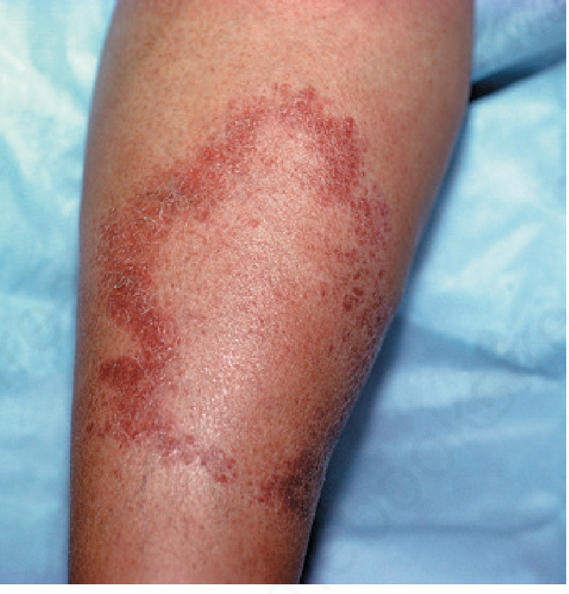
FeatureTTBTBBBLLL
Number1-33-10Several, variableManyInnumerable
SizeLargeLargeMediumSmallerSmall to diffuse
BorderWell-defined, raisedDefined outer; vague satellitePunched-out inner/vague outerIrregularIll-defined, merges
SurfaceDry, scaly, hairlessDry, slightly scalyLess dryShiny earlyWaxy/varnished
ColorHypopigmented/erythematousHypopig/erythematousMixedPale/erythematousPale macules or diffuse
DistributionAsymmetricAsymmetricMay be symmetricSymmetricBilaterally symmetric
AnhidrosisMarkedModeratePartialMinimalAbsent late
Hair loss in lesionMarkedModerateVariableMinimalAbsent early
Central healingCommon ("saucer right-side up")PresentAnnular ("Swiss cheese")OccasionalAbsent
SensationAbsentReducedModerately reducedMildly reducedLate loss
BB - "Swiss cheese" / "Punched-out" appearance: Fitzpatrick's and Dermatology 2-Volume Set (5e) describe the mid-borderline (BB) lesion as having annular plaques with a "Swiss cheese" or punched-out appearance due to holes of apparently normal skin within the lesion ring. This is characteristic of BB.
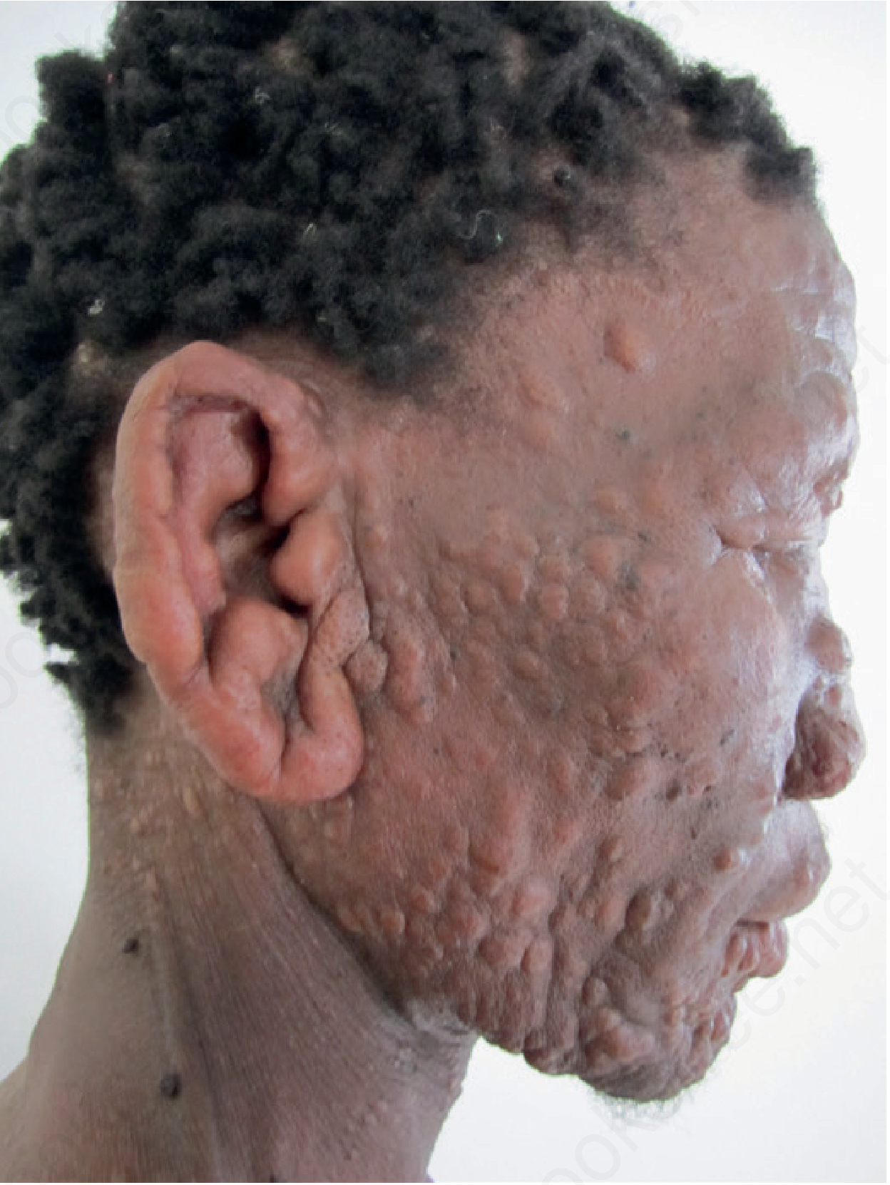
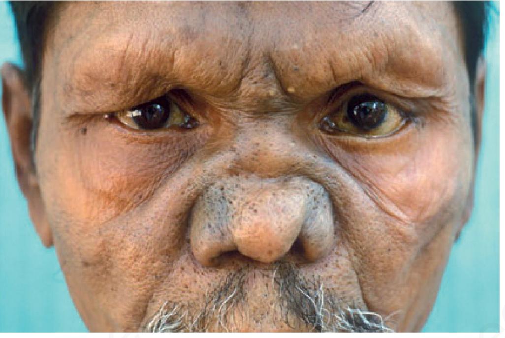
LL - "Leonine facies": Diffuse infiltration of the forehead, cheeks, earlobes, and periorbital skin creates a lion-like thickening. Also "varnished" skin appearance. - Dermatology 2-Volume Set 5e

Step 2: Immune (Spared) Sites - Always Check

The following areas are spared because M. leprae favors cooler areas:
  • Scalp
  • Axillae
  • Groin and perineum
  • Inner aspects of elbows and knees
  • Dorsal midline of back (warmer, over spine)
Finding a lesion in these sites should prompt re-evaluation of the diagnosis - these are almost never affected in leprosy.

Step 3: Sensory Testing of Lesions

IAL/Park's recommended technique - Ballpoint pen test:
  1. Use a light ballpoint pen (plastic body, without cap)
  2. Explain and demonstrate to the patient while their eyes are open
  3. Touch normal skin first - ask patient to point to where they feel the touch (eyes open, practice)
  4. Ask patient to close eyes
  5. Touch the lesion and normal skin alternately - randomize timing and location
  6. Ask: "Can you feel this?" and "Where is it?" (patient points to location)
  7. For inaccessible areas (back, buttocks): ask patient to count each touch
Do NOT use: pins, cotton wool, feathers - these are less standardized. The ballpoint pen (Park's/IAL recommended method) gives reproducible results.
Sequence of sensory modalities to test:
  1. Light touch (ballpoint pen or cotton wisp)
  2. Temperature (hot and cold test tubes, 40°C and 5°C)
  3. Pain/pinprick (sterile pin)
Temperature is lost first → then light touch → then pain.

Step 4: Madarosis Assessment

  • Inspect eyebrows: outer 1/3 loss first in LL (madarosis)
  • Inspect eyelashes: loss later in LL
  • Inspect body hair on arms/legs: gradual loss in LL
  • Inspect beard area in males
Nerve examination sites in leprosy - Park's Textbook diagram showing facial, radial, median, ulnar, lateral popliteal (common peroneal), and posterior tibial nerves
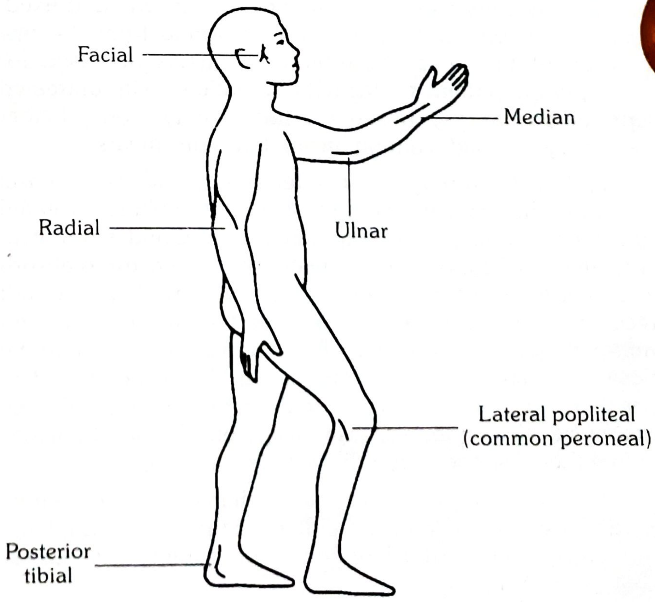
Sites of nerve involvement in leprosy. Source: Park's Textbook of Preventive & Social Medicine

C. DETAILED NERVE EXAMINATION

Per IAL Practical Manual and Park's Textbook, nerve examination has two components:
  1. Palpation - for thickening, tenderness, consistency
  2. Nerve function assessment - sensory AND motor
Technique of palpation (IAL standard):
  • Use pads of index and middle fingers
  • Palpate the nerve over its most superficial location for at least 5-10 cm of its length
  • Detect: beading, nodularity, fusiform thickening
  • Compare both sides
  • Apply mild to firm pressure and watch for patient response (wincing = tenderness)

GRADING OF NERVE THICKNESS (IAL Practical Manual - 4-grade scale)

GradeDescription
0Normal - not thickened
1Slightly thickened - palpable, just detectable above normal
2Moderately thickened - clearly palpable, firm cord
3Grossly thickened - visibly enlarged; may be beaded or nodular

GRADING OF NERVE TENDERNESS (IAL Practical Manual - 4-grade scale)

GradeDescription
0No tenderness
1Mild - slight discomfort on firm pressure
2Moderate - obvious pain, patient verbalizes discomfort
3Severe - patient winces, withdraws limb; spontaneous pain also present
Clinical significance of grading: Grading nerve thickness and tenderness is used for: (a) baseline documentation, (b) communication between leprosy workers, (c) monitoring response to steroids in reactions, (d) detecting silent neuritis. A sudden increase in tenderness grade signals acute neuritis - requires urgent steroid intervention.

NERVE-BY-NERVE EXAMINATION

1. GREAT AURICULAR NERVE (GAN)
Course: Emerges from C2-C3 root, winds around the posterior border of the SCM, runs obliquely across the surface of the muscle upward toward the parotid gland and auricle. Supplies sensation to the parotid region, angle of mandible, and earlobe.
Palpation:
  • Ask patient to turn head to the opposite side - this tightens the SCM and makes the GAN more prominent
  • Palpate the posterior surface of the SCM at the junction of upper and middle third
  • The nerve runs obliquely (posteroinferior to anterosuperior) across the SCM surface
  • Normal: barely palpable or impalpable
  • Abnormal: firm cord, may be nodular, beaded, visibly enlarged
Sensory test: Skin over angle of jaw, parotid area, earlobe - light touch and temperature
Clinical pearl (IAL): GAN is the most commonly visibly enlarged nerve in leprosy. Its enlargement is almost pathognomonic when accompanied by a skin lesion. It can be enlarged in TT, BT, and sometimes LL.

2. SUPRA-ORBITAL NERVE (Branch of V1, Trigeminal)
Course: Exits through supra-orbital notch/foramen at the superior orbital rim, approximately at the mid-pupillary line (junction of medial and middle thirds of supraorbital rim).
Palpation: Palpate along the entire superior orbital rim for tenderness and any nodular thickening at the notch.
Sensory test: Touch the forehead and anterior scalp - compare both sides.
Motor (associated facial nerve): Zygomatic branch supplies orbicularis oculi - test separately (see below).

3. FACIAL NERVE
Course: Exits stylomastoid foramen, enters parotid gland, and divides into:
  • Temporal branch: Frontalis (forehead wrinkling), upper orbicularis oculi
  • Zygomatic branch: Orbicularis oculi (lower, eye closure) - most affected in leprosy
  • Buccal branch: Orbicularis oris, zygomaticus, nasalis
  • Marginal mandibular branch: Depressor anguli oris
  • Cervical branch: Platysma
Palpation: Not routinely palpable; assess by function.
Motor testing (sequence):
  1. Eye closure (lagophthalmos test): Ask patient to close eyes gently. Observe gap between lids - measure in mm. Check Bell's phenomenon (eye rolls upward when patient tries to close; protective reflex - if absent, corneal risk is even higher).
  2. Forceful eye closure: Ask to close eyes tightly - assesses residual orbicularis oculi function.
  3. Forehead wrinkling: Ask to raise eyebrows - temporal branch.
  4. Smile and show teeth: Buccal/zygomatic.
  5. Puff cheeks: Buccal branch.
  6. Lower lip depression: Marginal mandibular.
IAL/WHO grading for eye closure:
  • Grade 0: Complete eye closure (normal)
  • Grade 1: Incomplete closure - gap <3mm (monitor)
  • Grade 2: Gap >3mm with corneal exposure (disability grade 2 if corneal damage)
Associated trigeminal (V1) test: Corneal reflex - touch cornea gently with cotton wisp from the side. Response = blink. Absent/reduced corneal reflex (V1 damage) + lagophthalmos (VII damage) = catastrophic combination for corneal ulceration.

4. ULNAR NERVE
Course: Medial cord of brachial plexus (C8, T1). Passes medially down arm, enters the ulnar groove posterior to the medial epicondyle (cubital tunnel), passes between heads of flexor carpi ulnaris (FCU), runs down medial forearm with ulnar artery, passes through Guyon's canal at the wrist, divides into superficial (sensory) and deep (motor) branches.
Most common site of leprosy involvement: The nerve as it passes through the cubital tunnel (cool, superficial, mechanical pressure point).
Palpation (primary site):
  • Patient's elbow flexed to 90-110° (this opens the cubital tunnel and makes the nerve more palpable)
  • Palpate in the groove between medial epicondyle and olecranon
  • Roll the nerve gently against the bony floor
  • Then palpate proximally up the medial upper arm along the medial intermuscular septum
Motor testing - Ulnar nerve:
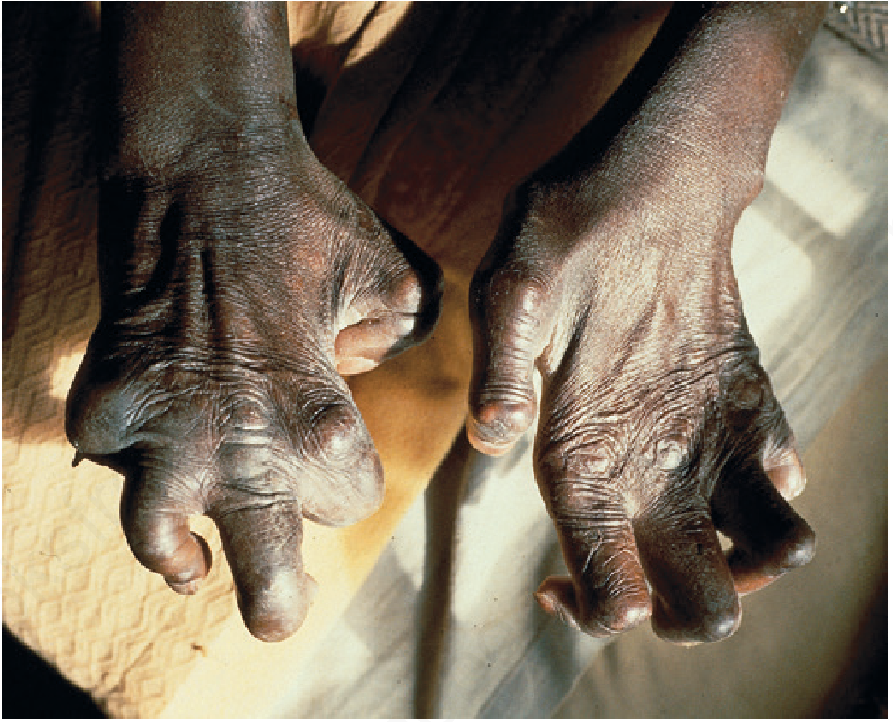
  • Claw hand test: Ask patient to straighten all fingers. Ulnar claw = inability to fully extend ring (4th) and little (5th) fingers at IP joints (due to loss of 3rd and 4th lumbricals).
  • Card test (interosseous test): Place card between adjacent fingers; patient holds it while you pull. Weakness of interossei = card slides out easily.
  • Froment's sign: Hold a piece of paper between thumb and index finger; patient grips while examiner pulls. If adductor pollicis (ulnar) is weak, the thumb IP joint flexes (FPL compensates) = positive Froment's sign.
  • Little finger abduction: Ask to spread little finger out (abductor digiti minimi - ulnar).
  • Inspect for interosseous guttering (dorsum of hand between metacarpals) and hypothenar wasting.
Sensory testing:
  • Medial 1.5 fingers: little finger + medial half of ring finger (dorsal and palmar)
  • Medial border of hand
  • Use ballpoint pen or monofilament

5. MEDIAN NERVE
Course: C5-T1, lateral and medial cords. Runs medial to brachial artery, enters cubital fossa medial to biceps tendon, passes between pronator teres heads, runs between FDS and FDP, passes through carpal tunnel under flexor retinaculum. Divides into recurrent motor branch (thenar muscles) and common/proper digital sensory branches.
Site most affected in leprosy: Carpal tunnel (at wrist).
Palpation:
  • At the wrist: palpate medial to FCR tendon, lateral to palmaris longus, at the proximal wrist crease
  • Can also be palpated in the mid-forearm between FDS and FDP
Motor testing:
  • Thumb abduction (APB test): Ask patient to point thumb perpendicular to the palm (toward ceiling). Resist downward pressure. Weakness/absence = thenar wasting from median palsy.
  • Pen cap test / "OK sign": Weakness of FPL and FDP index (anterior interosseous nerve) causes inability to make a circle; patient makes a rectangular/rectangular pinch instead.
  • Bottle sign: Cannot fully oppose thumb and index around a cylindrical bottle.
  • Inspect thenar eminence for wasting (flattening = "ape hand" when combined with thenar + hypothenar wasting).
Sensory testing:
  • Radial 3.5 fingers: thumb, index, middle, radial half of ring (dorsal and palmar distal)
  • Thenar eminence (palmar cutaneous branch - outside carpal tunnel)

6. RADIAL NERVE (Superficial/Cutaneous Branch)
Course: The superficial (sensory) branch of the radial nerve leaves the main trunk approximately 8-10 cm above the wrist, passes deep to brachioradialis, emerges at the lateral forearm to supply the dorsum of the radial 3.5 fingers and dorsal thenar area.
Palpation site:
  • Radial aspect of the distal forearm, just lateral to the radial artery at the wrist
  • Palpate under the brachioradialis tendon, rolling the nerve over the radial styloid
Motor testing (if main radial trunk involved - less common in leprosy):
  • Wrist extension, finger extension, thumb extension
  • Wrist drop if radial nerve proper is involved
Sensory testing:
  • Dorsum of first web space (between thumb and index) - most reliable
  • Dorsal thumb and radial 3.5 fingers

7. COMMON PERONEAL NERVE (Lateral Popliteal Nerve)
Course: Branch of the sciatic nerve. Passes from the posterior knee, winds around the neck of the fibula (very superficial here - most vulnerable to leprosy), pierces the peroneus longus, divides into:
  • Deep peroneal: Tibialis anterior, extensor digitorum longus, extensor hallucis longus; sensory: first web space of foot
  • Superficial peroneal: Peroneus longus and brevis; sensory: dorsum of foot
Palpation:
  • Stand behind/beside the patient
  • Identify the fibular head (prominent bony prominence on lateral knee)
  • The nerve wraps around the neck of the fibula, approximately 2-3 cm below the fibular head on the posterior-lateral aspect
  • Roll the nerve against the fibula - it should feel like a cord crossing the bone
  • Palpate both distally (as it enters lateral compartment) and posteriorly (in the popliteal fossa above the fibular head)
Motor testing:
  • Foot drop: Ask patient to walk; look for high-stepping gait and slapping of foot
  • Dorsiflexion: "Pull your toes and foot up toward your shin" against resistance - tests tibialis anterior (deep peroneal)
  • Toe extension: Extend toes against resistance - extensor digitorum longus
  • Eversion: "Turn your foot outward (sole faces out)" against resistance - peroneus longus and brevis (superficial peroneal)
Sensory testing:
  • Dorsum of foot (superficial peroneal)
  • First web space (deep peroneal)

8. POSTERIOR TIBIAL NERVE
Course: Terminal branch of the tibial nerve. Passes behind the medial malleolus with the posterior tibial artery, through the tarsal tunnel (under flexor retinaculum), divides into medial plantar, lateral plantar, and calcaneal branches.
Memory aid for structures behind medial malleolus (anterior to posterior): "Tom, Dick and Harry" = Tibialis posterior → flexor Digitorum longus → posterior tibial Artery and Nerve → flexor Hallucis longus
Palpation:
  • Find the medial malleolus
  • Palpate the groove between the medial malleolus and the Achilles tendon
  • The nerve lies posterior to the pulsating posterior tibial artery
  • Roll the nerve gently; it is softer and less prominent than the artery
Motor testing:
  • Intrinsic foot muscles (abductors/flexors of toes): Ask patient to flex and spread toes
  • Observe for claw toes (intrinsic minus foot)
  • Plantar flexion (S1 primarily, but tibial nerve contributes)
Sensory testing:
  • Entire plantar surface of the foot (medial and lateral plantar nerves)
  • Medial and lateral toes (plantar aspect)
  • Heel (medial calcaneal branch)
Clinical significance: Posterior tibial nerve damage = loss of plantar sensation = painless plantar ulcers - the most common cause of morbidity and limb loss in leprosy. Per IAL Practical Manual, this is why regular plantar sensory testing is mandatory.

9. SURAL NERVE
Course: Purely sensory; formed by medial sural cutaneous nerve (from tibial) and peroneal communicating branch (from common peroneal). Runs subcutaneously along the lateral border of the Achilles tendon to the lateral malleolus and lateral foot.
Palpation: Along the lateral border of the Achilles tendon, approximately 3-5 cm proximal to the lateral malleolus.
Sensory test: Lateral aspect of foot and little toe.
Significance: The sural nerve is the most commonly biopsied nerve in leprosy for nerve biopsy specimens (accessible, purely sensory = no motor deficit from biopsy). Enlargement with lateral foot anesthesia points to sural nerve leprosy.

NERVE FUNCTION ASSESSMENT - QUICK CLINICAL PROTOCOL

Per WHO/IAL standard for field and clinic use, test these 4 key motor functions and 3 sensory areas:
Motor (4 nerves - WHO minimum):
  1. Facial nerve: Eye closure (complete/incomplete)
  2. Ulnar nerve: Little finger abduction (spread 5th finger out)
  3. Median nerve: Thumb abduction (point thumb up from palm)
  4. Common peroneal (lateral popliteal): Foot dorsiflexion (pull toes up)
Sensory (3 areas):
  1. Ulnar and median sensory territory in hand (palmar surface at little finger and index finger)
  2. Plantar surface of foot (posterior tibial territory)
  3. Over skin lesions (using ballpoint pen)
Grade each as: Normal / Impaired / Absent.

D. EYE EXAMINATION

Systematic, urgent - leprosy is a leading preventable cause of blindness:
  1. Visual acuity - Snellen chart; finger counting if severe
  2. Lagophthalmos - measure gap in mm; test Bell's phenomenon
  3. Cornea - clarity (ulcer, scarring, pannus); corneal anesthesia (V1 testing with cotton wisp from lateral approach)
  4. Iris/pupil - irregular pupil, posterior synechiae, Koeppe nodules (iris lepromas), constricted/sluggish pupil
  5. Conjunctiva - perilimbal injection (ciliary flush in iritis); tarsal nodules
  6. Sclera - scleritis (ENL)
  7. Eyelids - madarosis (loss of brows/lashes), ectropion/entropion

E. NOSE AND THROAT EXAMINATION

  • Nose: Saddle deformity (nasal bridge collapse - septal destruction in LL), nostril atrophy, epistaxis
  • Throat: Hoarseness (laryngeal leprosy - LL), palpate neck for GAN

F. HAND AND FOOT EXAMINATION

Hands:
  • Inspect dorsum: claw (ulnar/combined), wrist drop (radial), interosseous guttering, trophic changes
  • Inspect palm: thenar and hypothenar wasting, callus, fissures, trophic ulcers
  • Test all digits for sensation
  • Assess grip strength
Feet:
  • Inspect dorsum: foot drop, trophic changes, digital deformities
  • Inspect sole: plantar ulcers (at pressure points - metatarsal heads, heel, ball of foot); callus, fissures
  • Assess plantar sensation (posterior tibial territory)
  • Check pulses

PART 3: INVESTIGATIONS (SEQUENCE AND SIGNIFICANCE)

INVESTIGATION 1: SLIT-SKIN SMEAR (SSS)

Sites (IAL recommended): Both earlobes (× 2), both active lesion edges (× 2), chin, forehead = minimum 4-6 sites for MB; for PB, may include only active lesion edge and earlobes.
Technique (step-by-step):
  1. Clean site with spirit, allow to dry completely
  2. Pinch skin firmly between thumb and forefinger to blanch - reduces bleeding and concentrates tissue fluid
  3. Make a 5mm incision, 2-3mm deep (dermis only)
  4. Scrape incision walls firmly 3-4 times with the blade edge
  5. Spread material as a uniform circular smear (5-7mm diameter) on a grease-free glass slide
  6. Air-dry, fix with methanol
  7. Stain: Fite-Faraco (preferred) or modified ZN (5% H₂SO₄)
  8. Examine 100 oil-immersion fields (100×)
Reading:
  • Bacillary Index (BI): Ridley's scale 0-6+ (log scale)
  • Morphological Index (MI): % solid-staining (viable) bacilli
BI drops ~1 unit per year on effective MDT. Rising BI after release from treatment = relapse.
Per Dermatology 2-Volume Set (5e): Organisms found in 100% LL patients, 75% borderline, only 5% TT. For tuberculoid-suspected lesions, examine at least 6 histology sections before declaring negative.

INVESTIGATION 2: SKIN BIOPSY & HISTOPATHOLOGY

IAL recommendation for biopsy:
  • From the most active, representative lesion
  • Include the active edge (not the healed center)
  • Full-thickness punch biopsy (4-6mm) to subcutaneous fat
  • Request Fite-Faraco + H&E + S-100 stain
Why Fite-Faraco over ZN? - Dermatology 2-Volume Set (5e): "Fite-Faraco stain is more sensitive; Ziehl-Neelsen stain may also stain bacilli red, but is less reliable for M. leprae due to incomplete preservation of staining."
Histopathological Findings by Type (Ridley-Jopling):
TypeGranulomaGiant cellsGrenz zoneAFBNerves
TTWell-formed epithelioid; lymphocytes at peripheryLanghans' (prominent)Absent - reaches epidermisNone/rareDestroyed (perineural concentric fibrosis - S100 shows remnants)
BTSimilar to TT but less mature epithelioid cellsPresent, fewerNarrow but presentRareGrossly swollen nerve bundles; intraneural granuloma
BBIll-defined; immature epithelioid cellsAbsentPresentModerateNot prominently swollen
BLFoamy histiocytes predominant; lymphocytes dispersedAbsentPresentAbundant; clumpsPerineural lymphocytic infiltration
LLVirchow foam cells; lipid-laden, bacilli-laden macrophagesAbsentWide, prominentGlobi (6+)Laminated onion-skin perineurium
HistoidSpindle cells (histiocytes) in whorls/storiform patternAbsentAbsentVery abundant (bacilli along spindle cell long axis)Variable
IndeterminateNon-specific lymphohistiocytic; no granulomaAbsentVariableNone/rarePerineural mononuclear infiltrate
Jopling's original description (1966): In TT, "foci of well-developed epithelioid cells with or without Langhans giant cells, encompassed by a zone of dense lymphocyte infiltration. The granuloma extends up to the epidermis without a subepidermal zone." In BT, "the best point of distinction is that there is a clear subepidermal zone, although it may be very narrow."

INVESTIGATION 3: LEPROMIN (MITSUDA) TEST

Preparations:
  • Dharmendra antigen (integral, heat-killed suspension) - more standardized
  • Mitsuda antigen (crude, from infected tissue)
Technique:
  1. Inject 0.1 mL intradermally into the flexor forearm
  2. Fernandez reaction: Read at 48 hours - erythema and induration (measures sensitization similar to tuberculin)
  3. Mitsuda reaction: Read at 28 days - papule/nodule (measures granuloma-forming CMI capacity)
Mitsuda interpretation:
ResultIndurationSpectrum
Strongly positive (+++)>10mm, may ulcerateTT - maximal CMI
Strongly positive (++)5-10mm firm noduleBT - good CMI
Positive (+)3-5mmBB - moderate CMI
Weakly positive/negative<3mmBL
Strongly negativeNo reactionLL - absent CMI
NOT a diagnostic test - positive in BCG-vaccinated individuals and TB contacts. It is a prognostic and classification tool (Jopling).
IAL note: The Mitsuda reaction is used in India and Nepal for classification in ambiguous cases and for research; it is not routinely performed at district health facilities.

INVESTIGATION 4: SENSORY TESTING (FORMAL)

Semmes-Weinstein Monofilament Test (SWMT): Standard monofilaments (2g for hand, 10g for foot) applied until bending. Maps sensory loss areas precisely. The WHO recommends monofilament testing for all leprosy patients at diagnosis and every 3 months during MDT.
Ballpoint pen test (IAL/Park's): See examination section above.
Hot-Cold tubes: Formal temperature discrimination - especially important as it is the first modality lost.

INVESTIGATION 5: VOLUNTARY MUSCLE TESTING (VMT)

Graded per MRC scale (0-5) for all key muscles. Documented on a standard VMT chart:
Minimum set to test (WHO/IAL):
  • Orbicularis oculi (facial/zygomatic)
  • APB = Abductor pollicis brevis (median)
  • ADM = Abductor digiti minimi + 1st dorsal interosseous (ulnar)
  • Tibialis anterior (common peroneal)
  • EHL = Extensor hallucis longus (deep peroneal)
Documented at: diagnosis, monthly during MDT, at completion, and at each follow-up.

INVESTIGATION 6: WHO DISABILITY GRADING

GradeEyesHandsFeet
0No problem due to leprosyNo anesthesia, no deformityNo anesthesia, no deformity
1Eye problems due to leprosy but vision not severely affected (VA ≥6/60); no lagophthalmosAnesthesia present but NO visible deformityAnesthesia present but NO visible deformity
2Severe visual impairment (VA <6/60) OR lagophthalmos, corneal diseaseVisible deformity/damage (claw, wrist drop, absorption of digits, ulcer)Visible deformity/damage (foot drop, claw toes, plantar ulcer, absorption of digits)
Nepal NLS 2021-2025: New Grade 2 Disability (G2D) at diagnosis is a key program indicator. Nepal recorded 95 new G2D cases in FY 2020/21 - a measure of late case detection.

INVESTIGATION 7: ADDITIONAL INVESTIGATIONS

TestIndicationSignificance
CBCBefore MDTDapsone → hemolytic anemia; clofazimine → GI changes
G6PD levelsBefore dapsone, especially in male patientsG6PD deficiency → severe hemolysis with dapsone
LFTsBefore and during MDTRifampicin hepatotoxicity
RFTsBaselineRelevant for drug dosing
Blood glucoseAll patientsDiabetic neuropathy as co-morbidity/differential
Nerve conduction study (NCS/EMG)Pure neural leprosy; ambiguous neuropathyDocuments large-fiber neuropathy; useful in reactions - Fitzpatrick's 9th ed.
High-resolution ultrasound (HRUS) of nervesPure neural leprosy; nerve abscess suspicionEnlargement pattern; nerve cross-sectional area; vascularity [PMID 41773898]
MRI neurographyProximal nerve involvement assessmentShows nerve enlargement, increased T2 signal, nodular enhancement
PCR for M. lepraePB/pure neural/indeterminate with negative SSS and biopsyPositive in ~50% PB; higher in MB; reference centers
Anti-PGL-1 antibodyResearch/contact screening>90% positive in LL; ~50% TT; poor PB sensitivity
CXRBaseline before rifampicinRule out TB co-infection
Slit-lamp examinationENL/suspected iritisPosterior synechiae, Koeppe nodules, iris lepromas

PART 4: DIFFERENTIAL DIAGNOSIS

(See previous comprehensive table; additional IAL-referenced differentials:)
Maculoanesthetic leprosy (IAL category) vs. Vitiligo: IAL groups maculoanesthetic leprosy with TT. The key distinguishing feature from vitiligo: maculoanesthetic patches have impaired sensation and may have subtle nerve thickening. Vitiligo is chalk-white depigmentation (not just hypopigmentation), normal sensation, and normal nerve examination.
Post-kala-azar dermal leishmaniasis (PKDL) - Nepal-specific differential: PKDL is endemic in Nepal's Terai (Province 2 - same as leprosy hotspot). Presents with hypopigmented macules and nodules. Distinguishing: NO nerve involvement, NO sensory loss; skin smear/biopsy shows Leishmania amastigotes; history of prior kala-azar. This differential is especially important in Province 2.
Mycosis Fungoides (Hypopigmented variant): Hypopigmented patches; may have mild hypoesthesia confusing clinicians; biopsy shows Pautrier microabscesses and CD8+ T-cell epidermotropism. No nerve thickening.

PART 5: MANAGEMENT AS PER NEPAL GUIDELINES

(Updated with IAL/WHO 2024 guidance)

A. CLASSIFICATION AND TREATMENT

Nepal follows WHO classification for treatment decisions aligned with the National Leprosy Strategy 2021-2025.
Key update (2023-2025): WHO now recommends uniform 3-drug MDT for both PB and MB cases (previously PB received only 2 drugs). Nepal's National Program is implementing this. The only difference is duration (6 months vs. 12 months).
MDT Regimens - Nepal/WHO (Current):

PAUCIBACILLARY (PB) - 6 months

DrugAdultChild (10-14 yr)Child (<10 yr or <40 kg)Route
Rifampicin600 mg once monthly (supervised)450 mg once monthly10 mg/kg monthlyOral
Dapsone100 mg daily50 mg daily2 mg/kg/dayOral
Clofazimine300 mg monthly + 50 mg daily150 mg monthly + 50 mg alt. days100 mg monthly + 50 mg twice weeklyOral

MULTIBACILLARY (MB) - 12 months

DrugAdultChild (10-14 yr)Child (<10 yr or <40 kg)Route
Rifampicin600 mg once monthly (supervised)450 mg once monthly10 mg/kg monthlyOral
Clofazimine300 mg monthly + 50 mg daily150 mg monthly + 50 mg alt. days6 mg/kg/month + 1 mg/kg/dayOral
Dapsone100 mg daily50 mg daily2 mg/kg/dayOral
MDT is free at all public health facilities in Nepal. Drugs come pre-packed in color-coded blister packs (maroon for MB, brown for PB).

B. MANAGEMENT OF REACTIONS

Jopling's principle: "Reactions are a common reason why patients seek consultation. If a patient believes the chemotherapy is triggering the reaction, the patient will tend to discontinue treatment, leading to treatment failure." MDT must NEVER be stopped.

Type 1 (Reversal) Reaction

  • Prednisolone 40-60 mg/day (1 mg/kg/day), minimum 6-month tapering course
  • Starting dose: 40 mg/day for 2 weeks, then gradually taper (reduce by 5 mg every 2-4 weeks based on response)
  • Steroid-sparing: methotrexate 7.5-15 mg/week if prolonged course or steroid dependence
  • Analgesics for nerve pain
  • Splint affected limb in acute neuritis (prevents contracture)
  • Continue MDT throughout

Type 2 (ENL) Reaction

  • Mild: NSAIDs + chloroquine 300 mg/day
  • Moderate-severe: Prednisolone 40-60 mg/day (shorter taper than Type 1, typically 4-8 weeks)
  • Severe/recurrent/chronic ENL: Thalidomide 100-400 mg/day (start at 400 mg for 4 days, taper; NOT in women of childbearing age)
  • Clofazimine 300 mg/day (anti-inflammatory in ENL; takes 4-6 weeks; reduces steroid dependence in chronic ENL)
  • Treat orchitis: bed rest, scrotal support, NSAIDs
  • Iritis: Urgent ophthalmology + topical prednisolone + atropine
  • Continue MDT throughout

Silent Neuritis

IAL Textbook emphasizes: Silent neuritis (nerve function impairment without pain or active reaction) must be detected by regular VMT/sensory testing - it is managed the same way as overt neuritis with prednisolone.

C. DISABILITY PREVENTION AND SELF-CARE (Nepal Program)

Per Nepal National Leprosy Strategy 2021-2025 Pillar 3:
  1. Eye care: Artificial tears 4-6 times/day; protective glasses; visual inspection daily
  2. Hand care: Warm water soaking + oil massage (prevents dryness from clofazimine); wound inspection daily; protective gloves for hot objects
  3. Foot care: Custom molded footwear; daily plantar inspection with a mirror; no barefoot walking
  4. Wound management: Regular debridement + dressings for plantar ulcers; complete offloading
  5. Physiotherapy: Active and passive exercises; splinting for acute neuritis
  6. Reconstructive surgery: Tendon transfers for claw hand (NLEP programs in Nepal); tarsal tunnel decompression; nerve abscess drainage

D. CONTACT MANAGEMENT - SDR-PEP (Nepal 2021-25 Strategy)

  • Screen all household and close contacts
  • Eligible contacts receive Single Dose Rifampicin (SDR-PEP):
    • Adults: 600 mg single oral dose
    • Children 6-14 years: 450 mg single oral dose
    • Children 2-5 years: 150-300 mg single oral dose
    • Exclude: Active TB, already on rifampicin, rifampicin allergy, children <2 years
  • Efficacy: ~57% risk reduction in contacts
  • Register in Nepal's Nikusth online platform
  • Re-screen contacts annually for 5 years

E. FOLLOW-UP

  • Monthly: supervised rifampicin; examine for reactions, new lesions; dispense daily drugs
  • VMT/sensory testing: every 3 months during MDT; at completion
  • Disability grading: at diagnosis and completion
  • Post-treatment: MB every 6-12 months for 5 years; PB for 2 years
  • BI monitoring: 6 monthly in MB (should fall ~1 unit/year)

PART 6: VIVA QUESTIONS & QUIZ (IAL/Jopling-Integrated)


VIVA QUESTIONS


Q1. What were the original criteria used by Ridley and Jopling (1966) to classify leprosy into 5 groups?
A: Ridley and Jopling (Int J Lepr, 1966;34(3):255-273) defined five groups based on four parameters simultaneously assessed:
  1. Clinical: Number, size, distribution, and character of skin lesions; nerve involvement pattern
  2. Bacteriological: Bacillary Index (BI) from SSS/biopsy; Morphological Index (MI)
  3. Immunological: Lepromin (Mitsuda) test result at 28 days
  4. Histopathological: Nature and organization of granuloma; presence/absence of grenz zone; nerve involvement pattern; giant cells
The key insight of Ridley-Jopling was that these four parameters correlate with each other because they are all driven by the same underlying variable: the host's cell-mediated immunity (CMI) against M. leprae. The classification is therefore an immune spectrum.

Q2. How does the IAL (New Indian) classification differ from Ridley-Jopling?
A: The New Indian Classification (IAL, 1981) simplifies R-J by:
  1. Consolidating borderline groups BT + BB + BL → single "Borderline (B)" category
  2. Including maculoanesthetic leprosy within tuberculoid (T) - previously a separate category in the older Indian classification (Madrid, 1953)
  3. Formally recognizing Polyneuritis (P) as a separate category = pure neuritic leprosy without skin lesions
  4. Adding Indeterminate (I) as a starting point
The 5 IAL groups: I (Indeterminate), T (Tuberculoid), B (Borderline), L (Lepromatous), P (Polyneuritis).
This classification is practical for field and clinic use in India and Nepal where simplified grouping is needed, but the R-J system remains the gold standard for research, reactions monitoring, and detailed clinical classification.

Q3. What is the significance of the "unstable" nature of the borderline group in Jopling's classification?
A: Jopling emphasized that borderline leprosy (BT, BB, BL) is immunologically unstable - patients in this spectrum can shift in either direction along the immune spectrum:
  • Upgrading (toward TT): Increasing CMI - causes Type 1 (Reversal) reaction. The sudden increase in CMI leads to inflammatory destruction of existing lesions and nerves. This is the mechanism of reversal reaction and is most severe in BL patients (greatest antigen load during upgrading).
  • Downgrading (toward LL): Decreasing CMI (due to continued antigen exposure, malnutrition, immunosuppression) - causes downgrading reaction (clinically similar to reversal).
The two polar forms (TT and LL) are stable - they rarely undergo reactions or shift in spectrum. This is why monitoring and steroid treatment is most important in borderline patients.

Q4. What are the classic nerve deformities produced by each nerve in leprosy? List the nerve and its deformity.
A:
NerveDeformity
UlnarClaw hand (4th and 5th fingers); interosseous wasting; hypothenar wasting
MedianApe thumb deformity (thenar wasting; thumb falls into same plane as fingers); loss of opposition
Ulnar + Median combinedComplete claw hand (all 4 fingers); combined thenar + hypothenar wasting
RadialWrist drop (extensor weakness)
Facial (zygomatic branch)Lagophthalmos (inability to close eye) → corneal ulcer → blindness
Common peroneal (lateral popliteal)Foot drop; high-stepping gait
Posterior tibialLoss of plantar sensation → plantar ulcers; claw toes; intrinsic foot muscle wasting

Q5. Explain the bacteriological and morphological indices and how they help in monitoring leprosy treatment.
A:
  • Bacillary Index (BI): Ridley's 0-6+ logarithmic scale measuring density of AFB in SSS. Used to confirm MB leprosy at diagnosis; monitor bacteriological response. BI falls approximately 1 unit per year on effective MDT. A BI that fails to fall or rises = drug resistance or relapse.
  • Morphological Index (MI): Percentage of solid-staining (intact) bacilli - represents viable, infective organisms. Normal bacteria stain solid; dead or dying bacteria fragment and stain as beaded/granular. On effective MDT, MI drops to zero within weeks to months - first evidence of bacteriological response. Rising MI (especially >1%) after apparent cure = relapse, often with drug-resistant organisms.
Combined monitoring: MI detects early treatment response; BI detects slow clearance and relapse.

Q6. A patient with leprosy develops sudden wrist drop, claw hand, and foot drop overnight during treatment. What is happening and what is the emergency management?
A: This is acute neuritis in the context of a Type 1 (Reversal) Reaction - a dermatological and neurological emergency. The sudden immune attack on nerves can cause irreversible damage within hours if untreated.
Immediate management:
  1. Do NOT stop MDT
  2. Prednisolone 1 mg/kg/day (40-60 mg) orally - START IMMEDIATELY - every hour of delay increases risk of permanent nerve damage
  3. Splint the affected limb in functional position to prevent contracture (wrist in neutral/slight dorsiflexion; foot in 90° dorsiflexion)
  4. Analgesia for nerve pain
  5. Admit if bilateral, severe, or if corneal exposure
  6. Arrange urgent ophthalmology if lagophthalmos
  7. Taper prednisolone slowly over minimum 6 months
  8. Document VMT baseline; repeat every 2 weeks
Per IAL Textbook: "Reactions should be treated aggressively to prevent peripheral nerve damage. The goal is to reverse any nerve function impairment."

Q7. What are the lepromin test results across the R-J spectrum and why does this occur?
A:
TypeMitsuda (28-day) resultReason
TT+++/++ (>5mm, may ulcerate)Maximum CMI; robust granuloma-forming capacity; IFN-γ and IL-2 dominant
BT++/+ (3-10mm)Good CMI
BB± (weakly positive or negative)Moderate CMI
BLNegativePoor CMI
LLStrongly negativeAbsent CMI to M. leprae; IL-4/IL-5/IL-10 (Th2) dominant; macrophages cannot kill bacilli
Key point (Jopling): The lepromin test is NOT a diagnostic test - it is a measure of CMI status and helps classify ambiguous cases along the spectrum. It is also positive in BCG-vaccinated individuals and TB-infected individuals, making it non-specific for diagnosis.

Q8. What is the "immune privilege" concept in leprosy and which body sites are spared?
A: M. leprae has an optimal growth temperature of 30-32°C (below core body temperature). It therefore grows preferentially in cooler areas of the body. Areas that are relatively warmer due to proximity to major vessels, hair insulation, or body folds are spared.
Spared "immune privileged" sites (warm areas):
  • Scalp
  • Axillae
  • Groin and perineum
  • Inner aspects of elbows and knees
  • Mid-back (over spine)
  • Webspaces of toes (in early disease)
Sites commonly affected (cool areas):
  • Face, ears, earlobes (especially LL)
  • Buttocks, upper arms, thighs
  • Hands, feet

Q9. Describe the histoid leprosy - its clinical features, histopathology, and significance.
A: Histoid leprosy is an atypical form of multibacillary (LL) leprosy, first described by Wade (1963). It most commonly occurs:
  • After dapsone monotherapy with resistance (hence called "histoid" due to histiocyte spindle cell pattern)
  • After irregular/incomplete MDT
  • Occasionally de novo
Clinical features:
  • Firm, shiny, dome-shaped papules and nodules (1-15mm)
  • Yellow-red to skin-colored
  • Appear on normal background skin (unlike regular LL where skin is diffusely infiltrated)
  • Favor buttocks, lower back, face, bony prominences
  • May resemble dermatofibromas or molluscum contagiosum
Histopathology (pathognomonic):
  • Spindle-shaped histiocytes arranged in whorls, storiform pattern, or tight circles
  • Bacilli extremely abundant, aligned along the long axis of spindle cells
  • BI 6+ (maximum)
  • Solid-staining bacilli (high MI) - indicates viable, potentially drug-resistant organisms
  • Distinct from surrounding tissue
Significance:
  • High bacillary load → highly infectious
  • Suggests dapsone resistance → test MI and folP1 gene mutation
  • Treat with full MB-MDT; if dapsone resistant, use alternative regimen (Rifampicin + Ofloxacin + Minocycline - ROM)

Q10. What is Lucio phenomenon and how does it differ from ENL?
A: Both are reactional states in LL, but they are distinct:
FeatureENL (Type 2)Lucio Phenomenon
Form of leprosyBL/LL (any subtype)Diffuse Lepromatous Leprosy (Lucio leprosy = non-nodular, diffuse form)
Geographic distributionGlobalMainly Mexico and Central America
MechanismImmune complex-mediated (Type III)Endarteritis obliterans with vascular occlusion and necrosis
Skin lesionsTender red nodules (ENL) on normal skinIrregular hemorrhagic infarcts; angular punched-out ulcerations on extremities
Systemic featuresFever, arthralgia, orchitis commonSevere; high mortality
HistologyNeutrophilic infiltrate; leukocytoclastic vasculitis with AFBEndothelial proliferation and thrombus in dermal vessels; bacilli inside endothelial cells
TreatmentPrednisolone/thalidomideDifficult; may need wound care, antibiotics; MDT

MCQ QUIZ (IAL/Jopling Additions)


Q1. The Ridley-Jopling classification was published in which journal and year?
  • A) Lancet, 1962
  • B) Int J Leprosy, 1966
  • C) BMJ, 1970
  • D) JAMA, 1968
Answer: B. Ridley DS, Jopling WH. Classification of leprosy according to immunity. A five-group system. Int J Lepr. 1966;34(3):255-273. The preliminary version was published in Lepr Rev 1962 but the definitive 5-group paper appeared in 1966.

Q2. In the IAL New Indian Classification (1981), pure neuritic leprosy without skin lesions falls under which group?
  • A) Indeterminate (I)
  • B) Tuberculoid (T)
  • C) Borderline (B)
  • D) Polyneuritis (P)
Answer: D - Polyneuritis (P). This is the unique contribution of the IAL classification - it formally recognizes pure neuritic leprosy as a distinct group, which is especially relevant for Nepal where PNL is common.

Q3. In Ridley-Jopling classification, which form of leprosy is described as "immunologically unstable" and prone to reactions?
  • A) TT and LL
  • B) Borderline (BT, BB, BL)
  • C) Indeterminate only
  • D) LL only
Answer: B - Borderline (BT, BB, BL). Jopling's core principle: the two polar forms (TT and LL) are immunologically stable. The borderline group is inherently unstable and can shift in either direction, manifesting as Type 1 reactions during upgrading.

Q4. The Mitsuda reaction in lepromin test is READ at:
  • A) 24 hours (Fernandez reaction)
  • B) 72 hours
  • C) 7 days
  • D) 28 days
Answer: D - 28 days. The Fernandez (early) reaction is at 24-48 hours. The Mitsuda (late) reaction at 28 days is the clinically relevant reading, reflecting the granuloma-forming capacity of CMI.

Q5. Which statement about the grenz zone is CORRECT according to Jopling's 1966 classification?
  • A) The grenz zone is absent in lepromatous leprosy
  • B) The grenz zone is present in TT because the infiltrate does not reach the epidermis
  • C) In TT, granulomas extend to the epidermis WITHOUT a grenz zone; in BT there is a clear (though narrow) subepidermal zone
  • D) The grenz zone is only seen in histoid leprosy
Answer: C. Per Jopling's original description: TT has granulomas that "extend up to the epidermis without a subepidermal zone." The grenz zone (subepidermal clear zone) appears from BT onward and widens toward the lepromatous pole.

Q6. Histoid leprosy is associated with:
  • A) Clofazimine monotherapy failure
  • B) Rifampicin resistance
  • C) Dapsone monotherapy or dapsone resistance
  • D) Treatment with thalidomide
Answer: C. Histoid leprosy (Wade, 1963) is classically described after dapsone monotherapy (pre-MDT era) or in patients with dapsone resistance. It can also occur de novo but dapsone resistance must always be considered. The folP1 gene mutation in M. leprae is the molecular mechanism of dapsone resistance.

Q7. In lepromatous leprosy, the principal cytokines that suppress CMI are:
  • A) IFN-γ and TNF-α
  • B) IL-4, IL-5, IL-10 (Th2 dominant)
  • C) IL-12 and IL-17
  • D) IL-2 and IL-23
Answer: B. LL represents a classic Th2 response. IL-4 and IL-10 downregulate CMI, suppress macrophage activation (macrophages cannot kill M. leprae), and enhance antibody production (polyclonal hypergammaglobulinemia, false-positive VDRL, RF, ANA). TT is a Th1 response (IFN-γ, IL-2, IL-12).

Q8. Which nerve is palpated at the neck of the fibula in leprosy examination?
  • A) Posterior tibial nerve
  • B) Sural nerve
  • C) Saphenous nerve
  • D) Common peroneal (lateral popliteal) nerve
Answer: D - Common peroneal (lateral popliteal) nerve. It wraps around the neck of the fibula (approximately 2-3 cm below the fibular head), which is its most superficial point and the site of leprosy involvement. This is the nerve responsible for foot drop when damaged.

Q9. A 4-grade scale for grading nerve thickness in leprosy is used in which textbook/manual?
  • A) Fitzpatrick's only
  • B) IAL Practical Manual of Leprosy
  • C) Park's only
  • D) Red Book
Answer: B - IAL Practical Manual of Leprosy. The 4-grade scale (0-3) for both nerve thickness and nerve tenderness is described in the Practical Manual of Leprosy (based on IAL guidelines). Grade 0 = normal; Grade 1 = slightly thickened; Grade 2 = moderately thickened; Grade 3 = grossly thickened/visible.

Q10. The "onion skin" (laminated) perineurium on nerve histology is characteristic of:
  • A) Tuberculoid leprosy
  • B) Lepromatous leprosy
  • C) Borderline tuberculoid leprosy
  • D) Histoid leprosy
Answer: B - Lepromatous leprosy. In LL, the perineural cells undergo repeated rounds of proliferation and fibrosis in response to chronic bacillary presence, producing the characteristic "onion-skin" or lamellar lamination of the perineurium. This contrasts with TT, where nerves show perineural concentric fibrosis and are destroyed within the granuloma.

Q11. Post-kala-azar dermal leishmaniasis (PKDL) is an important differential for leprosy in Nepal because:
  • A) Both are caused by acid-fast bacilli
  • B) Both affect the same Province 2 (Terai) of Nepal and produce hypopigmented patches
  • C) PKDL also causes nerve thickening
  • D) PKDL has positive Mitsuda test
Answer: B. Province 2 (Madhesh Province, Terai) is the highest burden area for BOTH leprosy and kala-azar/PKDL in Nepal. PKDL presents with hypopigmented macules and nodules similar to leprosy. The key distinguishing features: PKDL has NO sensory loss and NO nerve thickening; skin biopsy/smear shows Leishmania amastigotes (Donovan bodies).

Q12. The "saucer right-side up" appearance is a classic description for which type of leprosy lesion?
  • A) Lepromatous plaque
  • B) Tuberculoid plaque
  • C) ENL nodule
  • D) Histoid nodule
Answer: B - Tuberculoid (TT) plaque. The TT lesion has a raised, defined peripheral border that slopes down to a flattened, atrophic, hypopigmented center - like a saucer placed right-side up. The BB lesion is described as "saucer upside-down" (outer border less defined, inner edge punched-out/cleared). These classical descriptions are from Jopling.

Q13. The IAL classification formally uses "Polyneuritis (P)" to represent:
  • A) Patients with polyneuropathy from dapsone
  • B) Patients with ENL causing multiple nerve involvement
  • C) Pure neuritic leprosy - peripheral nerve involvement without skin lesions
  • D) Patients with leprosy and co-existing diabetic neuropathy
Answer: C - Pure neuritic leprosy (PNL). This is a form in which the peripheral nerve trunks are involved without any skin lesions. Per IAL, it may be TT or BT depending on the number of nerves involved. It is classified under group P in the IAL system. Diagnosis requires nerve biopsy or PCR. Nepal has the highest burden of PNL globally, alongside Brazil and India.

QUICK REFERENCE: JOPLING'S KEY MNEMONICS & PEARLS

PearlSource
"Temperature is lost first, before touch and pain"Jopling - dissociation of sensation pathognomonic for leprosy
"The borderline group is unstable; TT and LL are stable"Ridley-Jopling 1966
"Saucer right-side up = TT; upside-down = BB"Jopling clinical description
"Granuloma extends to epidermis in TT (no grenz zone); grenz zone present from BT onward"Ridley-Jopling histology 1966
"Never skip nerve examination"IAL Practical Manual
"MDT must never be stopped during reactions"IAL + WHO
"Histoid leprosy = dapsone resistance until proven otherwise"IAL Textbook
"GAN is the most visibly enlarged nerve"IAL/Andrews'
"BI falls 1 unit/year on MDT; MI falls first (weeks)"Ridley's scale
"PKDL in Province 2 Nepal = key differential for leprosy"Nepal NLS 2021-25

References:
  1. Ridley DS, Jopling WH. Classification of leprosy according to immunity: a five-group system. Int J Lepr. 1966;34(3):255-273.
  2. IAL - All India Leprosy Workers Conference. New Indian Classification. Lepr India. 1982;54:22-32.
  3. IAL Practical Manual of Leprosy (Kar HK, Kumar B eds.). Chapter on nerve examination and grading.
  4. International Textbook of Leprosy - Clinical Diagnosis chapter (internationaltextbookofleprosy.org)
  5. Andrews' Diseases of the Skin, 13th ed. (Clinical Dermatology), Chapter 17.
  6. Dermatology 2-Volume Set, 5e (Bourke et al.), Chapter 75.
  7. Fitzpatrick's Dermatology, 9th ed., Chapter 159.
  8. Park's Textbook of Preventive & Social Medicine, 26th ed., Chapter on Leprosy.
  9. Nepal National Leprosy Strategy 2021-2025.
  10. WHO Leprosy Guidelines & Fact Sheet (who.int).
  11. An update of the diagnosis, treatment, and prevention of leprosy (PMC11346855)
  12. High-Resolution Ultrasonography for Detecting Peripheral Neuropathy in Leprosy: A Systematic Review and Meta-Analysis (PMID 41773898)

Is muscle and other examination included.

Finding Sources
Finding Sources
Searching the Web

leprosy complete muscle examination VMT voluntary muscle testing ulnar median peroneal facial nerve grading IAL

Reading File
Finding Sources
Finding Sources
Reading File
Finding Sources
Loading Image
Loading Image
Reading Web Page

https://leprosyreview.org/admin/public/api/lepra/website/get…

I now have all the material needed. Here is the complete, dedicated muscle and systemic examination section to complement the long case:

LEPROSY - COMPLETE MUSCLE EXAMINATION & SYSTEMIC EXAMINATION

Supplementary Section for the Long Case

Sources: Andrews' Diseases of the Skin 13e | Dermatology 2-Volume Set 5e | Fitzpatrick's 9e | IAL Practical Manual | Leprosy Review (VMT/INFIR Study, 2008)

SECTION A: VOLUNTARY MUSCLE TESTING (VMT) - COMPLETE GUIDE

Background and Purpose

VMT is the standard method for monitoring motor nerve function in leprosy. Per the INFIR Cohort Study (Leprosy Review, 2008; van Brakel et al.), VMT is used to monitor changes in motor function in facial, ulnar, median, radial, and lateral popliteal nerves. Where there is evidence of recent change, corticosteroid treatment is started. Early detection and treatment is the recommended strategy for preserving and restoring nerve function.
When to perform VMT:
  • At diagnosis (baseline)
  • Every month during MDT (monthly supervised dose visit)
  • At every 3 months as minimum (WHO)
  • At treatment completion
  • At any point when patient reports new weakness, pain, or reaction
  • Post-treatment follow-up every 6-12 months (MB: 5 years; PB: 2 years)
Key principle: Sensory loss precedes motor loss in leprosy neuropathy. By the time VMT shows a deficit, sensory damage is already established. Therefore VMT change signals advanced or worsening nerve damage - act urgently.

MRC GRADING SCALE (Modified for Leprosy VMT)

GradeDescriptionClinical Meaning
5Full movement against full resistance - NormalNo weakness
4Full movement against partial (reduced) resistanceMild weakness
3Full movement against gravity only (no added resistance)Moderate weakness
2Movement only with gravity eliminated (horizontal plane)Severe weakness
1Visible/palpable contraction but no joint movementTrace contraction
0No contraction whatsoeverComplete paralysis
For leprosy monitoring:
  • Any grade change of 1 or more = significant nerve function impairment (NFI)
  • A drop from 5 to 4 is significant - do NOT dismiss it
  • A drop from 4 to 3 = urgent intervention needed (steroid)
  • Grade 0 or 1 = permanent damage likely if not treated immediately

MUSCLE TESTING NERVE BY NERVE


1. FACIAL NERVE - ORBICULARIS OCULI (Zygomatic Branch)

Why it matters: Lagophthalmos → exposure keratitis → corneal ulcer → blindness. The zygomatic branch of CN VII is the most commonly affected facial nerve branch in leprosy.
Muscle tested: Orbicularis oculi (eye closure)
Test method (step-by-step):
  1. Inspect at rest: Look for asymmetry of eyelid position, any visible gap between lids
  2. Gentle closure test: Ask patient: "Close your eyes gently as if sleeping"
    • Normal: lids meet completely with no gap
    • Abnormal: gap visible between upper and lower lid = lagophthalmos
  3. Measure the gap: Use a millimeter ruler to measure the inter-lid gap
  4. Forced closure test: Ask: "Now close your eyes as tightly as you can"
    • Grade 5: complete tight closure (lashes buried)
    • Grade 4: complete closure but not tight
    • Grade 3: eyelids close but with visible effort/asymmetry
    • Grade 2: partial closure only; significant gap remains
    • Grade 1: minimal movement of lids
    • Grade 0: no movement
  5. Bell's phenomenon: While asking patient to close eyes, observe eye movement
    • Normal Bell's: eye rolls upward (protective - cornea moves away)
    • Absent Bell's: cornea remains exposed even when trying to close = catastrophic risk of ulceration
  6. Test both sides and compare
Associated tests (always do with facial nerve exam):
  • Corneal reflex (V1/trigeminal): Touch limbus with fine cotton wisp from lateral side (so patient cannot see it coming). Normal = blink. Absent/reduced = corneal anesthesia on top of lagophthalmos = EMERGENCY
  • Corneal fluorescein test: If ophthalmology equipment available - instill fluorescein, examine under blue light for corneal erosions
Grading eye closure for WHO disability:
  • Grade 0: Complete closure - no disability
  • Grade 1: Incomplete closure but no corneal damage (disability grade 1)
  • Grade 2: Lagophthalmos + corneal damage/ulcer/opacity/VA <6/60 (disability grade 2)
Other facial nerve branches to test:
BranchMuscleTest
TemporalFrontalisRaise eyebrows (wrinkle forehead)
TemporalUpper orbicularis oculiWink/forceful upper eyelid closure
Zygomatic (most affected)Lower orbicularis oculiEye closure (as above)
BuccalOrbicularis oris, zygomaticusShow teeth; smile; puff cheeks
Marginal mandibularDepressor anguli orisPull lower lip down

2. ULNAR NERVE - MOTOR TESTING

Muscles innervated (C8, T1):
  • Hypothenar group: Abductor digiti minimi (ADM), Flexor digiti minimi, Opponens digiti minimi
  • Interossei (all 4 dorsal, 3 palmar)
  • 3rd and 4th lumbricals
  • Adductor pollicis
  • Deep head of Flexor pollicis brevis
  • Palmaris brevis
Key VMT muscle for leprosy monitoring: Abductor Digiti Minimi (ADM)
Standard VMT test for Ulnar nerve (Watson, IAL standard):
"Spread the little finger" test:
  1. Ask patient to place hand flat on table, palm down
  2. Instruct: "Spread your little finger out to the side, away from the other fingers"
  3. Resist this movement by pressing the little finger back toward the ring finger
  4. Grade 5: full abduction against full resistance
  5. Grade 4: full abduction against reduced resistance
  6. Grade 3: full abduction against gravity alone (horizontal) but no added resistance
  7. Grade 2: barely moves little finger
  8. Grade 1: tendon flickers but no movement
  9. Grade 0: no movement
Additional ulnar motor tests:
a. Claw hand assessment (most visible sign of chronic ulnar palsy):
  • Ask patient to straighten all fingers simultaneously on a flat surface
  • Ulnar claw: Ring (4th) and little (5th) fingers cannot fully extend at PIP and DIP joints - they remain flexed
  • Caused by loss of 3rd and 4th lumbricals (which extend IP joints via extensor mechanism)
  • "Intrinsic minus" hand - loss of intrinsic muscle action
  • The index and middle fingers (with intact 1st and 2nd lumbricals from median nerve) are NOT clawed
b. Interosseous muscle testing:
  • Card test: Place a card between adjacent fingers; patient holds it while you pull the card
  • Alternate: Ask patient to abduct (spread) all fingers simultaneously against resistance
  • Visible guttering between metacarpals on dorsum of hand = interosseous wasting
c. Froment's sign (Adductor pollicis - deep ulnar branch):
  • Ask patient to grip a piece of paper between thumb and index finger (lateral pinch)
  • Examiner pulls the paper away
  • Positive: Thumb IP joint flexes (FPL from median compensates for weak adductor pollicis) = ulnar palsy
  • Negative: Thumb IP joint remains extended (adductor pollicis working normally)
d. Hypothenar wasting:
  • Inspect palmar surface: flattened hypothenar eminence
  • Palpate: soft, atrophic bulk compared to normal side
e. Interosseous guttering:
  • Inspect dorsum of hand: depression/hollowing between metacarpals (especially 1st web space - first dorsal interosseous wasting)
Leprosy-specific note (Andrews'): "The ulnar nerve near the medial condyle of the humerus may be as thick as the little finger, round, and stiff, and is often easily felt several centimeters above the elbow." The claw hand (Fig. 17.7 - Andrews') is the most common hand deformity in leprosy.
Claw hand of Hansen disease - bilateral severe clawing with trophic changes
Claw hand in advanced Hansen disease with trophic skin changes - Andrews' Diseases of the Skin

3. MEDIAN NERVE - MOTOR TESTING

Muscles innervated (C6-T1):
  • Thenar group: Abductor pollicis brevis (APB), Opponens pollicis, Flexor pollicis brevis (superficial head)
  • 1st and 2nd lumbricals
  • Pronator teres, Pronator quadratus
  • Flexor carpi radialis, Palmaris longus
  • Flexor digitorum superficialis (all 4)
  • Flexor digitorum profundus (index + middle - via anterior interosseous nerve)
  • Flexor pollicis longus (via anterior interosseous nerve)
Key VMT muscle for leprosy: Abductor Pollicis Brevis (APB)
Standard VMT test for Median nerve (Watson, IAL standard):
"Thumb-up" test (APB test):
  1. Ask patient to place hand flat on table, palm up
  2. Instruct: "Point your thumb straight up toward the ceiling, perpendicular to your palm"
  3. Resist downward pressure on the thumb nail/distal phalanx
  4. Grade 5: full elevation against full resistance
  5. Grade 4: full elevation against reduced resistance
  6. Grade 3: full elevation against gravity alone
  7. Grade 2: slight movement
  8. Grade 1: flicker of APB seen/palpated at thenar base
  9. Grade 0: no movement
Additional median motor tests:
a. Thenar wasting:
  • Inspect: flattened thenar eminence ("ape hand" = thenar flat + thumb falls back into plane of fingers)
  • Palpate: soft bulk over thenar area
b. Opposition test:
  • Ask patient to touch the tip of the little finger with the tip of the thumb (full opposition)
  • Weakness = cannot reach little finger or does so with IP flexion instead of true CMC rotation
c. "OK sign" / Pen cap test (Anterior Interosseous Nerve):
  • Ask patient to make the "OK" sign (circle with thumb and index finger)
  • Normal: perfect circle
  • AIN palsy: rectangular/triangular shape (cannot flex index DIP or thumb IP)
  • Less commonly affected in leprosy than APB
d. Bottle sign:
  • Patient cannot wrap thumb and index around a cylindrical bottle due to thenar weakness
Combined ulnar + median palsy signs:
  • "Ape hand": Thenar + hypothenar both flat, thumb in same plane as fingers, no opposition
  • Complete claw hand: All four fingers clawed (ulnar provides ring+little; median provides index+middle via 1st+2nd lumbricals)
  • Total intrinsic minus hand: Severe combined palsy

4. RADIAL NERVE - MOTOR TESTING

Muscles innervated:
  • Brachioradialis, Extensor carpi radialis longus (above spiral groove)
  • Extensor carpi radialis brevis, Extensor digitorum communis, Extensor carpi ulnaris (via posterior interosseous nerve/PIN)
  • Extensor pollicis longus, Extensor pollicis brevis, Abductor pollicis longus (PIN)
  • Extensor indicis proprius (PIN)
Note: Radial nerve trunk involvement is LESS common in leprosy than ulnar. The superficial radial nerve (sensory only) is more commonly affected.
VMT for radial nerve:
a. Wrist extension test:
  • Ask patient to extend wrist (cock up wrist) against resistance applied to dorsum of hand
  • Grade the movement 0-5 as per MRC scale
  • Wrist drop = inability to extend wrist (extensor carpi radialis/ulnaris paralysis)
b. Finger extension test:
  • Ask patient to extend fingers at MCP joints (not IP joints - tested by lumbricals/interossei)
  • Weakness or inability = extensor digitorum communis palsy
c. Thumb abduction/extension:
  • Ask to move thumb away from hand in the plane of the palm (abductor pollicis longus = radial)
  • Ask to extend thumb (extensor pollicis longus = PIN)
Visible deformity: Wrist drop - wrist hangs down in flexion at rest

5. COMMON PERONEAL (LATERAL POPLITEAL) NERVE - MOTOR TESTING

Muscles innervated:
  • Deep peroneal branch: Tibialis anterior, Extensor digitorum longus, Extensor hallucis longus (EHL), Peroneus tertius
  • Superficial peroneal branch: Peroneus longus, Peroneus brevis
Key VMT muscle for leprosy: Tibialis Anterior (dorsiflexion)
Standard VMT test (Watson/IAL standard):
"Foot-up" / Dorsiflexion test:
  1. Patient seated, foot hanging freely
  2. Instruct: "Pull your foot and toes up toward your shin"
  3. Resist by pressing down on the dorsum of the foot
  4. Grade 0-5 per MRC scale
Additional peroneal motor tests:
a. Eversion test (Superficial peroneal - peronei):
  • Ask patient to turn sole of foot outward (evert) against resistance
  • Weakness = superficial peroneal nerve; foot tends toward inversion at rest if peronei paralyzed
b. Toe extension (Deep peroneal - EHL, EDL):
  • Ask patient to extend big toe and all toes upward against resistance
  • EHL tested separately: "point big toe toward ceiling"
c. Foot drop assessment:
  • Ask patient to walk: observe for high-stepping gait (patient lifts knee high to clear the dropped foot)
  • Listen for foot slap (foot slaps ground due to loss of controlled plantarflexion at heel-strike)
  • At rest: foot hangs in plantarflexion and inversion (tibialis anterior + extensors paralyzed; unopposed gastrocnemius + tibialis posterior)
Visible deformity: Foot drop, steppage gait, claw toes (if intrinsics also involved)

6. POSTERIOR TIBIAL NERVE - MOTOR TESTING

Muscles innervated:
  • Intrinsic foot muscles: Flexor digitorum brevis, Abductor hallucis, Flexor hallucis brevis, Lumbricals, Interossei (via medial and lateral plantar nerves)
  • Flexor digitorum longus, Tibialis posterior, Flexor hallucis longus (more proximal branches)
VMT test:
a. Toe flexion / Intrinsic foot muscle test:
  • Ask patient to flex (curl) toes against resistance
  • Ask patient to abduct/adduct toes (intrinsic muscles)
  • Weakness = intrinsic muscle wasting → claw toes
b. Toe claw deformity assessment:
  • Inspect for clawing of all toes (intrinsic minus foot - similar to hand)
  • Hyperextension at MTP + flexion at IP joints
c. Plantar sensory map:
  • Always test plantar sensation at multiple sites using ballpoint pen or monofilament (10g SWMT)
  • Medial plantar: ball of foot, great toe
  • Lateral plantar: 5th metatarsal head, lateral toe
  • Calcaneal: heel
Visible complications: Plantar ulcer (mal perforans pedis) at pressure points due to insensate foot. This is the most common, preventable complication.

SUMMARY VMT CHART (For clinical documentation)

Record at each visit:
NerveMuscle TestedTestRightLeft
FacialOrbicularis oculiEye closure (mm gap)/5/5
UlnarAbductor digiti minimiSpread 5th finger/5/5
MedianAbductor pollicis brevisThumb perpendicular to palm/5/5
RadialExtensor carpi radialisWrist extension/5/5
Common peronealTibialis anteriorFoot dorsiflexion/5/5
Posterior tibialIntrinsic footToe flexion/spread/5/5
Significance of VMT change (action required):
  • Grade 5→4 on any test: increase frequency of monitoring to monthly; consider early steroid if progressive
  • Grade 5→3 or lower, or any acute drop: START PREDNISOLONE IMMEDIATELY
  • Any new weakness during reaction: EMERGENCY - same-day steroid

SECTION B: COMPLETE MUSCLE & WASTING ASSESSMENT (LOOK-FEEL-MOVE)

HANDS - SYSTEMATIC SEQUENCE

Step 1 - LOOK (Inspection)
Dorsum first, then palmar surface. Always compare both hands side by side.
FindingAnatomical basisNerve
Claw hand (4th, 5th fingers)Intrinsic minus: loss of 3rd & 4th lumbricals (extend IP joints) + interosseiUlnar
Claw hand (index, middle fingers)Loss of 1st & 2nd lumbricalsMedian
Complete claw (all fingers)Combined ulnar + median intrinsic palsyUlnar + Median
Wrist dropLoss of wrist extensorsRadial
Ape hand / Simian handThenar wasting, thumb flat in plane of palmMedian
Interosseous gutteringWasting of dorsal interossei; most visible in 1st web spaceUlnar
Hypothenar flatteningWasting of hypothenar eminenceUlnar
Thenar flatteningWasting of thenar eminenceMedian
Finger shorteningOsteolysis/bone resorption from repeated trauma + ischemiaLL (advanced)
ContracturesFibrosis from chronic clawing + trophic changesAny
Trophic changesDry, scaly, fissured skin (anhidrosis, autonomic neuropathy)Multiple
Ulcers/scarsRepeated painless traumaAny (anesthetic hand)
CallusesAbnormal pressure pointsAny
Step 2 - FEEL (Palpation)
  • Palpate thenar and hypothenar eminences: compare bulk, firmness, atrophy
  • Palpate first dorsal interosseous: thumb and index on either side of 1st metacarpal - compare bulk
  • Palpate dorsal interosseous gutters: run finger between metacarpals, note depression
  • Skin temperature: asymmetric coolness = autonomic fiber damage (sympathetic)
  • Skin moisture: anhidrosis = autonomic damage
  • Check pulses (radial, ulnar): vascular supply affects trophic changes
Step 3 - MOVE (Function)
Active movement:
  1. Full finger extension (spread on flat surface)
  2. Full finger flexion (make a fist)
  3. Thumb opposition to each finger tip
  4. Grip strength (ask to squeeze examiner's two fingers)
  5. Key pinch (hold paper between thumb and lateral index)
  6. Precision pinch (pulp to pulp - pick up small object)
Passive movement:
  • Check PIP and DIP joint range of motion
  • Assess for fixed flexion contracture (cannot passively extend)

FEET - SYSTEMATIC SEQUENCE

Step 1 - LOOK
FindingMechanismSignificance
Foot dropTibialis anterior + toe extensors paralyzedCommon peroneal nerve
High-stepping gaitCompensatory for foot dropObserve while walking
Claw toesIntrinsic foot muscle wasting (intrinsic minus foot)Posterior tibial
Equinovarus deformityFoot drop + eversion weaknessCommon peroneal
Plantar ulcer (Mal perforans pedis)Insensate plantar skin + repeated trauma; perforating ulcer at pressure pointsPosterior tibial (loss of plantar sensation)
Pressure sites for plantar ulcersBall of foot (1st-5th metatarsal heads), heel, lateral borderMap ulcer location
Trophic changes (plantar)Dry, scaly plantar skin; deep fissuresAutonomic
Shortening of toesOsteolysis in LLAdvanced LL
Charcot joint (neuropathic arthropathy)Repeated trauma to anesthetic joints → bone destruction, deformityAny anesthetic joint
Step 2 - FEEL
  • Palpate all plantar pressure points: look for callus over active ulcer (masked ulcer)
  • Probe any ulcer depth and track (sinus tract)
  • Palpate dorsal foot for tendon function
  • Check temperature asymmetry (autonomic neuropathy)
  • Check foot pulses (dorsalis pedis, posterior tibial)
Step 3 - MOVE
  • Dorsiflexion: ask to pull toes/foot up
  • Plantarflexion: ask to push foot down
  • Eversion and inversion
  • Toe flexion and extension
  • Walk: inspect gait pattern (steppage gait, foot slap, antalgic)

GAIT ANALYSIS IN LEPROSY

A brief gait assessment is essential:
Gait PatternNerve InvolvedMechanism
Steppage (high-stepping) gaitCommon peronealFoot drop - lifts knee high to clear foot
Foot slapCommon peronealLoss of controlled heel-strike (TA palsy)
Antalgic gaitAnyPain (neuritis, ulcer, neuropathic joint)
Broad-based gaitPosterior tibial + sensoryProprioceptive loss; like sensory ataxia
Equinovarus posture at restCommon peronealFoot inverted and plantar-flexed

SECTION C: SYSTEMIC EXAMINATION - ORGAN BY ORGAN

Per Andrews' Diseases of the Skin (13e): "In lepromatous leprosy, the body is diffusely involved and bacteremia occurs. Except for the gastrointestinal tract, lungs, and brain, virtually every organ can contain leprosy bacilli."

1. EYE EXAMINATION (In sequence)

Leprosy is a leading cause of preventable blindness. Per Andrews': 2.8-4.6% of MB patients are blind at diagnosis; 11% will have a potentially blinding process.
Systematic eye examination:
a. Visual acuity:
  • Snellen chart for each eye separately
  • If <6/60: count fingers, hand movement, light perception
  • Grade 2 disability if VA <6/60
b. Eyelids:
  • Madarosis: Outer 1/3 eyebrow loss (LL - earliest sign); progresses to lash loss; eventual total loss
  • Lagophthalmos: Measure inter-lid gap in mm (see VMT section above)
  • Entropion: Inward turning of lid (chronic inflammation)
  • Ectropion: Outward turning of lid (facial nerve palsy)
  • Ptosis: Drooping (rare)
  • Tarsal nodules: Palpate tarsal plate for nodular infiltration (LL)
c. Cornea:
  • Corneal sensation (V1): Fine cotton wisp from lateral approach (outside visual field); normal = blink; reduced/absent = corneal anesthesia
  • Corneal clarity: Look for opacity, ulcer, pannus (vascular infiltration from limbus), interstitial keratitis
  • Pearls: Bilateral milky-white corneal opacities (lepromas within corneal stroma) - pathognomonic of LL
  • Avascular keratitis: Infiltration without vessels in early leprosy
  • Exposure keratitis: Punctate epithelial erosions from lagophthalmos
d. Conjunctiva:
  • Perilimbal injection (ciliary flush) = active iritis/uveitis
  • Tarsal conjunctival nodules
  • Conjunctival anesthesia (V1)
e. Anterior chamber:
  • Slit-lamp examination ideally: Flare and cells (iritis), posterior synechiae (adhesions of iris to lens)
  • Iris pearls (Koeppe nodules): Miliary lepromas on iris surface - pathognomonic of LL; appear as tiny white dots on the iris near the pupil margin
  • Iris atrophy: From chronic iritis
  • Hypopyon: Pus in anterior chamber (severe iritis)
  • Pupil: Irregular, constricted, sluggish (from iritis); may be irregular due to posterior synechiae
f. Sclera:
  • Scleritis / Episcleritis: In ENL (Type 2 reaction) - sectoral or diffuse redness, tenderness
  • Scleral nodules: In LL
g. Posterior segment (ophthalmology referral):
  • Optic neuritis (rare)
  • Retinal changes
Grading Eye Disability (WHO):
GradeCriterion
0No eye problem due to leprosy
1Eye problems due to leprosy but vision not severely affected (VA ≥6/60) AND no lagophthalmos
2Severe visual impairment (VA <6/60) OR lagophthalmos present with/without corneal damage
Action: Any Grade 1 eye finding → ophthalmology referral within 1 week. Any Grade 2 finding or acute iritis → SAME DAY ophthalmology referral.

2. EAR EXAMINATION

  • Helix and earlobe: Earliest and most common site for lepromas in LL - inspect for nodules, thickening, soft rubbery swelling of the earlobe
  • Great auricular nerve: Palpate (see nerve examination section)
  • Hearing: Ask about tinnitus or hearing loss (ENL can cause inner ear involvement rarely)

3. NOSE EXAMINATION

External:
  • Saddle nose deformity: Collapse of nasal bridge due to destruction of nasal cartilage and bone in LL
  • Atrophied nostrils: Narrowed nostrils (advanced LL)
  • Loss of nasal spine: Loss of nasal tip support
Internal (anterior rhinoscopy):
  • Nodules/infiltrations: On nasal mucosa, septum (earliest internal manifestation in LL)
  • Septal perforation: From necrosis of septum (LL) - look for crusting, hole in septum
  • Active epistaxis: Friable, highly vascularized nodular mucosa in active LL
  • Atrophic rhinitis pattern: In advanced LL - dry, crusted, patchy mucosa
Lepromatous leprosy - saddle nose deformity and diffuse facial infiltration
Advanced lepromatous leprosy: saddle nose, facial infiltration, loss of eyebrows (madarosis) - Andrews' Diseases of the Skin

4. MOUTH AND THROAT EXAMINATION

  • Hard palate: Lepromatous nodules on hard palate (LL) - rare but recognized
  • Uvula and soft palate: Infiltration in advanced LL
  • Upper incisor teeth: Can be lost in advanced LL due to maxillary bone involvement
  • Larynx (indirect laryngoscopy or ENT referral):
    • Nodules on vocal cords → hoarseness (symptom of laryngeal leprosy in LL)
    • Epiglottic infiltration
    • Subglottic stenosis (rare, advanced)

5. LYMPH NODE EXAMINATION

  • Inguinal lymphadenopathy: Generalized in MB/LL (part of reticuloendothelial involvement)
  • Axillary lymphadenopathy: Less common
  • During ENL reactions: Lymphadenitis as part of systemic Type 2 reaction
  • Characteristics: Usually soft, non-tender in LL infiltration; tender during ENL

6. TESTICULAR AND GENITAL EXAMINATION

Crucial in male patients with LL or MB leprosy:
Per Andrews' (13e): "The lymph nodes, bone marrow, liver, spleen, and testicles are most heavily infected... Testicular atrophy with loss of androgens can result in gynaecomastia (gynecolhelia - hypertrophy of nipple) and/or gynecomastia, or premature osteoporosis."
SignMechanismWhen seen
Testicular tendernessOrchitis during ENL (acute)ENL (Type 2 reaction)
Testicular swellingAcute orchitis in ENLENL
Testicular atrophyChronic infiltration → fibrosis → atrophyChronic/advanced LL
Loss of libido/impotenceAndrogen deficiency from testicular damageAdvanced LL
GynecomastiaAndrogen/estrogen imbalance from testicular destructionAdvanced LL
Scrotal infiltrationLepromatous infiltration of scrotal skinLL
InfertilityTesticular atrophy → azoospermiaAdvanced LL
Examination:
  • Inspect genitalia for skin lesions (leprosy can involve scrotal skin)
  • Palpate both testes: size, consistency, tenderness
  • Check for epididymo-orchitis (ENL)
  • Inspect for gynecomastia (bilateral tender breast tissue in males)

7. MUSCULOSKELETAL EXAMINATION

a. Joints:
FindingMechanismType of Leprosy
Leprous arthritis (during reactions)Inflammatory synovitis (direct invasion or immune complex deposition)BL/LL during ENL
Neuropathic (Charcot) jointRepeated trauma to anesthetic joint → bone destruction, disorganizationAny anesthetic joint; advanced
PeriostitisDirect lepromatous infiltration of periosteumLL
Septic arthritisSecondary infection of neuropathic jointAdvanced
Examine all major joints for:
  • Swelling, redness, warmth (active synovitis)
  • Range of motion (reduced = contracture, arthritis)
  • Stability (Charcot joint = hypermobile, disorganized)
b. Bones:
FindingMechanismType
Osteolysis / Absorption of phalangesResorption from repeated trauma + avascular necrosisLL (chronic)
"Pencil-in-cup" deformity of phalangesProgressive osteolysis of distal phalanges; proximal end becomes cup-shapedLL (advanced)
Pathological fractureBone destruction from infiltration or CharcotLL
OsteoporosisAndrogen deficiency (testicular atrophy) + disuseLL
Periosteal reactionLepromatous periostitisLL
X-ray findings in advanced LL:
  • Moth-eaten trabecular pattern in phalanges
  • Tapered, pencil-like terminal phalanges
  • Concentric absorption (whole digit shortens)
  • Articular destruction in Charcot joints
c. Muscle examination (formal):
Beyond the nerve-specific VMT above, perform a general muscle examination:
  • Thenar bulk: Palpate at rest; pinch thickness
  • Hypothenar bulk: As above
  • Intrinsic hand muscles (dorsal interossei): Ask patient to spread fingers while you feel the 1st dorsal interosseous between thumb and index
  • Intrinsic foot muscles: Palpate plantar foot for flesh bulk
  • Tibialis anterior: Palpate lateral aspect of shin; ask to dorsiflex - feel belly contract
  • Peroneal muscles: Lateral compartment; ask to evert - feel belly contract
  • Calf (gastrocnemius/soleus): Assess for wasting (posterior tibial nerve)
  • Thenar, hypothenar, and interosseous wasting graded 0-3: Grade 0 = normal bulk; Grade 1 = slight wasting; Grade 2 = moderate wasting; Grade 3 = severe wasting with guttering

8. SKIN TROPHIC CHANGES - COMPLETE ASSESSMENT

Trophic changes result from combined sensory neuropathy (no protective sensation), autonomic neuropathy (no sweating, poor circulation), and motor neuropathy (abnormal pressure distribution). Per Andrews' (13e): "Subsequent to nerve damage, ulceration, hyperkeratosis, bullae, alopecia, anhidrosis, and mal perforans pedis can develop."
Trophic ChangeMechanismWhere
Dry, scaly skin (xerosis)Anhidrosis from autonomic damageHands, feet, lesional skin
Deep fissuresSevere dryness + pressureHeels, plantar surface, digits
Hyperkeratosis / CallusAbnormal pressure distribution on anesthetic skinMetatarsal heads, heel
Plantar ulcer (mal perforans)Repeated unrecognized trauma on insensate soleBall of foot (MTH 1-5), heel
BullaeTrophic blisters from unrecognized burnsHands, feet
Digital ulcersPressure necrosis, burnsFingertips, toes
Alopecia in skin lesionsDestruction of hair follicle by granulomaWithin TT/BT lesions
Anhidrosis in lesionsAutonomic fiber destructionWithin TT/BT lesions
Absorption of digitsCombined osteolysis + soft tissue lossLL (advanced)
Plantar ulcer grading (used in leprosy disability assessment):
  • Grade 0: No ulcer
  • Grade 1: Open ulcer (superficial - dermis/subcutaneous)
  • Grade 2: Deep ulcer (to fascia, tendon, joint, or bone)
  • Grade 3: Healed ulcer with scar (inactive)

9. AUTONOMIC NERVOUS SYSTEM EXAMINATION

Autonomic fibers are among the earliest to be damaged in leprosy (both sympathetic and parasympathetic postganglionic fibers run with peripheral nerves).
SignTestFinding in LeprosySignificance
SweatingStarch-iodine test over lesion; or Minor's testAbsent (anhidrosis in lesion)Postganglionic sympathetic damage (pilomotor, secretomotor fibers)
PiloerectionApply ice or scratch skin adjacent to lesionAbsent piloerection within lesionSame sympathetic damage
Skin temperatureTouch dorsum of hand symmetricallyCooler in affected hand/footLoss of vasomotor tone
Blood pressureLying and standing BPPostural hypotension (rare, severe LL)Autonomic neuropathy
Heart rate variabilityDeep breathing testReduced in LLCardiovascular autonomic neuropathy
Intradermal histamine testInject 0.1 mL 1:10,000 histamine within lesionNormal skin: red flare + wheal + papule (triple response). Leprosy lesion: only wheal, NO flareLoss of axon reflex (sensory C fibers destroyed) - confirms neurogenic involvement of lesion
Histamine (Nicotinic acid) test: This test helps confirm that a hypopigmented patch is neurologically involved (leprosy) versus vitiligo or pityriasis alba (which retain the normal triple response including flare). In leprosy, the flare response (axon reflex - depends on intact sensory C-fibers) is absent within the lesion even when the wheal (direct vascular response) is intact.

10. DEEP TENDON REFLEXES

In leprosy neuropathy:
  • Reflexes may be reduced or absent in affected nerve distribution (axonal neuropathy)
  • Ankle jerk (S1): affected if posterior tibial nerve involved
  • Knee jerk: usually preserved unless femoral nerve involved (uncommon)
  • Reflexes are typically NORMAL or only mildly reduced in leprosy - profound areflexia should prompt reconsideration of the diagnosis (consider Guillain-Barré, CMT, etc.)
  • Upper limb reflexes: generally preserved; biceps/triceps normal

11. LYMPH NODE, LIVER, AND SPLEEN

Lymph nodes: Examine axillary, inguinal, cervical nodes:
  • Generalized, soft lymphadenopathy in MB/LL (lepromatous infiltration of nodes)
  • Tender lymphadenopathy during ENL reactions (lymphadenitis)
Liver and Spleen (in MB/LL):
  • Mild hepatomegaly: occurs from reticuloendothelial infiltration; usually asymptomatic
  • Mild splenomegaly: from same mechanism
  • Secondary amyloidosis (chronic MB leprosy) → hepatosplenomegaly + proteinuria/renal failure
Examine for:
  • Hepatomegaly: percuss and palpate liver
  • Splenomegaly: ballottement and palpation
  • Signs of liver disease (jaundice from rifampicin hepatotoxicity)

12. RENAL SYSTEM

  • Secondary amyloidosis: From chronic lepromatous leprosy → renal amyloid
  • Clinical sign: Peripheral edema, proteinuria, nephrotic syndrome
  • Glomerulonephritis: Per Andrews': "Occurs in more than 5% and perhaps as many as 50% of Hansen disease patients; not correlated with bacillary load or ENL"
  • Examine for: pedal edema, periorbital edema, frothy urine history
  • Dipstick urine: test for proteinuria at baseline in all MB patients

SECTION D: COMPLETE EXAMINATION CHECKLIST FOR LEPROSY LONG CASE

Use this sequential checklist at the bedside:

GENERAL (5 min)

  • General appearance, nutritional status
  • Vitals (fever in ENL)
  • Gait (observe entering the room)
  • Gynecomastia
  • Lymphadenopathy

SKIN (10 min)

  • Count all lesions; map on body diagram
  • Describe each: color, size, border, surface, distribution, sweating, hair
  • Test sensation over each lesion (ballpoint pen, temperature, pin)
  • Histamine test if needed (confirm neurological involvement)
  • Trophic changes (hands, feet, lesional areas)

NERVES (15 min)

  • GAN (both sides)
  • Supra-orbital nerve (both sides)
  • Ulnar nerve at medial epicondyle (both sides)
  • Median nerve at wrist (both sides)
  • Superficial radial nerve at wrist (both sides)
  • Common peroneal at fibular neck (both sides)
  • Posterior tibial at medial malleolus (both sides)
  • Sural nerve along Achilles (both sides)
  • Grade thickness (0-3) and tenderness (0-3) for each

MOTOR/MUSCLE VMT (10 min)

  • Facial: eye closure (both sides, measure gap)
  • Ulnar: 5th finger spread (grade 0-5, both sides)
  • Median: thumb perpendicular (APB, grade 0-5, both sides)
  • Radial: wrist extension (grade 0-5, both sides)
  • Common peroneal: foot dorsiflexion (grade 0-5, both sides)
  • Posterior tibial: toe flexion (grade 0-5, both sides)
  • Froment's sign
  • Claw hand assessment
  • Foot drop assessment

HANDS (5 min)

  • Claw deformity (which fingers, bilateral?)
  • Thenar wasting
  • Hypothenar wasting
  • Interosseous guttering
  • Trophic changes, ulcers, scars, contractures
  • Grip strength, pinch strength

FEET (5 min)

  • Foot drop, toe deformities
  • Plantar ulcers (site, depth, size, discharge)
  • Plantar sensation map (monofilament/ballpoint pen)
  • Callus, fissures, trophic changes

EYE (5 min)

  • VA both eyes
  • Lagophthalmos (measure gap)
  • Bell's phenomenon
  • Corneal reflex (V1)
  • Corneal clarity
  • Anterior chamber (pupil, iris, synechiae)
  • Madarosis
  • ENL: iritis, scleritis

SYSTEMIC (5 min)

  • Nose: saddle deformity, septal perforation, nodules
  • Ears: earlobe nodules, GAN
  • Throat: hoarseness, laryngeal involvement
  • Testes: size, tenderness (males, especially LL)
  • Gynecomastia
  • Lymph nodes
  • Liver/spleen
  • Feet: pulses, Charcot joints
  • Reflexes

DISABILITY GRADING (2 min)

  • Grade eyes: 0 / 1 / 2
  • Grade hands: 0 / 1 / 2
  • Grade feet: 0 / 1 / 2
  • Record maximum disability grade (worst of three)

SECTION E: ADDITIONAL VIVA QUESTIONS ON MUSCLE/SYSTEMIC EXAMINATION


Q. What is the "intrinsic minus" hand and why does it occur in leprosy?
A: The intrinsic minus hand is the result of loss of all intrinsic hand muscles (lumbricals + interossei). In leprosy:
  • Ulnar nerve damage → loss of 3rd & 4th lumbricals + all interossei + hypothenars
  • Median nerve damage → loss of 1st & 2nd lumbricals + thenaars
Lumbricals and interossei normally extend the IP joints via the extensor mechanism (lateral bands of extensor hood). Without them, the extrinsic finger flexors (FDS, FDP) are unopposed at IP joints → clawing. The extrinsic extensors (EDC) are also unopposed at MCP joints → MCP hyperextension. Result: MCP hyperextension + IP flexion = intrinsic minus or claw hand.
In partial ulnar palsy: only 4th and 5th fingers claw (ring and little). The index and middle are NOT clawed because their lumbricals (1st and 2nd) are innervated by the median nerve.

Q. Why is the ring finger clawed more severely than the little finger in ulnar palsy?
A: This is the "ulnar paradox." In high ulnar nerve lesions (above the elbow), FDP to ring and little fingers is also paralyzed. Since there is no active IP flexor, the ring and little fingers cannot flex fully. In low ulnar nerve lesions (below the elbow), FDP is intact → it strongly flexes the IP joints → produces worse clawing. Paradoxically, a lower (distal) ulnar lesion produces MORE clawing than a high lesion. Important for understanding and grading disability.

Q. What are the causes of blindness in leprosy and how do you prevent them?
A (Andrews', Dermatology 5e): 2.8-4.6% of MB patients are blind at diagnosis; 11% will develop a potentially blinding process.
Causes and Prevention:
CauseTypePrevention/Treatment
Exposure keratitis/corneal ulcerLagophthalmos (VII palsy)Artificial tears; lid taping; tarsorrhaphy
IridocyclitisENL (Type 2 reaction)Topical steroids + atropine; treat ENL
Corneal pearls (lepromas)LL - direct infiltrationMDT reduces bacillary load
Interstitial keratitisLeprous immune reaction in corneaMDT + steroids
PannusChronic inflammationMDT; treat reactions
Secondary cataractFrom chronic iritisTreat iritis; cataract surgery
Corneal anesthesiaV1 (supra-orbital/trigeminal) damageProtect from trauma; lubricants; scleral lens
Key teaching: The combination of lagophthalmos (VII) + corneal anesthesia (V) is the most dangerous - patient cannot feel or see corneal damage and cannot blink to protect the cornea.

Q. What visceral organs are involved in lepromatous leprosy and what are the clinical consequences?
A (Andrews' 13e): In LL, bacteremia occurs and virtually every organ except the GI tract, lungs, and brain can contain bacilli. The reticuloendothelial system is most heavily involved:
OrganFindingClinical consequence
TestesLepromatous infiltration → orchitis → atrophyAndrogen loss, gynecomastia, infertility, impotence, osteoporosis
Lymph nodesLepromatous infiltrationGeneralized lymphadenopathy
LiverLepromatous infiltration of Kupffer cellsMild hepatomegaly; LFTs usually normal
SpleenInfiltrationMild splenomegaly
Bone marrowInfiltrationAnemia; rarely significant hematological dysfunction
KidneyAmyloidosis; glomerulonephritisNephrotic syndrome; renal failure
Nasal mucosaLepromatous infiltration (earliest internal site)Rhinitis, epistaxis, septal perforation, saddle nose
LarynxNodules on vocal cordsHoarseness
EyeDirect infiltration + immune reactionIris pearls, iridocyclitis, blindness
BonesPeriostitis; osteolysisPain, pathological fracture, phalanx absorption

Q. What is mal perforans pedis and how does it develop in leprosy?
A: Mal perforans pedis (plantar perforating ulcer) is a deep, painless, punched-out ulcer on the plantar surface, typically under pressure points (metatarsal heads, heel, ball of foot).
Mechanism (three-step pathogenesis):
  1. Sensory loss (posterior tibial nerve): Patient cannot feel trauma, heat, or pressure on plantar surface
  2. Repeated microtrauma and hyperkeratosis: Abnormal gait from motor changes + daily weight-bearing on insensate foot → callus buildup → pressure underneath callus increases
  3. Ulceration and deep penetration: Pressure necrosis under callus → ulcer tracks deep to fascia, tendon, bone → osteomyelitis → Charcot joint → digit or foot loss if untreated
Characteristics of mal perforans:
  • Painless (patient often unaware)
  • Located at maximum pressure point
  • Surrounded by hyperkeratotic callus
  • May be covered by callus - probe the callus to find hidden ulcer
  • Often infected secondarily (diabetic foot mimicry)
Management: Complete offloading (total contact cast, crutches), debridement, wound dressings, treat secondary infection, custom footwear after healing, patient education.

References: Andrews' Diseases of the Skin 13e (Chapter 17, pp. 396-416) | Dermatology 2-Volume Set 5e (Chapter 75) | Fitzpatrick's Dermatology 9e (Chapter 159) | IAL Practical Manual of Leprosy | VMT in Leprosy: Suresh M et al., Leprosy Review 2008;79:277-294 | Park's Textbook of Preventive & Social Medicine (Chapter on Leprosy)

Now give a complete history of 40 years old Ram thapa from Bhaktapur with 3 anaesthatic patch over trunk since 6 months

This is a clinical simulation/history writing task - no tools needed. I have all the patient details and leprosy knowledge from the extensive previous sessions. Let me write the complete simulated history.

COMPLETE CASE HISTORY

Long Case - Dermatology


Date of Examination: 4th July, 2026 Examined by: PGY-2 Resident, Department of Dermatology Examined at: Dermatology OPD, Bhaktapur Hospital

PATIENT IDENTIFICATION DATA

NameRam Thapa
Age40 years
SexMale
AddressSrijana Nagar, Ward No. 7, Bhaktapur Municipality, Bagmati Province
OccupationFarmer / Agricultural labourer
ReligionHindu
EthnicityJanajati (Thapa)
Marital statusMarried
EducationLiterate (up to class 8)
Date of admission/consultation4th July, 2026
InformantPatient himself (reliable)
ReferenceSelf-referral via PHC

CHIEF COMPLAINTS

  1. Three hypopigmented patches over the trunk - 6 months
  2. Reduced/absent sensation over the patches - 5 months
  3. Mild itching absent; no pain over the patches
(Complaints presented in order of onset. The patient noticed the patches first and later realized he could not feel touch over them.)

HISTORY OF PRESENT ILLNESS

Ram Thapa, a 40-year-old farmer from Bhaktapur, was apparently well 6 months ago when he first noticed a single hypopigmented (lighter than his normal skin) patch over his left upper back, approximately the size of his palm. He describes it as a whitish discoloration compared to the surrounding normal brown skin. He initially attributed it to a fungal infection ("daad" in Nepali) and applied local antifungal cream (clotrimazole, purchased from a local pharmacy) for approximately 6 weeks, without any improvement or change in the patch.
About 5 months ago, he noticed that when his wife touched the patch while applying oil, he could not feel her touch over the patch, although he could feel it clearly on the surrounding normal skin. He found this "strange" but did not seek medical attention immediately, thinking it would resolve on its own.
4 months ago, two more similar patches appeared - one over the right side of the chest (below the right nipple, approximately 4cm × 3cm) and another over the anterior abdomen, just above the umbilicus (approximately 5cm × 4cm). All three patches shared the same characteristics: hypopigmented, dry to touch, and with absent or markedly reduced sensation. He confirms the patches are non-itchy, painless, non-scaly on examination (though slightly dry), and have shown no central clearing or spontaneous resolution.
He denies any new patch appearing over the face, limbs, scalp, axillae, or groin.
He visited a local health post 2 months ago. The health worker suspected leprosy, performed a preliminary examination and referred him to this hospital for further evaluation. He has received no treatment so far.

DETAILED QUESTIONNAIRE ON NEURAL SYMPTOMS

On careful and directed questioning:
Sensory symptoms:
  • He admits to occasional mild numbness and tingling sensation in the right hand (ulnar side - little and ring fingers) since approximately 3 months, which he initially dismissed as "pressure from sleeping on his arm at night." The tingling is intermittent and not severe.
  • He denies any burning, shooting, or electric-shock-like pain in the hands or feet.
  • He denies any difficulty distinguishing hot from cold with hands or feet, though on directed questioning he recalls "once or twice not feeling a hot vessel properly" while cooking.
  • He has not burned or injured himself without feeling it (no significant history of unnoticed trauma).
  • He denies numbness over feet or plantar surface.
Motor symptoms:
  • He denies any weakness of hand grip.
  • He denies difficulty spreading fingers or picking up small objects.
  • He denies wrist drop or inability to extend the wrist.
  • He denies foot drop or difficulty walking or lifting his foot.
  • He denies inability to close either eye fully.
  • He has not noticed any deformity of the fingers, toes, hands, or feet.
Nerve pain:
  • He denies direct pain or tenderness along any nerve in the arms, legs, or face.
  • He has not noticed any swelling or thickening of any nerve in the arm or neck himself.

REGARDING LEPROSY REACTIONS

Type 1 (Reversal) reaction features:
  • He denies any sudden redness, swelling, or warmth of the existing patches.
  • He denies appearance of new, inflamed patches suddenly.
  • He denies sudden worsening of any previously described numbness.
Type 2 (ENL) reaction features:
  • He denies appearance of new, painful, red raised nodules over normal-looking skin.
  • He denies fever, joint pains, or body aches accompanying the skin condition.
  • He denies painful swelling of testes.
  • He denies redness or pain in eyes.

REVIEW OF SYSTEMS

Nose: He occasionally has a mild blocked nose but attributes it to seasonal cold. He denies recurrent epistaxis, change in nose shape, or loss of smell.
Eyes: He denies redness of eyes, watering, blurred vision, or inability to close eyes.
Ears: He denies tinnitus, discharge, or hearing loss. He has not noticed any swelling of the earlobe or over the neck.
Voice and throat: He denies hoarseness of voice or difficulty swallowing.
Eyebrows and eyelashes: He has not noticed thinning of eyebrows or eyelashes.
Body hair: No noticeable loss of body hair reported.
Genitourinary: He has no difficulty passing urine, no decreased urine output, no frothy urine, no genital lesions. He is sexually active and reports no change in libido. No testicular pain or swelling. No gynaecomastia noticed.
Musculoskeletal: No joint swelling, no bone pain. No pathological fracture. No shortening of fingers or toes.
Skin elsewhere: He confirms the patches are confined to the trunk. No lesions on palms, soles, face, or scalp.
Neuropsychiatric: He is anxious about the diagnosis since he heard the word "leprosy" at the health post. He denies formal depression but states he has been withdrawn from family gatherings for the past month, fearing stigma. He has not informed his neighbours. He denies suicidal ideation.

PAST HISTORY

Similar illness: No previous episode of skin patches or nerve-related complaints in the past.
Previous treatment for current illness: Only topical antifungal cream (clotrimazole), self-applied for 6 weeks, with no response.
Medical history:
  • Diabetes mellitus: Denied. No history of polyuria, polydipsia, or polyphagia. No family history of diabetes.
  • Tuberculosis: Denied. No history of prolonged cough, evening fever, night sweats, or significant weight loss. Not on any TB treatment. BCG vaccination scar present on left upper arm (childhood vaccination per Nepal EPI programme).
  • Hypertension: Denied.
  • Leprosy (previous episode): Denied.
  • Other chronic illness: Denied.
Surgical history: Appendicectomy 8 years ago at a private hospital (uneventful recovery).
Hospitalization: Only for above appendicectomy.

DRUG HISTORY

  • Current medications: None regular.
  • Past medications: Self-applied clotrimazole cream (OTC) for 6 weeks - no improvement.
  • Drug allergies: No known drug allergies. No known allergy to sulfonamides (important before prescribing dapsone). He has never taken rifampicin, dapsone, or clofazimine.
  • Traditional/herbal medicine: He applied mustard oil (तोरीको तेल) over the patches, believing it would help; no change noted.
  • Alcohol and tobacco: He drinks locally brewed alcohol (tongba/rakshi) approximately 1-2 times per week - moderate social use. He smokes 5-6 cigarettes per day (bidi) - 10 pack-years. Both are relevant for compliance and peripheral neuropathy co-morbidity.

FAMILY HISTORY

  • Father: Alive, 68 years. Healthy. No skin patches or nerve thickening known.
  • Mother: Alive, 63 years. Has type 2 diabetes (on oral hypoglycaemics). No skin problems.
  • Siblings: Two brothers. Elder brother (45 years) - healthy. Younger brother (35 years) - lives in the same household. On directed questioning, Ram mentions his younger brother had a "whitish patch" over his left forearm about 18 months ago which "went away on its own." This is highly suspicious for prior indeterminate or paucibacillary leprosy or contact exposure - needs screening urgently.
  • Wife (35 years): No skin complaints. Lives in same household. Contact screening mandatory.
  • Children: Two children (son 12 years, daughter 8 years). No skin complaints reported. Both must be screened.
  • Family history of leprosy: No confirmed history. However, the younger brother's history is suspicious.

PERSONAL HISTORY

DietMixed (rice, lentils, vegetables, occasional meat). Nutritional status appears adequate clinically.
AppetiteNormal
WeightNo significant change over past 6 months
SleepDisturbed - anxious since knowing about possible leprosy diagnosis
Bowel habitsRegular
UrineNormal colour, frequency, and amount
Menstrual historyNot applicable (male)
Marital historyMarried for 14 years; one wife; two children

SOCIAL HISTORY

Residence and living conditions:
  • Lives in a 3-room house with wife, two children, and younger brother (total 5 members) in Ward No. 7, Bhaktapur.
  • House is semi-permanent structure (brick and mud). Ventilation is moderate - windows present but small.
  • Household crowding present (5 members in 3 rooms) - relevant for transmission risk.
  • Has access to piped water and basic sanitation.
  • Bhaktapur is NOT a high-endemic district (Province 2/Terai is the highest burden area in Nepal). However, leprosy cases do occur in Bagmati Province.
Occupation:
  • Farmer and agricultural labourer. Works with soil and plants daily.
  • Also does occasional daily-wage construction work in Bhaktapur.
  • No occupational exposure to known leprosy sources beyond community.
Travel history:
  • Travelled to Saptari (Province 2, Terai - endemic area) for agricultural work for approximately 3 months, 2 years ago. This is highly significant - Province 2 has the highest leprosy burden in Nepal. He stayed in crowded worker accommodations.
  • No travel outside Nepal.
Stigma and psychosocial:
  • He is very concerned about stigma. He is aware that "kustharog" (the Nepali colloquial term for leprosy) carries severe social stigma, including fear of abandonment, discrimination in the community, and impact on his children's marriage prospects.
  • He has not told anyone except his wife about the suspected diagnosis.
  • He is willing to take treatment but fears side effects of "strong medicines."
  • His wife is supportive.
  • He has moderate health literacy. He understands the disease is curable but needs reassurance.

SOCIOECONOMIC HISTORY

  • Income: He earns approximately NPR 500-700/day as daily-wage agricultural/construction labour. Income is irregular.
  • Socioeconomic class: Lower-middle class.
  • Health insurance: Enrolled under Nepal's Rastriya Swasthya Bima Karyakram (National Health Insurance), though coverage for some diagnostics may be limited.
  • Access to MDT: He is within reach of Bhaktapur Hospital (within 5 km). MDT is free under Nepal's National Leprosy Programme.
  • Treatment compliance risk factors: Irregular income, alcohol use, anxiety about stigma, poor health literacy regarding leprosy reactions. Will need enhanced counselling.

IMMUNIZATION HISTORY

  • BCG: Received in childhood (scar present left deltoid). This is relevant as BCG provides 28-60% protection against leprosy.
  • Other childhood vaccines received as per Nepal's EPI programme.

PRELIMINARY SUMMARY (Clinical Summary for Presenting the Case)

Ram Thapa, a 40-year-old male farmer from Bhaktapur, Bagmati Province, with a history of travel to leprosy-endemic Province 2 (Saptari) 2 years ago, and household crowding with a younger brother who had a suspicious self-resolving skin patch 18 months ago, presents with a 6-month history of three hypopigmented, anaesthetic, non-itchy, non-scaly patches over the trunk - left upper back, right chest, and anterior abdomen.
He has mild, intermittent ulnar-distribution tingling in the right hand for 3 months, no overt motor deficit, no facial nerve involvement, and no signs or symptoms of leprosy reactions (Type 1 or Type 2).
He has received no anti-leprosy treatment. There is significant psychosocial distress and stigma anxiety.

PROVISIONAL DIAGNOSIS

Leprosy (Hansen's Disease) - Borderline Tuberculoid (BT) type / Paucibacillary (PB) type
Reasoning:
  • 3 skin lesions on the trunk (1-5 lesions = PB by WHO classification)
  • Anaesthesia in the patches (cardinal sign of leprosy)
  • Possible mild ulnar nerve involvement (tingling - right hand)
  • No systemic features, no reaction, no MB features (no nodules, no madarosis, no leonine facies)
  • Travel history to endemic Province 2
  • Household contact with suspicious skin patch history (younger brother)
  • Failure to respond to antifungals confirms it is not tinea versicolor/pityriasis versicolor
(Final classification will depend on: SSS result, nerve examination findings, nerve function testing, and skin biopsy)

DIFFERENTIAL DIAGNOSES

DiagnosisForAgainst
Leprosy BT (most likely)3 anaesthetic hypopigmented patches, non-itchy, dry, failed antifungal, endemic area travel, household contactNeeds confirmation with SSS and biopsy
Indeterminate leprosySingle or few hypopigmented patches with minimal sensory changeUsually only 1 patch; minimal neural involvement; this patient has 3 patches with clear anaesthesia
Pityriasis versicolorHypopigmented patches on trunkItchy, fine powdery scale, responds to antifungals, KOH positive, NORMAL sensation
Pityriasis albaHypopigmented patches, drySeen in children/adolescents, atopic tendency, normal sensation, ill-defined
VitiligoHypopigmented/depigmented patchesChalk-white (not just hypopigmented), normal sensation, Wood's lamp fluorescence, no nerve involvement
Hypopigmented mycosis fungoidesHypopigmented patches, some anaesthesiaBiopsy: CD8+ T-cell epidermotropism, Pautrier microabscesses; no nerve thickening
Nevus depigmentosusHypopigmented patchCongenital, stable, single, normal sensation, no nerve changes

PLAN FOR INVESTIGATIONS

(In sequence of priority)
  1. Slit-skin smear (SSS) - from all 3 lesion edges + both earlobes + chin. Stain with Fite-Faraco. Report BI and MI. (Expected: likely negative = PB, but if positive, reclassify as MB)
  2. Skin biopsy - from the most active, well-defined border of the left upper back lesion. Request: H&E + Fite-Faraco + S-100 stain.
  3. Nerve function assessment:
    • Sensory: ballpoint pen test over all lesions AND hands AND feet
    • VMT: facial (eye closure), ulnar (5th finger spread), median (APB), peroneal (dorsiflexion)
    • Nerve palpation (GAN, ulnar, median, radial, common peroneal, posterior tibial)
  4. WHO Disability Grading (eyes, hands, feet) at baseline
  5. Blood investigations before MDT:
    • CBC (baseline; dapsone monitoring)
    • G6PD levels (male patient - risk of haemolytic anaemia with dapsone)
    • LFTs (rifampicin hepatotoxicity monitoring)
    • RFTs (baseline)
    • Fasting blood sugar (rule out diabetic neuropathy as co-morbidity; mother has T2DM)
    • CXR (rule out TB before starting rifampicin; BCG scar present)
  6. Urine dipstick (baseline proteinuria screening)

COUNSELLING PLAN

  1. Explain the diagnosis in simple Nepali: "Timro yeh kustharog ho, joh upachar garna sakiyo. Ausadhi liera yeh ramro huna sakcha." ("This is leprosy, which is curable. With medicine it can be healed.")
  2. Reassure: Leprosy is NOT highly contagious; routine household contact is generally safe; once treatment starts, patient is not significantly infectious within days.
  3. Explain MDT: 3 drugs, 6 months (PB), free at hospital.
  4. Warn about side effects: orange-red urine (rifampicin - normal), skin darkening (clofazimine - temporary), possible mild anaemia (dapsone - test G6PD first).
  5. Explain reactions: what to watch for (sudden redness/swelling of patches, new painful nodules, sudden weakness, eye redness) and that reactions need URGENT medical attention.
  6. Stress: come monthly for supervised rifampicin dose; do NOT miss doses.
  7. Organize contact screening for: wife, son (12 yr), daughter (8 yr), younger brother (suspicious past skin lesion).
  8. Offer younger brother SDR-PEP (Rifampicin single dose) if eligible and if current leprosy diagnosis confirmed.
  9. Register case in Nepal's Nikusth digital surveillance platform.
  10. Address stigma actively; involve ANM/health worker for community support if needed; maintain strict confidentiality.

Case documented by: PGY-2 Resident, Dermatology Supervised by: Consultant Dermatologist References: Nepal National Leprosy Strategy 2021-2025 | IAL Practical Manual | Andrews' Diseases of the Skin 13e | WHO Leprosy Guidelines
This is a shared conversation. Sign in to Orris to start your own chat.