
Acute respiratory distress syndrome
acute respiratory distress syndrome ARDS chest X-ray CT pathology
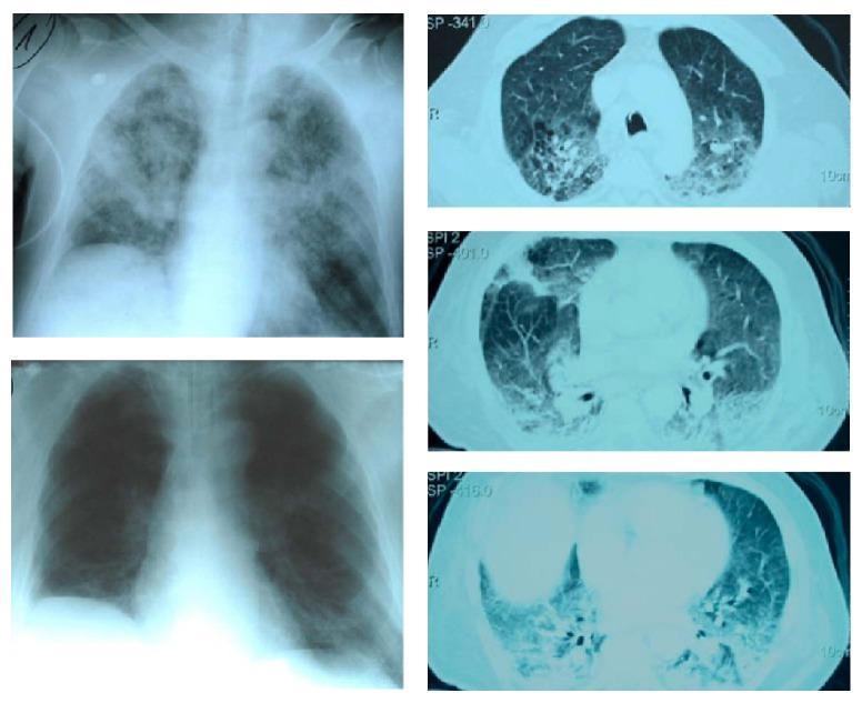
This composite diagnostic image includes chest X-rays and axial CT scans illustrating the progression and resolution of Acute Respiratory Distress Syndrome (ARDS). The top-left anterior-posterior chest X-ray shows diffuse, patchy bilateral alveolar infiltrates and ground-glass opacities in a postoperative patient, characteristic of pulmonary edema. To the right, three axial CT scan slices (cranial to caudal) demonstrate significant bilateral dorsal consolidation and dependent infiltrates with air bronchograms, typical of ARDS morphology. The bottom-left chest X-ray shows the same patient following Continuous Extrathoracic Negative Pressure Ventilation (CENPV), displaying marked regression of the bilateral infiltrates, improved lung aeration, and a reduction in previously noted pleural effusions. The educational focus is on the radiographic monitoring of lung recruitment and the resolution of pulmonary consolidations in critical care. This material is relevant for pulmonary medicine, anesthesia, and intensive care specialties.
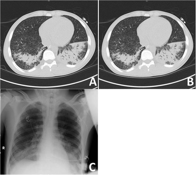
A multi-panel clinical imaging figure demonstrating severe lung pathology in a patient with Acute Respiratory Distress Syndrome (ARDS) secondary to autoimmune disease. Panels A and B are axial non-contrast CT scans of the chest at the level of the lower lobes, illustrating recruitment potential under varying Positive End-Expiratory Pressure (PEEP) levels. The lung parenchyma shows bilateral, heterogeneous inflammatory consolidations and ground-glass opacities, predominantly in the dependent (posterior) segments of the lower lobes. Panel C is a corresponding anteroposterior conventional chest radiograph (X-ray) showing bilateral basal opacities and increased density in the lower lung fields, consistent with the consolidations seen on CT. The images demonstrate the visual presentation of small airway disease and diffuse alveolar damage, characterized by airspace filling that obscures normal pulmonary markings. Clinical relevance includes the assessment of lung recruitment in critically ill patients and the radiographic manifestation of pulmonary hemorrhage or inflammation in systemic autoimmune conditions.
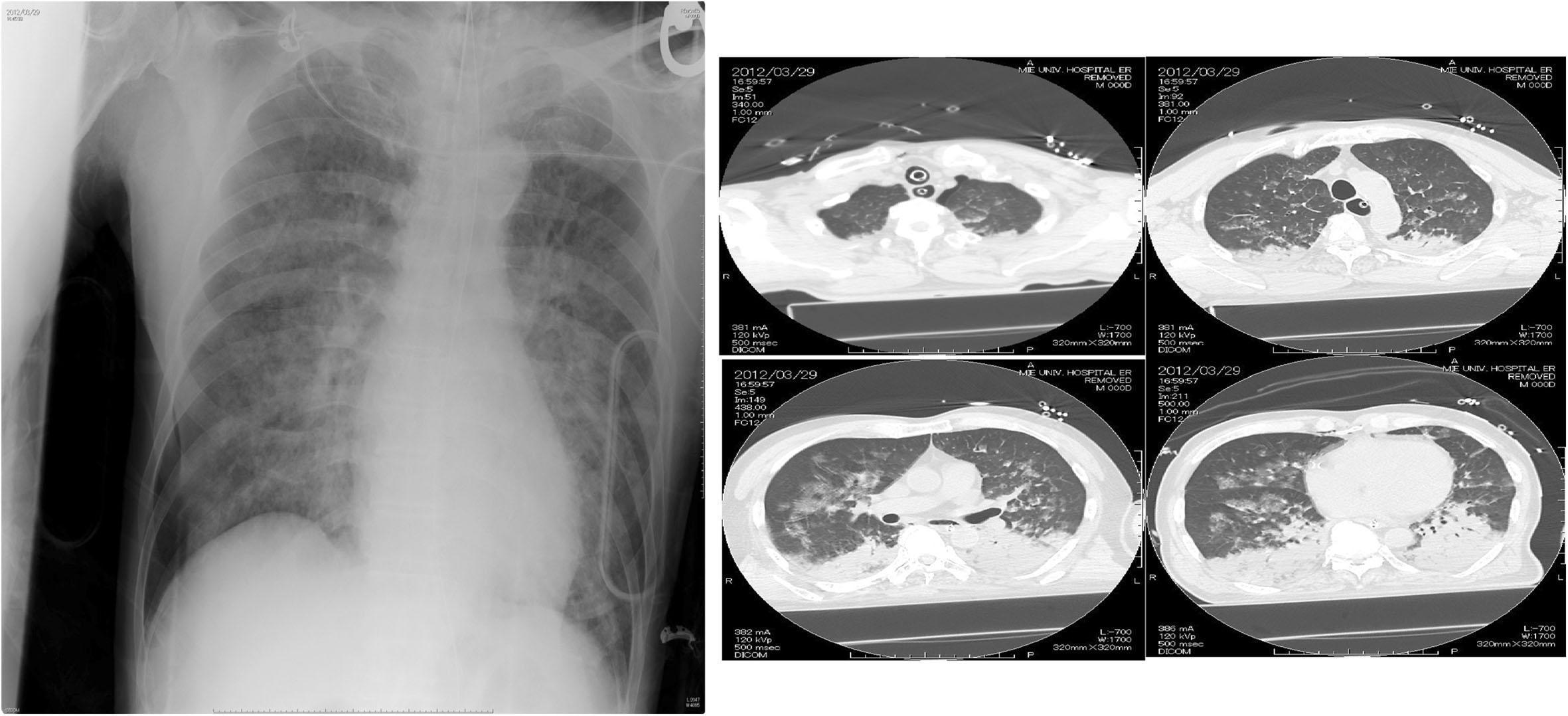
A composite diagnostic image featuring a frontal chest radiograph and multiple axial CT scans of the thorax. The chest X-ray demonstrates diffuse, patchy, bilateral pulmonary opacities and infiltrates involving all lung zones, characteristic of Acute Respiratory Distress Syndrome (ARDS) or extensive pulmonary edema. Normal vascular markings are obscured. The axial CT scan frames confirm widespread bilateral consolidation and ground-glass opacities, localized predominantly in the dependent (posterior) regions of the lungs in a supine patient. Additionally, the CT images reveal pathology in the left hemithorax, including multiple rib fractures and signs of a pneumohemothorax, characterized by air and fluid accumulation in the pleural space. Monitoring equipment, including an endotracheal tube and ECG leads, is visible. This clinical imaging set illustrates complex thoracic trauma and secondary respiratory failure, likely following chest compressions or acute lung injury.
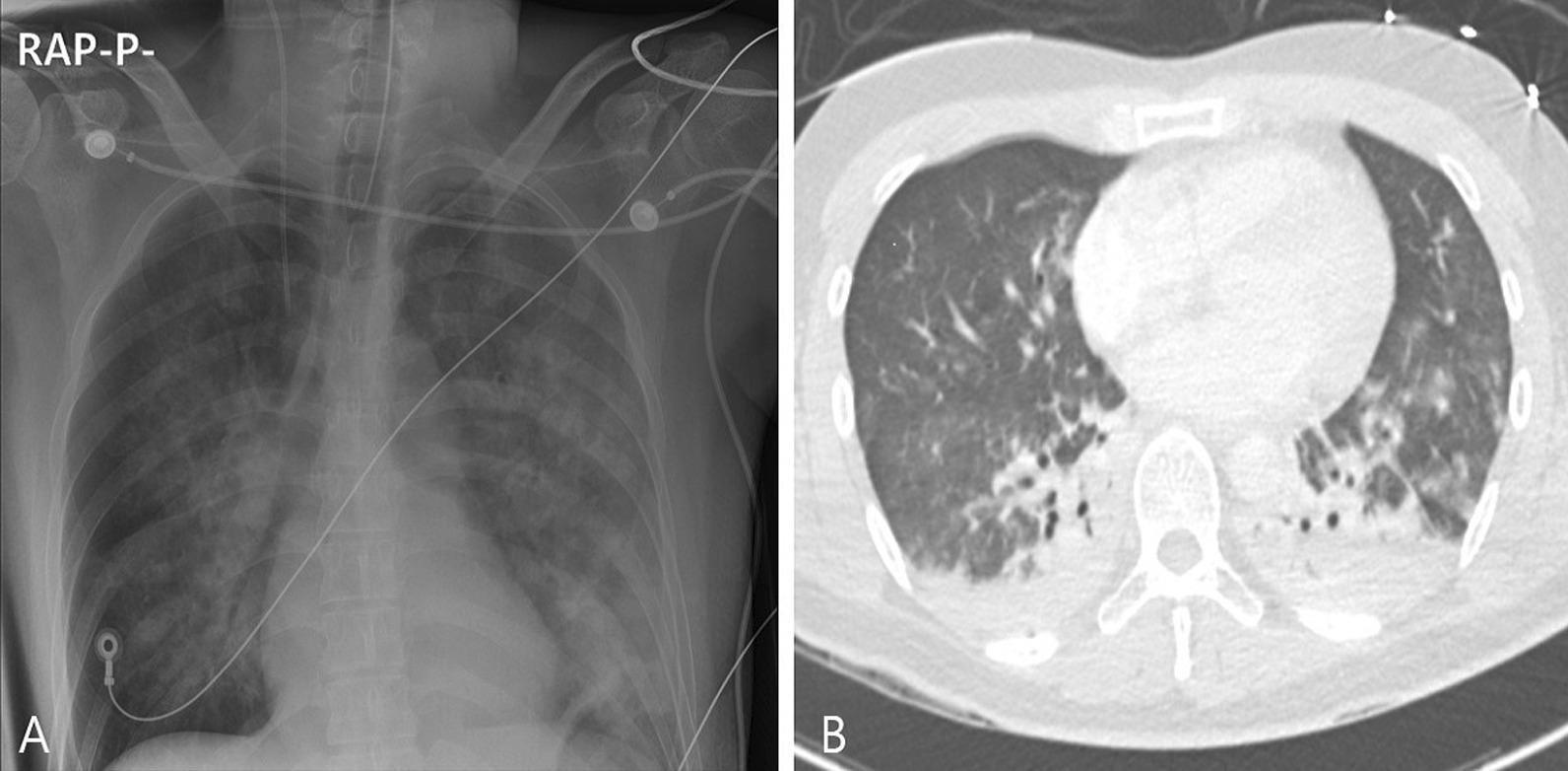
This composite diagnostic image displays acute pulmonary pathology through two imaging modalities. Image A is an anteroposterior chest X-ray demonstrating diffuse, bilateral pulmonary infiltrates and increased opacification throughout both lung fields, consistent with severe parenchymal lung disease or acute respiratory distress syndrome (ARDS). Medical support devices, including ECG leads and an endotracheal tube, are visible. Image B is an axial non-contrast chest CT scan (lung window) showing a distinct anterior-posterior density gradient. The dependent (posterior) regions of the lungs exhibit dense consolidation and atelectasis, while the non-dependent (anterior) regions show diffuse ground-glass opacities. Small bilateral pleural effusions are also suggested. The findings are characteristic of the early exudative phase of ARDS or severe inhalation injury, where fluid and inflammatory debris accumulate in the most dependent portions of the lungs.
"acute respiratory distress syndrome"[MeSH Terms] AND management
Acute Respiratory Distress Syndrome (ARDS)
Definition and Diagnostic Criteria (Berlin Definition)
| Criterion | Detail |
|---|---|
| Timing | Acute onset within 7 days of a known clinical insult or new/worsening respiratory symptoms |
| Chest imaging | Bilateral infiltrates on CXR or CT not fully explained by effusions, collapse, or nodules |
| Oxygenation | PaO₂/FiO₂ (P/F ratio) < 300 mm Hg on PEEP ≥ 5 cm H₂O |
| Origin of edema | Respiratory failure not fully explained by cardiac failure or fluid overload (use objective assessment, e.g., echo) |
- Mild: 200 < P/F ≤ 300 mm Hg
- Moderate: 100 < P/F ≤ 200 mm Hg
- Severe: P/F ≤ 100 mm Hg
Etiology and Precipitating Causes
- Pneumonia (most common overall)
- Aspiration of gastric contents
- Pulmonary contusion
- Inhalation injury
- Near-drowning
- Sepsis (most common indirect cause)
- Severe pancreatitis (develops in ~15-20% of cases, responsible for up to 60% of pancreatitis deaths)
- Major trauma/hemorrhage
- Burns
- Blood product transfusion (TRALI)
- Cardiopulmonary bypass
Pathophysiology
Diffuse Alveolar Damage (DAD)
-
Exudative phase (0-7 days): Disruption of the alveolar-capillary barrier leads to flooding of alveoli with protein-rich fluid, red cells, and fibrin. Hyaline membranes form. Type I pneumocyte necrosis occurs.
-
Proliferative phase (7-21 days): Type II pneumocyte proliferation attempts repair. Fibroblast influx begins. Organizing pneumonia pattern emerges.
-
Fibrotic phase (>21 days): Some patients develop progressive fibrosis with collagen deposition and loss of normal lung architecture.
Key Mechanisms
- Dependent zones: consolidated/fluid-filled or atelectatic but potentially recruitable
- Nondependent zones: relatively preserved, appear near-normal on CT
- Middle zones: poorly aerated but recruitable
Imaging
Management
1. Lung-Protective Mechanical Ventilation (Cornerstone of Treatment)
| Target | Value |
|---|---|
| Tidal volume | 6 mL/kg predicted body weight (PBW); can reduce to 4 mL/kg if needed |
| Plateau pressure | ≤ 30 cm H₂O |
| PaO₂ target | 55-80 mm Hg |
| SpO₂ target | 88-95% |
| pH | 7.30-7.45 |
| Permissive hypercapnia | Accepted to achieve low Vt/Pplat goals |
2. PEEP
- Some PEEP is mandatory to recruit alveoli and prevent de-recruitment
- Higher PEEP (~12-13 cm H₂O) may be better than lower PEEP (~8 cm H₂O) in moderate-severe ARDS (P/F < 200)
- In mild ARDS (P/F > 200), higher PEEP has not shown mortality benefit
- Very high PEEP with aggressive recruitment maneuvers can be harmful (the ART trial showed increased mortality)
- PEEP can be titrated using the PEEP/FiO₂ tables from ARDSNet or guided by esophageal pressure monitoring
3. Prone Positioning
- Improves oxygenation by redistributing pleural pressures, improving V/Q matching, and reducing VILI
- Reduces mortality by ~15% in patients with P/F < 150 mm Hg (PROSEVA trial)
- Should be initiated early (within 36 hours of diagnosis) in moderate-severe ARDS
- Requires experienced personnel; significant logistical considerations (line/tube management)
- Should be used regardless of its isolated oxygenation effect in severe ARDS
4. Fluid Management
- Conservative fluid strategy after initial resuscitation is preferred
- Diuresis to reduce extravascular lung water improves oxygenation (FACTT trial: conservative arm had more ventilator-free days)
- Avoid fluid overload; targeted euvolemia or slight negative balance
5. Neuromuscular Blockade
- Routine use is NOT recommended in moderate-severe ARDS (ACURASYS benefit not replicated by ROSE trial)
- Judicious use in selected patients: patient-ventilator asynchrony with breath stacking, very large spontaneous inspiratory efforts ("P-SILI")
6. Corticosteroids
- May reduce ongoing inflammatory damage and duration of mechanical ventilation
- Meta-analyses support methylprednisolone (1-2 mg/kg/day) especially in persistent/unresolving ARDS or when there is an inflammatory cause (e.g., COVID-19 - dexamethasone 6 mg/day demonstrated mortality benefit)
- No role in late fibroproliferative phase (>2 weeks)
7. Adjunctive Therapies - Limited/No Mortality Benefit
| Therapy | Effect |
|---|---|
| Inhaled nitric oxide | Improves oxygenation only; no mortality benefit |
| Inhaled epoprostenol | Improves oxygenation only; no mortality benefit |
| High-frequency oscillatory ventilation (HFOV) | No mortality benefit; potentially harmful in some patients |
| Recruitment maneuvers with very high PEEP | Can be harmful |
8. ECMO (Extracorporeal Membrane Oxygenation)
- Last-resort option for refractory ARDS (P/F < 80 despite optimal conventional management)
- CESAR trial: improved survival for severe ARDS when referred to ECMO centers
- EOLIA trial: no statistically significant benefit (trial stopped early; remains debated)
- Best outcomes at high-volume specialized centers
- VV-ECMO (venovenous) is used for isolated respiratory failure
9. Non-Invasive Support
- High-flow nasal cannula (HFNC): May benefit mild-moderate ARDS (P/F 150-300); FLORALI subgroup showed greatest benefit at P/F ≤ 200
- Non-invasive ventilation (NIV/BiPAP): Risk of generating large, harmful tidal volumes in moderate-severe ARDS; higher mortality seen in patients with P/F < 150 using NIV (LUNG-SAFE study)
- NIV and HFNC are generally reserved for mild ARDS or as a bridge; close monitoring is required
Complications and Outcomes
- Mortality commonly exceeds 30-40% in moderate-severe ARDS
- Survivors frequently have impaired quality of life and persistent functional disability
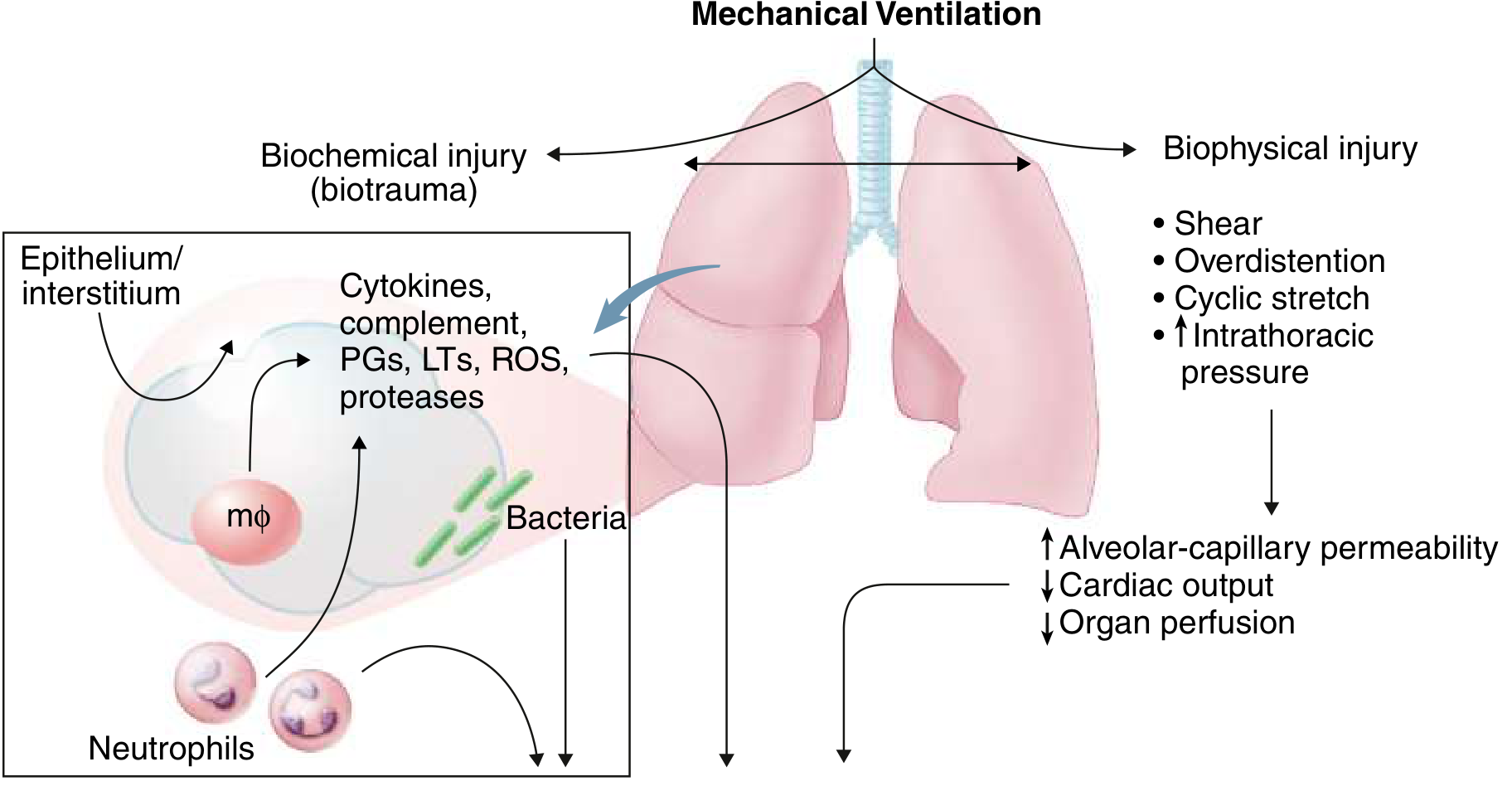
- Multi-organ dysfunction syndrome (MODS) is a major cause of death, partly driven by ventilator-induced cytokine release (biotrauma)
- No therapy directly reverses diffuse alveolar damage - mechanical ventilation is supportive until lung recovery
Summary Algorithm
Suspected ARDS
    ↓
Confirm Berlin criteria (bilateral infiltrates, P/F <300, non-cardiogenic, acute onset)
    ↓
Classify severity → Mild (P/F 200-300) | Moderate (100-200) | Severe (<100)
    ↓
Low tidal volume ventilation (6 mL/kg PBW), Pplat ≤30 cmH₂O
    ↓
Titrate PEEP (higher PEEP for P/F <200)
    ↓
Moderate-Severe: Consider prone positioning (≥16 hrs/day) if P/F <150
    ↓
Conservative fluid management
    ↓
Refractory (P/F <80): Consider ECMO referral
- Goldman-Cecil Medicine, pp. 1066-1068
- Murray & Nadel's Textbook of Respiratory Medicine, pp. 3301-3326
- Sabiston Textbook of Surgery, pp. 940-941
- Rosen's Emergency Medicine, pp. 1234-1240