
Anxiety and anxiety disorders
"anxiety disorders"[MeSH Terms] AND treatment
anxiety disorders brain pathways fear response amygdala diagram
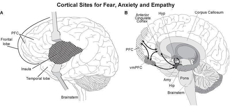
This medical illustration presents two anatomical diagrams of the human brain focused on the neurobiology of fear, anxiety, and empathy. Diagram A is a lateral view showing the retraction of the frontal and temporal lobes to reveal the insula, which is highlighted with cross-hatching. The Prefrontal Cortex (PFC) and brainstem are also labeled. Diagram B is a mid-sagittal section illustrating deep cortical and subcortical structures. It highlights the Anterior Cingulate Cortex (ACC) and the ventromedial Prefrontal Cortex (vmPFC) with cross-hatching. Key labeled subcortical structures include the corpus callosum, hypothalamus (Hyp), amygdala (Amy), hippocampus (Hip), pons, and brainstem. Black bidirectional arrows indicate functional connectivity and reciprocal pathways between the PFC, vmPFC, amygdala, and hippocampus. This anatomical diagram serves to educate on the limbic system's circuitry and the integration of cortical sites with subcortical emotional processing centers.
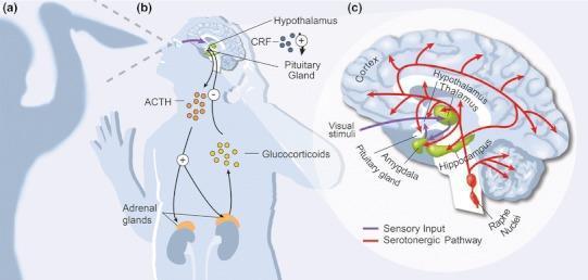
A multi-part medical illustration detailing the neurobiology of the mammalian fear and anxiety response. Section (a) depicts a subject reacting to a threatening stimulus. Section (b) is a pathophysiology diagram of the Hypothalamic-Pituitary-Adrenal (HPA) axis. It illustrates visual stimuli activating the hypothalamus to release Corticotrophin-Releasing Factor (CRF), which stimulates the pituitary gland to release Adrenocorticotrophin-Releasing Hormone (ACTH). ACTH travels to the adrenal glands (positioned above the kidneys), triggering glucocorticoid release. A negative feedback loop is shown where glucocorticoids inhibit the hypothalamus and pituitary. Section (c) provides an enlarged anatomical diagram of brain circuitry. It highlights the amygdala (green) as a central hub, receiving sensory input (purple arrows) from the retina via the thalamus. The diagram also maps the serotonergic pathway (red arrows) originating from the Raphe nuclei in the brainstem and innervating the hippocampus, thalamus, hypothalamus, amygdala, and cortex. This visual summary integrates endocrine and neurological systems involved in stress and emotional regulation.
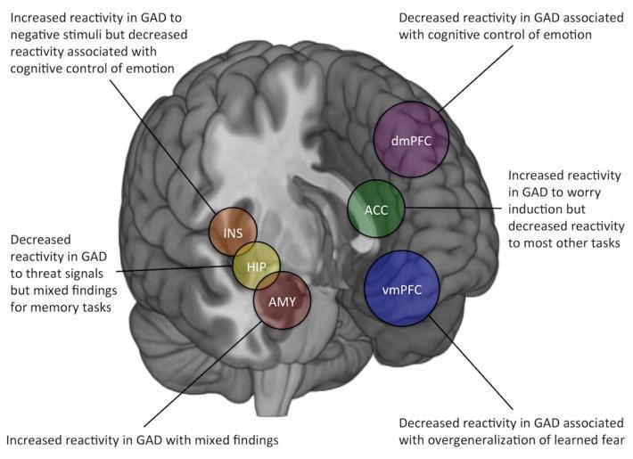
This medical illustration presents a medial sagittal view of the human brain, serving as an anatomical diagram of the corticolimbic circuit with functional annotations related to generalized anxiety disorder (GAD). Key regions are highlighted with color-coded circular overlays: the dorsomedial prefrontal cortex (dmPFC, purple), anterior cingulate cortex (ACC, green), ventromedial prefrontal cortex (vmPFC, blue), insula (INS, brown), hippocampus (HIP, yellow), and amygdala (AMY, red). The diagram summarizes findings from task-based fMRI studies, indicating how reactivity changes in these regions correlate with GAD symptoms. Specific annotations note increased amygdala (AMY) reactivity, decreased dmPFC and vmPFC reactivity associated with cognitive control of emotion and fear overgeneralization, and task-dependent reactivity patterns in the ACC, INS, and HIP. This educational resource is designed to illustrate the neurobiological mechanisms of anxiety disorders, highlighting the relationship between structural neuroanatomy and functional BOLD signal abnormalities in psychiatric pathophysiology.
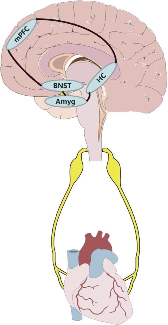
This pathophysiology diagram illustrates the neural circuit link between anxiety-related brain regions and the cardiovascular system via the autonomic nervous system. The upper portion shows a sagittal view of the human brain with highlighted limbic and cortical structures, including the medial prefrontal cortex (mPFC), hippocampus (HC), bed nucleus of the stria terminalis (BNST), and amygdala (Amyg). Interconnecting lines demonstrate the functional pathways between these nuclei, specifically highlighting the influence of the mPFC and amygdala on downstream autonomic regulation. Below the brainstem, two thick yellow pathways represent the autonomic efferent fibers (sympathetic and parasympathetic) descending to innervate the heart. The heart is depicted inferiorly with visible coronary vasculature and major vessels. This illustration is used in medical education to explain the psychosomatic connection between psychiatric disorders, such as anxiety, and cardiovascular disease (CVD) morbidity, highlighting how neural modulation of these circuits could impact heart health.
Anxiety and Anxiety Disorders
Classification (DSM-5-TR)
| Disorder | Key Feature |
|---|---|
| Panic Disorder | Recurrent unexpected panic attacks |
| Agoraphobia | Fear/avoidance of situations where escape seems difficult |
| Generalized Anxiety Disorder (GAD) | Persistent, excessive, unrealistic worry |
| Social Anxiety Disorder | Fear of social/performance situations |
| Specific Phobia | Marked fear of a specific object/situation |
| Separation Anxiety Disorder | Excessive fear of separation from attachment figures |
| Selective Mutism | Failure to speak in specific social situations |
Note: OCD, PTSD, and acute stress disorder are no longer classified as anxiety disorders in DSM-5.
Neurobiology of Anxiety
Panic Disorder
- Involves increased noradrenergic discharge from the locus coeruleus
- Sodium lactate infusion provokes attacks in 2/3 of patients; so does yohimbine (α2-antagonist), CCK-4, and CO₂ inhalation
- Resting-state fMRI shows abnormalities in the default mode network
- Genetic predisposition: concordance 30-45% in monozygotic twins
GAD
- Most anxiogenic and anxiolytic agents act on the GABA receptor/chloride ion channel complex
- Benzodiazepines bind two GABA receptor sites: Type I (broad distribution) and Type II (hippocampus, striatum, neocortex)
- Anti-anxiety effects relate to binding at alpha-2 and alpha-3 GABA subunits; sedation and memory impairment relate to the alpha-1 subunit
- Serotonin (5-HT) and 3α-reduced neuroactive steroids (allosteric GABA modulators) also play a role
- Buspirone (partial 5-HT1A agonist) and certain 5-HT antagonists (mirtazapine, nefazodone) have beneficial effects
Individual Anxiety Disorders
1. Panic Disorder
- Recurrent, unpredictable panic attacks: discrete episodes of intense fear with palpitations, sweating, trembling, shortness of breath, chest pain, dizziness, paresthesias, GI distress, feelings of unreality, and fear of impending doom/death
- Sudden onset within 10 min, usually resolving within 1 hour
- Diagnostic criteria: at least 1 month of concern about attacks or behavioral change related to them
- Lifetime prevalence: 2-3%; onset typically in late adolescence/early adulthood
- Agoraphobia develops in many patients - irrational fear of places where escape seems impossible, leading to progressive restriction of lifestyle
- Cardiovascular: paroxysmal atrial tachycardia, mitral valve prolapse
- Endocrine: pheochromocytoma, thyrotoxicosis, hypoglycemia
- Pulmonary and GI conditions can mimic panic
- Panic disorder is the primary diagnosis in 43% of patients with chest pain and normal coronary angiograms
- 75% of panic disorder patients will also meet criteria for major depression at some point
- High comorbidity with alcohol and benzodiazepine abuse (self-medication)
- First-line pharmacotherapy: SSRIs and SNRIs (e.g., escitalopram, paroxetine, venlafaxine, sertraline) - these require several weeks to take effect
- Benzodiazepines (alprazolam, clonazepam - FDA approved) are useful early for rapid relief but should not exceed 4-6 weeks due to tolerance and dependence risk
- Psychotherapy: CBT with breathing techniques, psychoeducation, and graduated exposure spanning 12-15 sessions
- Combined pharmacotherapy and psychotherapy is most effective
- Maintain drug treatment for 1-2 years after remission to prevent relapse
- Success rate: 75-85% (complete remission somewhat lower)
2. Generalized Anxiety Disorder (GAD)
- Persistent, excessive, and/or unrealistic worry
- Associated with: muscle tension, impaired concentration, autonomic arousal, feeling "on edge" or restless, and insomnia
- Onset usually before age 20; history of childhood fears and social inhibition common
- Lifetime prevalence: 5-6%; higher in first-degree relatives
- Restlessness or feeling keyed up or on edge
- Being easily fatigued
- Difficulty concentrating or mind going blank
- Irritability
- Muscle tension
- Sleep disturbance
- Combined pharmacotherapy and psychotherapy is most effective (complete symptomatic relief is rare)
- FDA-approved drugs: Escitalopram, paroxetine, duloxetine, venlafaxine; buspirone (partial 5-HT1A agonist) - requires weeks to work but has no abuse potential
- Benzodiazepines: Short course (lorazepam, oxazepam, clonazepam, or alprazolam); lorazepam and oxazepam are preferred in elderly/liver disease (metabolized by conjugation, not oxidation); contraindicated in pregnancy and breastfeeding
- GABAergic anticonvulsants (off-label): Gabapentin, pregabalin, oxcarbazepine, tiagabine, divalproex
3. Phobic Disorders
- Marked, persistent fear of specific objects or situations
- Immediate anxiety reaction on exposure; behavioral avoidance
- The individual recognizes the fear as excessive and unreasonable
- Prevalence: 7-9% of population; females affected 2x more than males
- Twin studies: concordance 23% (monozygotic) vs 15% (dizygotic) for agoraphobia, social phobia, and animal phobia
- Specific phobia: Fear of specific stimuli (heights, blood, flying, enclosed spaces, animals)
- Social phobia (Social Anxiety Disorder): Fear of social or performance situations involving exposure to unfamiliar people or evaluation by others (e.g., conversations at parties, public restrooms, meeting strangers)
- Agoraphobia: Fear of situations where escape would be difficult
- CBT with graduated exposure is the most effective treatment - behavioral techniques, particularly systematic desensitization and exposure therapy, have the strongest evidence
- SSRIs/SNRIs for social anxiety disorder (paroxetine, venlafaxine, sertraline FDA-approved)
- Beta-blockers (propranolol) and benzodiazepines for situational/performance anxiety
- Monoamine oxidase inhibitors (MAOIs) such as phenelzine can be effective for social anxiety
Medical Causes of Anxiety (Must Exclude)
| System | Conditions |
|---|---|
| Endocrine | Hyperthyroidism, pheochromocytoma, hypoglycemia, Cushing's disease |
| Cardiovascular | Paroxysmal arrhythmias, mitral valve prolapse, heart failure |
| Neurological | Temporal lobe epilepsy, vestibular dysfunction |
| Respiratory | Asthma, COPD, pulmonary embolism |
| Drug-related | Caffeine intoxication, stimulants, alcohol/benzodiazepine withdrawal |
Caffeine is especially notable - caffeine intoxication should always be considered when patients present with anxiety or insomnia, as symptoms may completely resolve with caffeine avoidance. Patients with panic disorder consume significantly more caffeine than controls, and greater consumption triggers panic attacks in panic disorder patients. (Maudsley Prescribing Guidelines, 15th ed.)
Pharmacotherapy Summary
| Drug Class | Drugs | Indication | Notes |
|---|---|---|---|
| SSRIs | Escitalopram, paroxetine, sertraline, fluoxetine | Panic, GAD, social phobia | First-line; weeks to onset |
| SNRIs | Venlafaxine, duloxetine | Panic, GAD | FDA-approved for GAD |
| Benzodiazepines | Alprazolam, clonazepam, lorazepam | Short-term/acute anxiety | Dependence risk; max 4-6 weeks |
| Buspirone | Buspirone | GAD | No abuse potential; slow onset |
| TCAs | Imipramine, desipramine | Panic disorder | Less preferred; side effects |
| MAOIs | Phenelzine | Social phobia, panic | Dietary restrictions required |
| Beta-blockers | Propranolol | Situational/performance anxiety | For somatic symptoms |
| Pregabalin/Gabapentin | Pregabalin, gabapentin | GAD (off-label) | GABAergic mechanism |
Psychotherapy
- Psychoeducation about the physiology of anxiety
- Cognitive restructuring (challenging catastrophic thoughts)
- Graduated exposure/desensitization (in vivo or imaginal)
- Breathing retraining and relaxation techniques
- Homework and compliance monitoring
Recent Evidence (PubMed, 2024-2026)
- A 2024 systematic review and meta-analysis found CBD has therapeutic potential in anxiety disorders, though clinical application remains cautious given regulatory status (PMID: 38924898)
- A 2025 meta-analysis confirmed aerobic and resistance exercise significantly reduce both anxiety and depression (PMID: 40432290)
- Harrison's Principles of Internal Medicine, 22nd ed. (2025), Chapter 463 - Psychiatric Disorders
- The Maudsley Prescribing Guidelines in Psychiatry, 15th ed.
- Goodman & Gilman's The Pharmacological Basis of Therapeutics
- Kaplan & Sadock's Comprehensive Textbook of Psychiatry