Pleural effusion

Reading File
Finding Sources
Searching PubMed

"pleural effusion" diagnosis management

Searching Images

pleural effusion chest X-ray and anatomy diagram

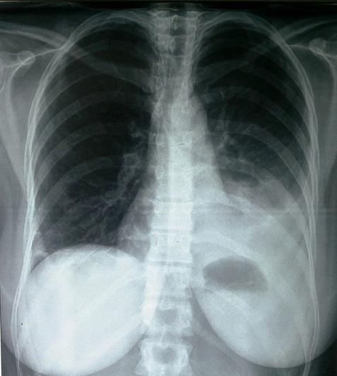
Frontal chest radiograph (X-ray) of a 37-year-old patient demonstrating a moderate-sized left-sided pleural effusion. Key diagnostic features include a homogeneous opacity in the lower third of the left hemithorax, resulting in the blunting of the left costophrenic angle and obscuring the left hemidiaphragm. The opacity exhibits a meniscus sign, characteristic of fluid accumulation in the pleural space. There is a subtle shift of the mediastinal structures toward the contralateral (right) side, indicating a mass effect from the effusion. The right lung field remains clear with normal lucency and preserved vascular markings. The right hemidiaphragm is well-defined and positioned normally. This imaging is clinically significant for diagnosing pleural fluid collections and assessing their secondary effects on thoracic anatomy. It is a classic representation of moderate pleurisy relevant for students and practitioners in pulmonology, radiology, and internal medicine.

Frontal chest radiograph (X-ray) of a 37-year-old patient demonstrating a moderate-sized left-sided pleural effusion. Key diagnostic features include a homogeneous opacity in the lower third of the left hemithorax, resulting in the blunting of the left costophrenic angle and obscuring the left hemidiaphragm. The opacity exhibits a meniscus sign, characteristic of fluid accumulation in the pleural space. There is a subtle shift of the mediastinal structures toward the contralateral (right) side, indicating a mass effect from the effusion. The right lung field remains clear with normal lucency and preserved vascular markings. The right hemidiaphragm is well-defined and positioned normally. This imaging is clinically significant for diagnosing pleural fluid collections and assessing their secondary effects on thoracic anatomy. It is a classic representation of moderate pleurisy relevant for students and practitioners in pulmonology, radiology, and internal medicine.

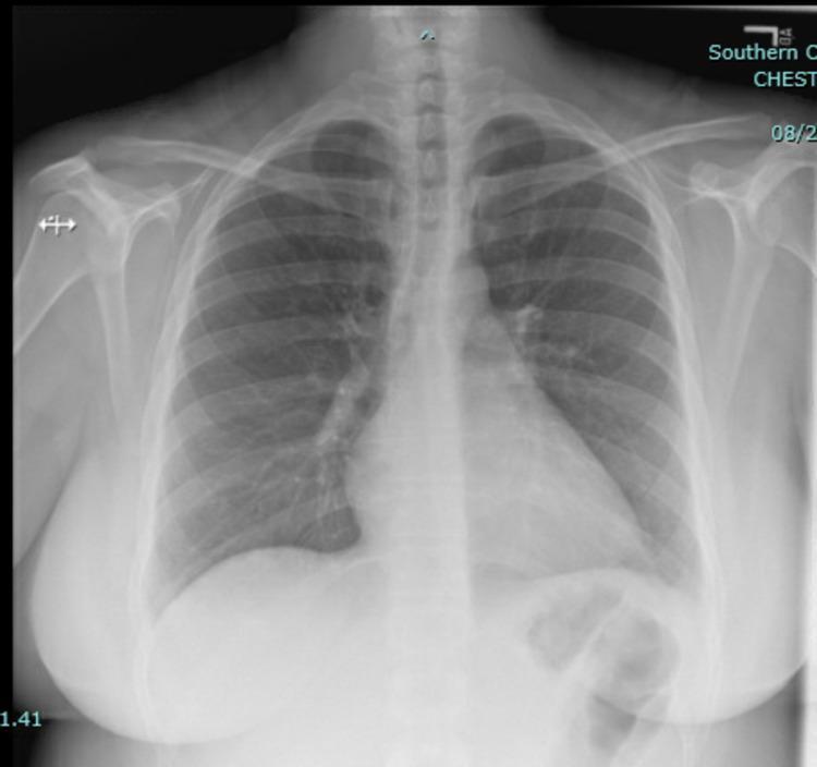
A posterior-anterior (PA) chest X-ray illustrating normal thoracic anatomy. The lung fields are clear and bilaterally symmetric, showing normal aeration without evidence of focal consolidation, opacities, or interstitial infiltrates. Both costophrenic and cardiophrenic angles are sharp, indicating the absence of pleural effusion. The cardiac silhouette is normal in size and shape, with a cardiothoracic ratio within physiological limits. The mediastinal contour and hila appear unremarkable. The bony structures, including the ribs, clavicles, and proximal humeri, are intact, and the overlying soft tissues are normal. This diagnostic image serves as a baseline comparison for clinical scenarios where pathology, such as pneumonia or heart failure, is suspected but radiographically absent, as seen in some cases of pulmonary embolism or intracardiac masses.

A posterior-anterior (PA) chest X-ray illustrating normal thoracic anatomy. The lung fields are clear and bilaterally symmetric, showing normal aeration without evidence of focal consolidation, opacities, or interstitial infiltrates. Both costophrenic and cardiophrenic angles are sharp, indicating the absence of pleural effusion. The cardiac silhouette is normal in size and shape, with a cardiothoracic ratio within physiological limits. The mediastinal contour and hila appear unremarkable. The bony structures, including the ribs, clavicles, and proximal humeri, are intact, and the overlying soft tissues are normal. This diagnostic image serves as a baseline comparison for clinical scenarios where pathology, such as pneumonia or heart failure, is suspected but radiographically absent, as seen in some cases of pulmonary embolism or intracardiac masses.

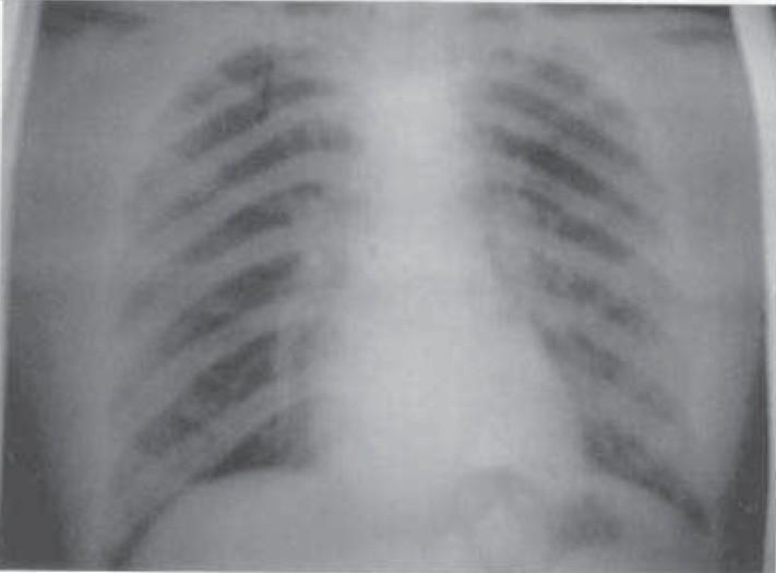
A posterior-anterior (PA) view chest X-ray illustrating normal thoracic anatomy. The radiographic image shows clear and well-aerated bilateral lung fields with no visible pulmonary opacities, infiltrates, or masses. The cardiac silhouette is of normal size and position within the mediastinum, and the trachea is observed at the midline. Both costophrenic angles are sharp, and the diaphragmatic domes are clearly defined and appropriately contoured, indicating the absence of pleural effusion. The skeletal structures, including the ribs and clavicles, appear intact without evidence of acute fracture or deformity. This diagnostic image serves as a baseline for a normal chest examination, illustrating healthy pulmonary vasculature radiating from the hilum toward the lung periphery.

A posterior-anterior (PA) view chest X-ray illustrating normal thoracic anatomy. The radiographic image shows clear and well-aerated bilateral lung fields with no visible pulmonary opacities, infiltrates, or masses. The cardiac silhouette is of normal size and position within the mediastinum, and the trachea is observed at the midline. Both costophrenic angles are sharp, and the diaphragmatic domes are clearly defined and appropriately contoured, indicating the absence of pleural effusion. The skeletal structures, including the ribs and clavicles, appear intact without evidence of acute fracture or deformity. This diagnostic image serves as a baseline for a normal chest examination, illustrating healthy pulmonary vasculature radiating from the hilum toward the lung periphery.

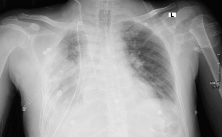
A frontal chest X-ray (radiograph) demonstrating a massive, dense, and homogeneous opacification of the right hemithorax. The opacity extends from the lung base to the apex, obliterating the right heart border, the right hemidiaphragm, and the costophrenic angle, which is indicative of a large pleural effusion. There is a notable mass effect causing a contralateral shift of the mediastinum, including the trachea and cardiac silhouette, toward the left. The left lung remains aerated but shows increased reticular markings in the mid-to-upper zones. Several medical devices and iatrogenic markers are visible: a central venous catheter is inserted via the left side and descends toward the superior vena cava; surgical staples are noted along the right lateral chest wall; and multiple ECG leads are present on the skin surface. An anatomical orientation marker 'L' is located in the upper left quadrant of the image frame, and a 60mm projection scale is visible on the right lateral margin. This clinical image illustrates the radiological presentation of extensive pleural fluid accumulation and its subsequent mechanical impact on thoracic anatomy.

A frontal chest X-ray (radiograph) demonstrating a massive, dense, and homogeneous opacification of the right hemithorax. The opacity extends from the lung base to the apex, obliterating the right heart border, the right hemidiaphragm, and the costophrenic angle, which is indicative of a large pleural effusion. There is a notable mass effect causing a contralateral shift of the mediastinum, including the trachea and cardiac silhouette, toward the left. The left lung remains aerated but shows increased reticular markings in the mid-to-upper zones. Several medical devices and iatrogenic markers are visible: a central venous catheter is inserted via the left side and descends toward the superior vena cava; surgical staples are noted along the right lateral chest wall; and multiple ECG leads are present on the skin surface. An anatomical orientation marker 'L' is located in the upper left quadrant of the image frame, and a 60mm projection scale is visible on the right lateral margin. This clinical image illustrates the radiological presentation of extensive pleural fluid accumulation and its subsequent mechanical impact on thoracic anatomy.

Reading File
Reading File
Reading File
Reading File
Reading File
Finding Sources
Reading File
Reading File
Reading File
Reading File
Loading Image
Loading Image
I now have comprehensive material to compile a thorough answer on pleural effusion. Let me compose the full response.

Pleural Effusion

Definition

A pleural effusion is an abnormal accumulation of fluid in the pleural space - the potential space between the visceral and parietal pleural layers. Under normal conditions, only 10-20 mL of fluid is present, facilitating frictionless lung movement. Any new pleural effusion is considered abnormal and warrants evaluation.

Pathophysiology

Fluid accumulates when the balance between fluid production and absorption is disrupted. Two broad mechanisms:
Transudates - result from an imbalance between hydrostatic and oncotic pressures, producing a protein-poor ultrafiltrate into the pleural space. The pleura itself is not diseased.
Exudates - result from pleural disease (inflammation or neoplasia), which causes active fluid secretion or leakage with high protein content.
  • Tintinalli's Emergency Medicine, p. 472

Causes (Differential Diagnosis)

TypeCommonLess Common
TransudatesHeart failureCirrhosis with ascites, peritoneal dialysis, nephrotic syndrome
ExudatesMalignancy (primary or metastatic), bacterial pneumonia (parapneumonic), pulmonary embolismViral/fungal/mycobacterial infection, SLE, rheumatoid arthritis, uremia, pancreatitis, postcardiac surgery, drugs (amiodarone)
Mixed featuresTransudates after diuretic therapyPulmonary embolism
  • Tintinalli's Emergency Medicine, p. 472
Hepatic hydrothorax is a specific cause in cirrhosis: affects ~5-10% of cirrhotic patients. Ascitic fluid passes through small diaphragmatic defects; 85% are right-sided; nearly all are transudative. - Murray & Nadel's Respiratory Medicine
Tuberculous effusion occurs early after MTB primary infection as pleurisy with effusion; 65% relapse rate if untreated. The fluid is an exudate with protein >50% of serum protein; AFB smears are rarely positive; cultures positive in only 25-30% of cases. - Rosen's Emergency Medicine

Clinical Features

  • May be clinically silent, or present with symptoms of the underlying disease
  • Dyspnea - as effusion volume increases
  • Pleuritic chest pain - if pleural inflammation is present
  • Physical examination findings:
    • Percussion dullness at the lung base
    • Decreased breath sounds at the base
    • Findings above the fluid level are relatively normal
    • With large/massive effusions, a fluid level may be impossible to distinguish clinically
  • Tintinalli's Emergency Medicine, p. 472
Hemodynamic effects: Large, mainly right-sided effusions can have hemodynamic effects similar to cardiac tamponade, affecting right ventricular filling. Hypoxemia is also seen (mildly elevated shunt ~6.9%), though often mild. - Murray & Nadel's Respiratory Medicine, p. 960

Imaging

Chest X-ray (PA erect):
  • Detects fluid at ≥150-200 mL
  • Meniscus sign - curved upper fluid border, concave toward the lung, higher laterally
  • Blunting of the costophrenic angle
  • Obscuration of the hemidiaphragm
  • Mediastinal shift toward contralateral side with large effusions
Moderate left-sided pleural effusion (PA CXR):
Moderate left-sided pleural effusion - PA chest radiograph showing homogeneous opacity in the lower left hemithorax with meniscus sign and costophrenic angle blunting
Massive right-sided pleural effusion with mediastinal shift:
Massive right-sided pleural effusion - PA CXR showing complete right hemithorax opacification with contralateral mediastinal and tracheal shift
Supine CXR: Fluid layers posteriorly, appearing as a hazy opacity over the lower hemithorax; costophrenic angles may not be blunted. - Grainger & Allison's Diagnostic Radiology
Subpulmonary (infrapulmonary) effusion: Mimics an elevated hemidiaphragm; on PA film, the diaphragm peaks more laterally than usual with a straight medial segment; on the left, stomach bubble separation >2 cm from the lung base is a clue. Confirm with ultrasound or CT.
Left lateral decubitus view: Detects small effusions (>10 mm fluid stripe is significant). Ultrasound is superior for bedside detection.
CT chest: Most sensitive; detects very small effusions missed on plain films; delineates loculated collections and underlying pathology.

Diagnosis

Thoracentesis - when to do it

  • Indicated for any clinically significant effusion (>1 cm on chest film) without a clear cause
  • Exception: If clinical suspicion for heart failure as the sole cause is high, a trial of treatment first is reasonable; thoracentesis is reserved if the effusion does not resolve in 3-4 days or the diagnosis becomes uncertain
  • Required to distinguish transudate from exudate
  • Tintinalli's Emergency Medicine, p. 472; Symptom to Diagnosis, p. 182

Light's Criteria (Exudate vs Transudate)

An effusion is classified as an exudate if ANY ONE of the following is present:
CriterionThreshold
Pleural fluid protein / serum protein> 0.5
Pleural fluid LDH / serum LDH> 0.6
Pleural fluid LDH> 2/3 of upper limit of normal serum LDH
  • Sensitivity 98%, Specificity 83% (LR+ 5.76; LR- 0.02)
  • If all three criteria are absent → transudate
  • Symptom to Diagnosis, p. 182; Tintinalli's Emergency Medicine
Albumin gradient correction: When a patient on diuretics has a transudate misclassified as an exudate by Light's criteria, a serum albumin - pleural albumin difference >1.2 g/dL supports a true transudate (LR+ 10.88 for exudate when <1.2 g/dL - the most specific test for exudate).

Pleural Fluid Analysis

TestInterpretation
pH < 7.2Empyema, malignant effusion, esophageal rupture
Glucose < 60 mg/dLEmpyema, TB, rheumatoid arthritis, SLE
Neutrophil predominanceAcute process: parapneumonic effusion (Sn 91%), PE, pancreatitis
Lymphocyte predominanceTB or malignancy (PPV 97%); cancer, postcardiac surgery
EosinophiliaNonspecific: inflammatory, pneumococcal, viral pleuritis, TB, repeated thoracentesis
Mesothelial cells <5%Highly suggestive of TB
Gram stain/culture positiveEmpyema
Cytology positiveMalignancy (specific, but sensitivity ~70% at best; lower for some cancers)
Triglycerides >110 mg/dLChylothorax (milky white fluid)
  • Symptom to Diagnosis, pp. 182-183; Tintinalli's Emergency Medicine

Tests for TB Pleural Effusion

TestSensitivitySpecificity
Pleural fluid culture42%-
Pleural biopsy culture64%-
Biopsy histology (caseating granulomas)70-80%-
Histology + tissue culture>90%-
Adenosine deaminase (ADA)92.2%92.2% (LR+ 11.82; LR- 0.08)
Interferon-gamma89%97% (LR+ 23.45; LR- 0.11)
  • Symptom to Diagnosis, p. 183

Management

General Principles

  • Always send standard pleural fluid tests: LDH, protein, albumin, pH, cell count with differential. Cytology is commonly added.
  • Treat the underlying cause.

Therapeutic Thoracentesis

  • Indicated for dyspnea at rest
  • Drain up to 1.0-1.5 L at a time
  • Draining >1.5 L acutely carries risk of re-expansion pulmonary edema - avoid large-volume rapid drainage
  • The effect of thoracentesis on arterial PO₂ is inconsistent (may increase, not change, or decrease) - Murray & Nadel's

Cause-Specific Management

CauseTreatment
Heart failureOptimize medical therapy (diuretics) - resolves >80% within 2 weeks
Parapneumonic/empyemaAntibiotics; chest drain for complicated parapneumonic/empyema; intrapleural fibrinolysis vs. surgery for complicated infections
Malignant effusionRepeated thoracentesis, indwelling pleural catheter, pleurodesis
Hepatic hydrothoraxSodium restriction + diuretics; TIPS (70-80% initial response); video-assisted thoracoscopic surgery (VATS) with pleurodesis; indwelling pleural catheter as bridge to transplant
TB effusionAnti-TB therapy
Hypothyroidism effusionThyroid hormone replacement; drain large symptomatic effusions
  • Murray & Nadel's Respiratory Medicine; Tintinalli's Emergency Medicine

Indwelling Pleural Catheter (IPC)

Used for recurrent malignant effusions or hepatic hydrothorax, allowing ambulatory drainage. Compared to repeated thoracentesis, avoids cumulative procedural risk.

Key Teaching Points

  1. Transudate = systemic pressure/oncotic problem; Exudate = local pleural pathology
  2. Light's criteria (98% sensitive) classify virtually all exudates - missing an exudate is the greater clinical risk
  3. Lymphocytic predominance in an exudate = TB or malignancy until proven otherwise (PPV 97%)
  4. Glucose <60 or pH <7.2 in an exudate = suspect empyema, TB, RA, or esophageal rupture
  5. Never drain >1.5 L at once (re-expansion pulmonary edema risk)
  6. ADA and interferon-gamma are the most accurate pleural fluid tests for TB diagnosis

Sources: Tintinalli's Emergency Medicine, 9e; Symptom to Diagnosis: An Evidence-Based Guide, 4e; Murray & Nadel's Textbook of Respiratory Medicine; Grainger & Allison's Diagnostic Radiology; Rosen's Emergency Medicine

Antibiotics spectrum

Reading File
Finding Sources
Finding Sources
Finding Sources
Reading File
Reading File
Finding Sources
Reading File
Reading File
Reading File
Reading File
Reading File
Loading Image
Finding Sources
Finding Sources
I now have all the material needed for a comprehensive, well-sourced answer. Let me compile it.

Antibiotic Spectrum of Activity

Classification Framework

Antibiotics can be classified by:
  1. Chemical structure (e.g., beta-lactams, aminoglycosides)
  2. Mechanism of action (e.g., cell wall inhibitors, protein synthesis inhibitors)
  3. Spectrum of activity (narrow, extended, or broad spectrum)
Spectrum terminology:
  • Narrow spectrum - covers a single or limited group of organisms
  • Extended spectrum - covers gram-positives and some gram-negatives
  • Broad spectrum - effective against a wide variety of species
  • Lippincott Illustrated Reviews: Pharmacology

Sites of Action - Overview

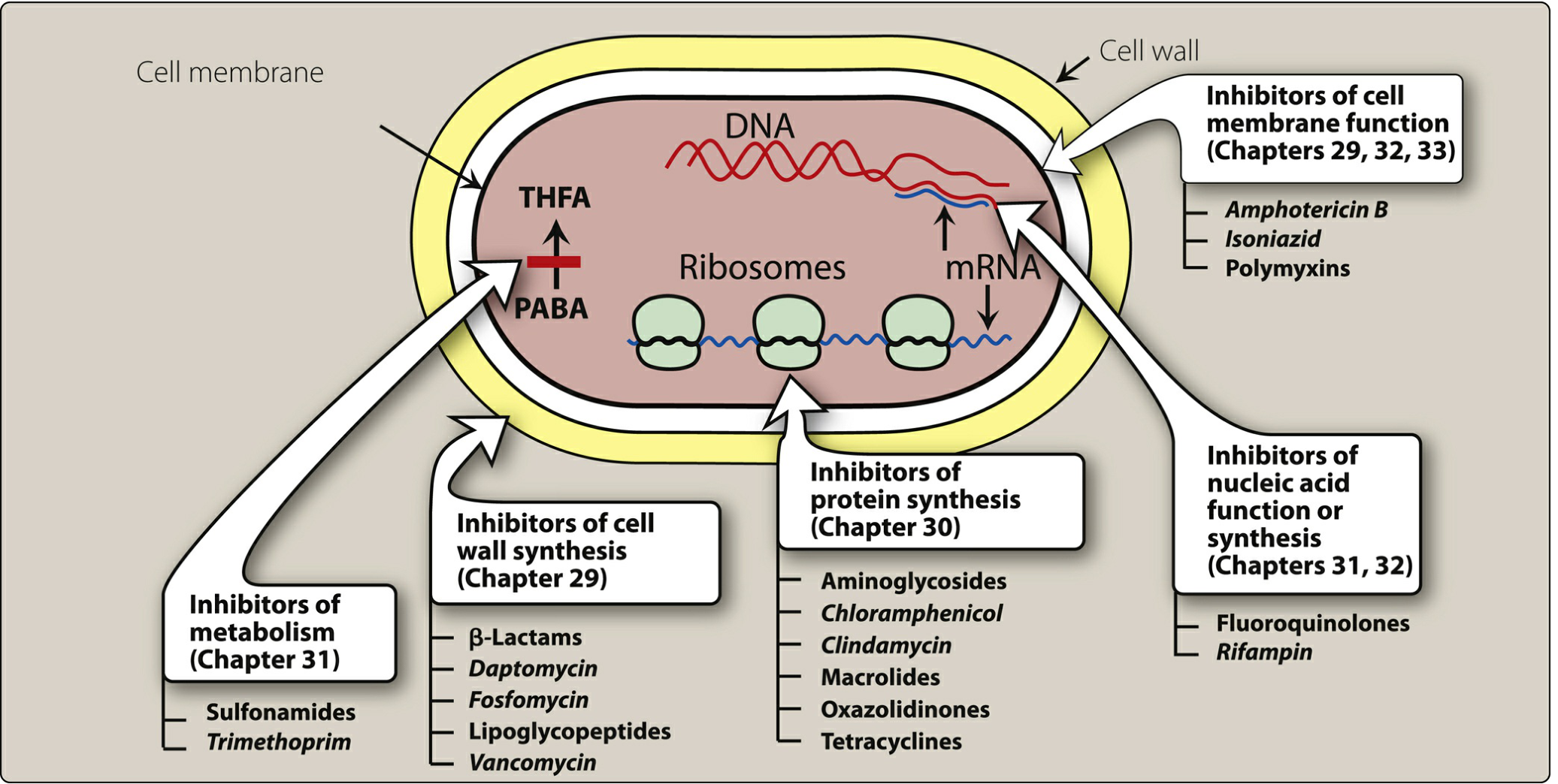
Classification of antimicrobial agents by site of action showing cell wall synthesis inhibitors (beta-lactams, vancomycin, daptomycin), protein synthesis inhibitors (aminoglycosides, macrolides, tetracyclines, clindamycin, oxazolidinones), nucleic acid inhibitors (fluoroquinolones, rifampin), cell membrane disruptors (polymyxins, isoniazid), and metabolism inhibitors (sulfonamides, trimethoprim)

1. Beta-Lactams

Mechanism

Inhibit bacterial cell wall synthesis by binding to penicillin-binding proteins (PBPs), blocking cross-linking of peptidoglycan. Bactericidal. Time-dependent killing.

A. Penicillins

ClassExamplesSpectrum
Natural penicillinsPenicillin G, Penicillin VAll beta-hemolytic streptococci; most gram-positive anaerobes; meningococci; limited vs. staphylococci; poor vs. gram-negative rods
Penicillinase-resistantMethicillin, Nafcillin, Oxacillin, CloxacillinSimilar to natural penicillins + enhanced activity vs. staphylococci (MSSA)
Broad-spectrum (aminopenicillins)Ampicillin, AmoxicillinGram-positive equivalent to natural penicillins + some gram-negative rods (E. coli, H. influenzae, Listeria)
Beta-lactam + Beta-lactamase inhibitorAmoxicillin-clavulanate, Ampicillin-sulbactam, Piperacillin-tazobactam, Ceftazidime-avibactamAbove + improved vs. beta-lactamase-producing staph and gram-negatives; piperacillin-tazobactam and ceftazidime-avibactam are the most active
  • Medical Microbiology 9e, Table 17.2

B. Cephalosporins (by generation)

GenerationExamplesKey Spectrum
1st (narrow)Cefazolin, Cephalexin, CephalothinGram-positives (MSSA, strep); limited gram-negatives (E. coli, Klebsiella, Proteus mirabilis)
2nd (expanded)Cefuroxime, Cefaclor1st gen + Haemophilus influenzae, Enterobacter, Citrobacter, additional Proteus
2nd - CephamycinsCefoxitin, CefotetanLike 2nd gen + anaerobes (Bacteroides fragilis); more beta-lactamase stable
3rd (broad)Ceftriaxone, Cefotaxime, CeftazidimeMost Enterobacteriaceae; Pseudomonas (ceftazidime); reduced gram-positive potency vs. 1st gen
4th (extended)CefepimeBroad: gram-positives + gram-negatives including Pseudomonas; more beta-lactamase stable
5th (anti-MRSA)CeftarolineAbove + MRSA activity
Key principle: As generation increases, gram-negative coverage increases; gram-positive coverage generally decreases (except cefepime and ceftaroline).
  • Medical Microbiology 9e, Table 17.3

C. Carbapenems and Monobactams

DrugSpectrum
Imipenem, Meropenem, DoripenemBroadest beta-lactam spectrum: most gram-positives (not MRSA), most gram-negatives including Pseudomonas, most anaerobes; resistant to most beta-lactamases
ErtapenemLike above but NO activity vs. Pseudomonas or Acinetobacter
Aztreonam (monobactam)Gram-negatives ONLY (including Pseudomonas); similar spectrum to aminoglycosides; safe in penicillin allergy (different ring structure)

2. Glycopeptides

DrugSpectrum
VancomycinGram-positives only: MRSA, MSSA, streptococci, enterococci, C. difficile (oral); NO gram-negative activity
TeicoplaninSimilar to vancomycin (available in Europe)
Dalbavancin, OritavancinNewer lipoglycopeptides - gram-positives including MRSA; longer half-life
Mechanism: Binds D-Ala-D-Ala terminus of peptidoglycan precursors, blocking cell wall synthesis. VRE has altered D-Ala-D-Lac or D-Ala-D-Ser terminus - glycopeptide cannot bind.

3. Protein Synthesis Inhibitors

A. Aminoglycosides

ExamplesSpectrum
Gentamicin, Tobramycin, Amikacin, Streptomycin, PlazomicinAerobic gram-negatives (including Pseudomonas); streptomycin used in TB/tularemia; synergistic with beta-lactams vs. gram-positives and enterococci; plazomicin has activity vs. CRE
Mechanism: Bind 30S ribosomal subunit irreversibly - bactericidal. Concentration-dependent killing + post-antibiotic effect (PAE). Key toxicities: ototoxicity, nephrotoxicity.
Note: Aminoglycosides have NO activity against anaerobes (require O₂ for uptake) and minimal intrinsic gram-positive activity alone.

B. Tetracyclines

DrugSpectrum
Tetracycline, Doxycycline, MinocyclineBroad bacteriostatic: gram-positives, gram-negatives, atypical (Mycoplasma, Chlamydia, Rickettsia, Brucella), spirochetes
Tigecycline (glycylcycline)Very broad: gram-positives (MRSA, VRE), gram-negatives, anaerobes; NOT Pseudomonas or Proteus
EravacyclineSimilar broad spectrum to tigecycline; unaffected by common tetracycline resistance mechanisms
Mechanism: Bind 30S ribosomal subunit - bacteriostatic. Key toxicities: photosensitivity, bone/teeth discoloration (avoid in children <8 years and pregnancy), direct bone/cartilage effects.

C. Macrolides

DrugSpectrum
ErythromycinGram-positives; atypicals (Mycoplasma, Legionella, Chlamydia, Campylobacter); NOT most gram-negatives
Azithromycin, ClarithromycinSimilar + improved gram-negative (H. influenzae); Mycobacterium avium complex (MAC)
Mechanism: Bind 23S rRNA of 50S subunit - bacteriostatic. Resistance via methylation of 23S rRNA (cross-resistance with clindamycin possible).

D. Clindamycin (Lincosamide)

Spectrum: Gram-positives (staph, strep, MRSA in some settings); anaerobic gram-negatives (Bacteroides); generally inactive against aerobic gram-negatives.
Mechanism: Binds 50S ribosome; blocks peptidyl transferase. Cross-resistance with erythromycin via 23S rRNA methylation.

E. Oxazolidinones

DrugSpectrum
LinezolidNarrow spectrum: Gram-positives ONLY - staphylococci (including MRSA), streptococci, enterococci (including VRE); reserved for multi-drug resistant organisms
Mechanism: Unique - inhibits formation of the 70S initiation complex; no cross-resistance with other protein synthesis inhibitors.

F. Chloramphenicol

Broad bacteriostatic spectrum (gram-positives + gram-negatives), but rarely used due to risk of aplastic anemia (bone marrow suppression). Mechanism: reversibly binds peptidyl transferase component of 50S subunit.

4. DNA/RNA Synthesis Inhibitors

A. Fluoroquinolones

GenerationExamplesSpectrum
1st (narrow)Nalidixic acidGram-negatives (UTI only); rapid resistance development
2nd-3rd (broad)Ciprofloxacin, LevofloxacinBroad: gram-positives + gram-negatives; ciprofloxacin has best Pseudomonas activity
4th (extended/"respiratory")MoxifloxacinBroad + enhanced gram-positives (Streptococcus pneumoniae, enterococci) + atypicals; similar gram-negative to ciprofloxacin; NO Pseudomonas coverage
Mechanism: Inhibit DNA topoisomerase II (gyrase - primary target in gram-negatives) and topoisomerase IV (primary target in gram-positives). Concentration-dependent killing. Key toxicities: tendinopathy/tendon rupture (especially with steroids), cartilage effects, QT prolongation (moxifloxacin), photosensitivity.

B. Rifampin

Spectrum: Bactericidal vs. M. tuberculosis; active vs. aerobic gram-positives (staphylococci, streptococci); gram-negatives resistant due to poor uptake. Also rifabutin (especially active vs. M. avium).
Mechanism: Binds DNA-dependent RNA polymerase - inhibits RNA synthesis initiation. Always use in combination (rapid resistance develops if used alone).

C. Metronidazole

Spectrum: Anaerobes (Bacteroides fragilis, Clostridium); protozoa (Trichomonas, Giardia, Entamoeba). No activity against aerobic or facultative bacteria.
Mechanism: Nitro group reduced by bacterial nitroreductase → cytotoxic compounds that disrupt DNA.

5. Cell Membrane Disruptors

Polymyxins (Colistin/Polymyxin B)

Spectrum: Gram-negatives ONLY - including MDR Pseudomonas, Acinetobacter, Klebsiella; used as "last resort" for carbapenem-resistant organisms. No gram-positive activity (gram-positives lack the lipopolysaccharide target).
Mechanism: Disrupt the outer membrane of gram-negative bacteria (bind LPS, causing leakage). Key toxicity: nephrotoxicity.

Daptomycin

Spectrum: Gram-positives ONLY - MRSA, VRE, enterococci; inactivated by lung surfactant (cannot use for pneumonia). Mechanism: disrupts gram-positive cell membrane.

6. Antimetabolites

Sulfonamides + Trimethoprim (TMP-SMX)

Spectrum: Broad - gram-positives, gram-negatives, Nocardia, Chlamydia, Pneumocystis jirovecii; TMP-SMX is drug of choice for P. jirovecii pneumonia (PCP), UTI, and MRSA in some community settings.
Mechanism: Sequential blockade of folic acid synthesis:
  • Sulfonamides compete with PABA → block dihydropteroate synthase
  • Trimethoprim inhibits dihydrofolate reductase → synergistic double block
Mammalian cells do not synthesize folate (obtain it exogenously), so selective toxicity is achieved.

Quick Reference Summary Table

ClassKey DrugsGram+Gram-AnaerobesAtypicalsMRSA
Natural penicillinsPen G, Pen V+++-+--
Penicillinase-resistantNafcillin, Oxacillin++ (MSSA)----
AminopenicillinsAmpicillin, Amoxicillin+++---
BL + BLIPip-tazo+++++++--
1st gen cephalosporinsCefazolin++++---
3rd gen cephalosporinsCeftriaxone, Ceftazidime++++---
4th gen cephalosporinsCefepime+++++---
5th gen cephalosporinsCeftaroline++++--+
CarbapenemsMeropenem, Imipenem+++++++++--
MonobactamsAztreonam-+++---
GlycopeptidesVancomycin+++---+
AminoglycosidesGentamicin, Tobramycinsynergy+++---
TetracyclinesDoxycycline+++-+++-
TigecyclineTigecycline++++++++++
MacrolidesAzithromycin+++-+++-
ClindamycinClindamycin++-+++-+/-
FluoroquinolonesCipro, Levo+++++-++-
MoxifloxacinMoxifloxacin++++++++-
OxazolidinonesLinezolid+++---+
PolymyxinsColistin-+++---
TMP-SMXCo-trimoxazole++++-++/-
MetronidazoleMetronidazole--+++--

Key Clinical Pearls

  1. MRSA coverage - vancomycin, linezolid, daptomycin, TMP-SMX (community), ceftaroline, tedizolid
  2. Pseudomonas coverage - piperacillin-tazobactam, ceftazidime, cefepime, meropenem/imipenem, ciprofloxacin, aztreonam; NOT ertapenem, moxifloxacin, or most oral agents
  3. Anaerobe coverage - metronidazole, clindamycin, carbapenems, piperacillin-tazobactam, cephamycins (cefoxitin)
  4. Atypical coverage - macrolides, doxycycline, fluoroquinolones (levofloxacin, moxifloxacin)
  5. Never use aminoglycosides alone for gram-positive infections; always combine with beta-lactams for synergy
  6. Rifampin must always be combined - monotherapy leads to rapid resistance
  7. Daptomycin cannot treat pneumonia - inactivated by pulmonary surfactant

Sources: Medical Microbiology 9e (Murray); Lippincott Illustrated Reviews: Pharmacology; Katzung's Basic and Clinical Pharmacology 16e; The Washington Manual of Medical Therapeutics; Harriet Lane Handbook 23e; Goodman & Gilman's Pharmacological Basis of Therapeutics
This is a shared conversation. Sign in to Orris to start your own chat.