
Neonatal injury
Which aspect of neonatal injury are you interested in? · What is your purpose?
"neonatal injury" OR "birth injury"
neonatal brachial plexus palsy Erb-Duchenne birth injury
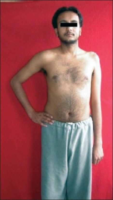
A clinical photograph of a male patient demonstrating the physical presentation of a left-sided upper brachial plexus palsy (Erb-Duchenne palsy), involving the C5 and C6 nerve roots. The image shows significant postural asymmetry between the upper limbs. The affected left arm exhibits classic clinical signs, including adduction at the shoulder and internal rotation of the humerus. There is visible muscle atrophy and a loss of rounded contour in the left deltoid and shoulder girdle compared to the healthy right side. Additionally, the left shoulder is positioned lower than the right. The patient's right arm displays a normal neutral position and muscle bulk. This visual serves as a primary clinical example of peripheral nerve injury manifestations, specifically highlighting the 'waiter's tip' position resulting from paralysis of the abductors and external rotators of the shoulder.
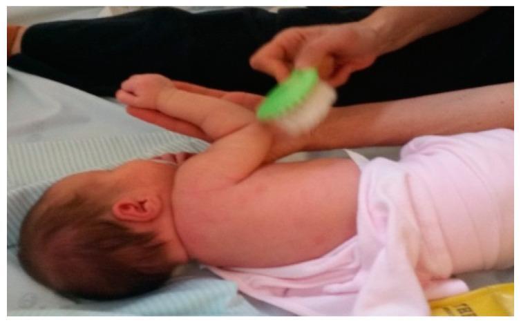
This clinical photograph illustrates a sensory stimulation technique used in neonatal rehabilitation, specifically for managing Neonatal Brachial Plexus Palsy (NBPP). The image depicts an infant lying in a lateral decubitus position on a padded surface. An adult caregiver or therapist is performing tactile and proprioceptive stimulation on the infant's affected upper limb. One hand stabilizes the infant's distal arm and hand in an elevated position, while the other hand applies a soft-bristled brush to the skin of the upper arm and shoulder region. This therapeutic intervention aims to provide varied sensorimotor input, promote central integration of the injured limb, and increase the infant's sensory perception of the arm. The use of different textures, such as the brush shown, is a key component of an early occupational therapy program designed to prevent sensory neglect and support the functional recovery of affected nerves following birth-related brachial plexus injury.
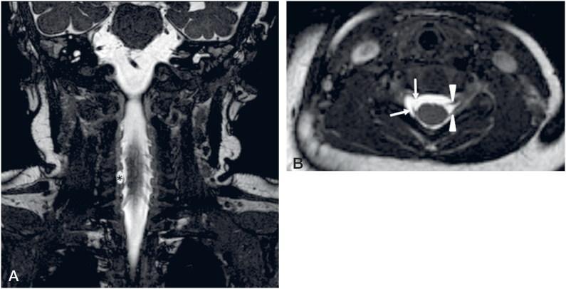
This diagnostic image consists of coronal (A) and axial (B) high-resolution Balanced Fast Field Echo (BFFE) MRI views of the cervical spine in a pediatric patient. The images demonstrate a right-sided brachial plexus birth injury involving a C6 root avulsion. In the coronal view (A), the spinal cord is surrounded by bright cerebrospinal fluid, with a distinctive hyperintense fluid collection (asterisk) on the right side indicating a traumatic pseudomeningocele (PMC). The axial view (B) provides a detailed comparison between the normal left side and the injured right side. On the left, the ventral and dorsal nerve roots are clearly visible as intact hypointense linear structures (arrowheads) emerging from the spinal cord. On the right, the ventral root is completely avulsed from the cord (upper arrow), and only a short remnant stump of the dorsal root is visible (lower arrow) within the CSF-filled pseudomeningocele. This imaging is characteristic of preganglionic nerve root injury in the context of neonatal brachial plexus palsy.
hypoxic ischemic encephalopathy neonatal MRI brain injury
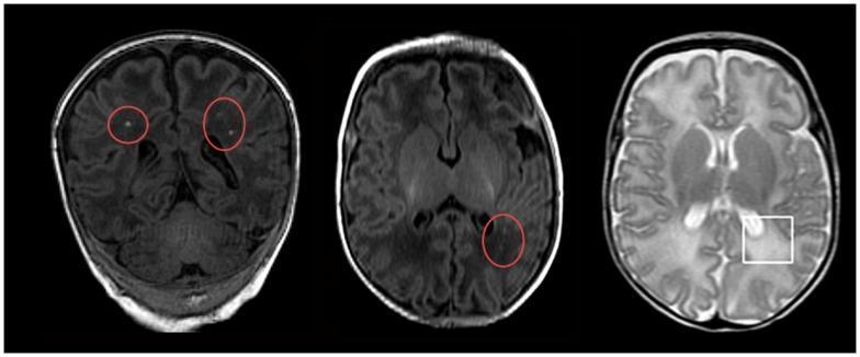
Diagnostic axial MRI images of a neonatal brain demonstrating pathological changes associated with hypoxic-ischemic encephalopathy (HIE) and white matter injury. The image panel contains two T1-weighted sequences and one T2-weighted sequence. On the T1-weighted images (left and center), red circles highlight multiple small, punctate hyperintensities located within the periventricular and subcortical white matter of the frontal and temporal lobes, characteristic of focal white matter lesions. On the T2-weighted image (right), a white square demarcates a region of diffuse excessive high signal intensity (DEHSI) in the posterior periventricular white matter, primarily involving the occipital region. This T2-hyperintensity is indicative of increased water content, such as vasogenic edema or dysmyelination. These imaging findings represent common neuroradiological markers for neonatal seizures and subsequent neurodevelopmental risks following hypoxic-ischemic events.
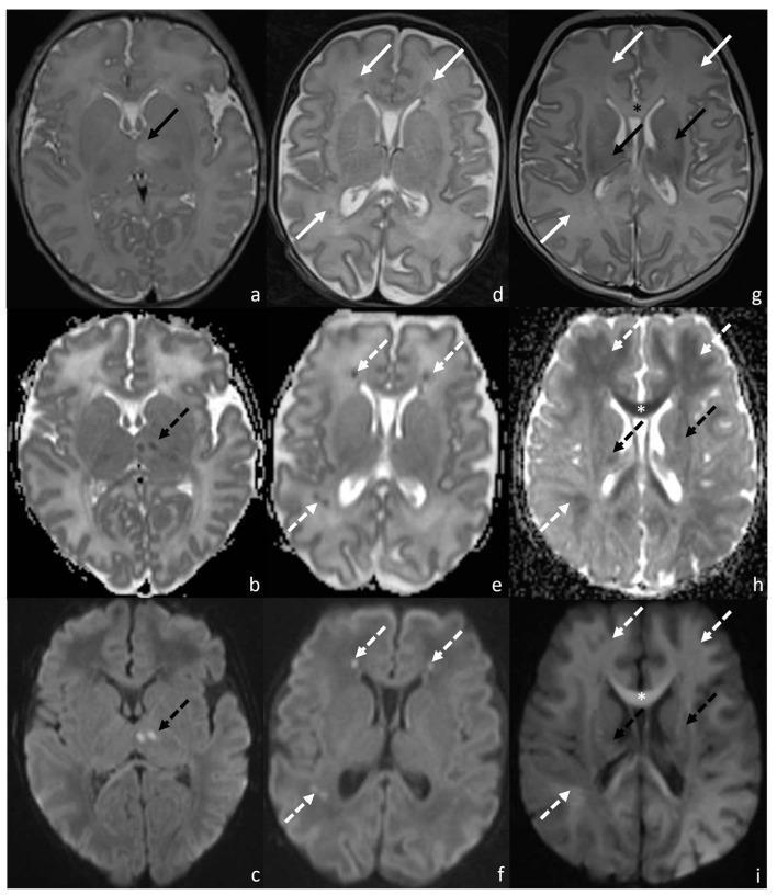
A multi-panel axial brain MRI compilation of neonatal patients demonstrating typical patterns of hypoxic-ischemic encephalopathy (HIE). The image is organized into three columns representing distinct injury patterns across T2-weighted (T2WI), Apparent Diffusion Coefficient (ADC), and Diffusion-Weighted Imaging (DWI) sequences. Pattern 1 (a-c) illustrates central involvement, highlighting focal hyperintensity on T2WI and diffusion restriction (low ADC, high DWI signal) in the left thalamus. Pattern 2 (d-f) demonstrates a watershed distribution, characterized by inhomogeneous signal alterations and diffusion restriction within the bilateral frontal and right parietal white matter. Pattern 3 (g-i) shows a mixed pattern involving both deep gray matter and peripheral structures, specifically showing restricted diffusion in the bilateral capsulo-lenticular regions, corpus callosum, and diffuse white matter areas. Arrows and asterisks facilitate the identification of pathological signal changes. This diagnostic image serves as a clinical reference for classifying neonatal HIE severity based on anatomical distribution (basal ganglia/thalamus vs. watershed areas).
Neonatal Injury - Comprehensive Overview
1. Risk Factors for Birth Injury
- Macrosomia / Large for gestational age (LGA) - especially in infants of diabetic mothers
- Shoulder dystocia - affects 0.3-0.5% of normal deliveries, but rises to 5-7% in infants >4000 g
- Operative vaginal delivery - forceps or vacuum extraction
- Breech presentation
- Prolonged second stage of labor
- Maternal diabetes - multiplies shoulder dystocia risk 5-fold at each birth-weight class
2. Peripheral Nerve Injuries
Neonatal Brachial Plexus Palsy (NBPP)
| Type | Roots | Muscles Affected | Features |
|---|---|---|---|
| Erb-Duchenne palsy (upper plexus) | C5-C6 | Shoulder abductors/external rotators, elbow flexors | "Waiter's tip" posture; hand spared; good prognosis if biceps function present early |
| Klumpke palsy (lower plexus) | C8-T1 | Intrinsic hand muscles | Finger deformities; ipsilateral Horner syndrome (ptosis, miosis, anhidrosis, enophthalmos) = preganglionic T1 injury; poor prognosis |
- Early (0-3 months): Gentle passive range-of-motion exercises to prevent contractures and preserve joint integrity while awaiting neurologic recovery
- Surgical threshold: No return of elbow flexion by 3 months of age → indicates poor prognosis → proceed to microsurgical repair (neurolysis, nerve transfer, or nerve grafts)
- Later reconstruction: Muscle rebalancing procedures to improve shoulder function
Facial Nerve Palsy
3. Head and Scalp Injuries
| Type | Tissue Plane | Key Features | Danger |
|---|---|---|---|
| Caput succedaneum | Skin → epicranial aponeurosis | Hemorrhagic edema; crosses suture lines; very common after vaginal delivery | Minimal |
| Subgaleal hemorrhage | Aponeurosis → periosteum | Associated with vacuum extraction; blood spreads under entire scalp | HIGH - hemorrhagic shock possible; urgent volume resuscitation needed |
| Cephalhematoma | Periosteum → cranial bone | Circumscribed, boggy, confined by sutures; from vacuum/forceps delivery | Low; resolves weeks-months; source of hyperbilirubinemia |
Skull Fractures
- Linear fractures (usually parietal) - bony continuity lost on imaging; clinical exam normal; no therapy needed. Watch for leptomeningeal cyst / "growing fracture"
- Depressed ("ping-pong") fractures - bone buckles inward without losing continuity; CT/MRI to rule out intracranial hemorrhage; may elevate spontaneously with skull molding, or by vacuum extractor
4. Hypoxic-Ischemic Encephalopathy (HIE)
Pathophysiology (Bradley and Daroff's Neurology in Clinical Practice)
- Excitotoxicity
- Apoptosis
- Reactive oxygen species
- Inflammation
Diagnosis - Sarnat Staging
| Stage | Clinical Features |
|---|---|
| Stage 1 (Mild) | Hyperalert, wide-open eyes, jittery, agitated |
| Stage 2 (Moderate) | Lethargy, low tone, decreased primitive reflexes, seizures |
| Stage 3 (Severe) | Stupor/coma, flaccid tone, decerebrate posturing, absent primitive reflexes, seizures |
Treatment - Therapeutic Hypothermia
- Gestational age ≥36 weeks
- Umbilical cord blood pH <7.0 OR base excess >16 mmol/L within 1 hour of delivery
- Moderate-to-severe encephalopathy on clinical exam
5. Spinal Cord Injury
- Breech delivery (excessive traction): Lower cervical / upper thoracic cord - causes urinary retention, hypotonia, weakness, areflexia of all limbs, evolving to spastic paraplegia/quadriplegia
- Vertex delivery (excessive torsion): Upper/mid-cervical cord - high lesions may cause stillbirth or rapid respiratory failure
- Lower thoracic/lumbar injuries: Usually from vascular occlusion due to umbilical artery catheterization or air embolism
6. Fractures
| Bone | Context | Notes |
|---|---|---|
| Clavicle | Most common birth fracture; shoulder dystocia | Usually heals spontaneously |
| Humerus | Macrosomia, difficult delivery | Arm immobilized to chest |
| Femur | Breech delivery | Gallows traction or Pavlik harness |
7. Other Neonatal Injuries
Visceral Injuries
- Liver/Spleen laceration: Seen with breech delivery or traumatic manipulation; presents with abdominal distension, pallor, and shock in first 24-48h
- Adrenal hemorrhage: From birth trauma or asphyxia; may present with flank mass and jaundice
Neonatal Stroke (Perinatal Arterial Ischemic Stroke - PAIS)
- MRI of choice; DWI restriction appears within hours (vs. days in HIE)
- Acute management is supportive: hydration, glucose homeostasis, seizure management
- No established role for thrombolysis
- Long-term sequelae: hemiparetic cerebral palsy (typically mild), epilepsy, cognitive/behavioral challenges
Cerebral Sinus Venous Thrombosis (CSVT)
- Most common sinuses: superior sagittal, transverse, deep sinuses
- Typically presents with seizures + encephalopathy
- Often associated with hemorrhagic infarction
Summary Table: Key Birth Injuries at a Glance
| Injury | Classic Mechanism | Key Sign | Management |
|---|---|---|---|
| Erb's palsy (C5-C6) | Shoulder dystocia | Waiter's tip | PT; surgery if no recovery by 3 months |
| Klumpke's palsy (C8-T1) | Arm hyperabduction | Horner's syndrome | Surgery; poor prognosis |
| Cephalhematoma | Vacuum/forceps | Suture-confined boggy mass | Observation |
| Subgaleal hemorrhage | Vacuum | Spreading scalp fullness | Volume resuscitation urgently |
| Caput succedaneum | Normal vaginal delivery | Crosses suture lines | Observation |
| HIE | Perinatal asphyxia | Sarnat staging; seizures | Therapeutic hypothermia 33-35°C × 72h |
| Clavicle fracture | Shoulder dystocia | Crepitus, limited arm movement | Immobilization |
| Depressed skull fracture | Forceps / compression | Ping-pong deformity | CT; elevation if brain impinged |