
Recurrent laryngeal nerve and parathyroid identification and preservation during thyroid surgery in detail in simplified manner for university ms ent exam
recurrent laryngeal nerve anatomy thyroid surgery identification
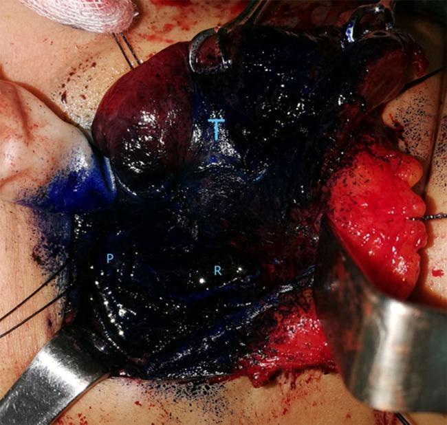
A clinical photograph of an intraoperative surgical field during a thyroidectomy, demonstrating the use of methylene blue spraying for tissue differentiation. The image illustrates the anatomical relationship between the thyroid gland (labeled 'T'), parathyroid gland ('P'), and the recurrent laryngeal nerve ('R'). The thyroid gland and surrounding perithyroidal connective tissues appear heavily stained with dark blue/black dye. In contrast, the parathyroid gland ('P') is visible as a relatively unstained, lighter-colored nodule, facilitating its identification and preservation. The recurrent laryngeal nerve ('R') is located within the deeply stained operative bed. Surgical retractors and stay sutures are visible, providing exposure of the neck anatomy. This image serves as an educational example of vital staining techniques used in endocrine surgery to distinguish parathyroid tissue from thyroid and adipose tissues, minimizing the risk of accidental excision or nerve injury.
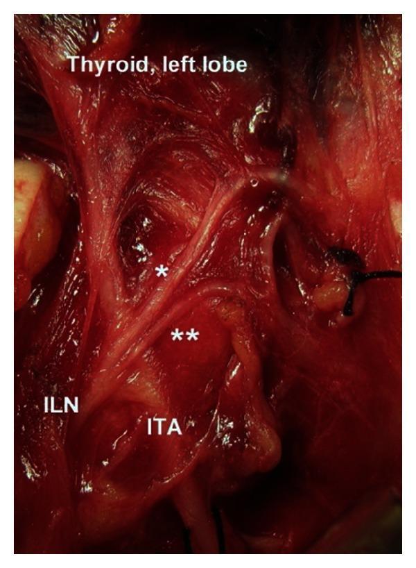
This clinical intraoperative photograph displays the surgical anatomy of the neck, specifically highlighting the neurovascular relationship between the left inferior laryngeal nerve (ILN) and the inferior thyroid artery (ITA). The ILN is seen coursing superiorly and undergoing extralaryngeal terminal division. The bifurcation occurs at the arterial segment, categorized as a Type 1 variation. Two distinct branches are visible: the anterior branch (marked with a single asterisk) and the posterior branch (marked with a double asterisk). The ITA is positioned inferior and lateral to the bifurcation point. Superiorly, the left lobe of the thyroid gland is partially visible, demonstrating the proximity of the anterior ILN branch as it approaches the laryngeal entry point. This image provides critical anatomical context for endocrine surgery, particularly thyroidectomy and parathyroidectomy, to avoid iatrogenic injury to the recurrent laryngeal nerve. The surgical field shows exposed muscular and glandular tissues with clear identification of the nerve's bifurcation pattern relative to fixed vascular landmarks.
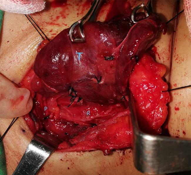
This clinical photograph displays an intraoperative view of the neck during a thyroidectomy procedure, focusing on the surgical anatomy of the thyroid bed before the application of methylene blue. The primary anatomical structures are labeled for educational identification: the thyroid gland (T) is visible as a superiorly located, reddish-brown vascular mass held by surgical forceps; the recurrent laryngeal nerve (R) is identified as a thin, pale, linear structure in the tracheoesophageal groove inferior to the thyroid lobe; and a parathyroid gland (P) is shown as a small, yellowish-tan nodule situated adjacent to the nerve and posterior thyroid capsule. The surgical field includes various instruments, such as retractors and forceps used for tissue manipulation, and black silk sutures are visible throughout the dissection site. This image serves as a clinical anatomical guide for identifying critical structures that must be preserved during endocrine surgery to prevent vocal cord paralysis or hypocalcemia. It illustrates the standard surgical topography and the natural appearance of these tissues in an active surgical field.
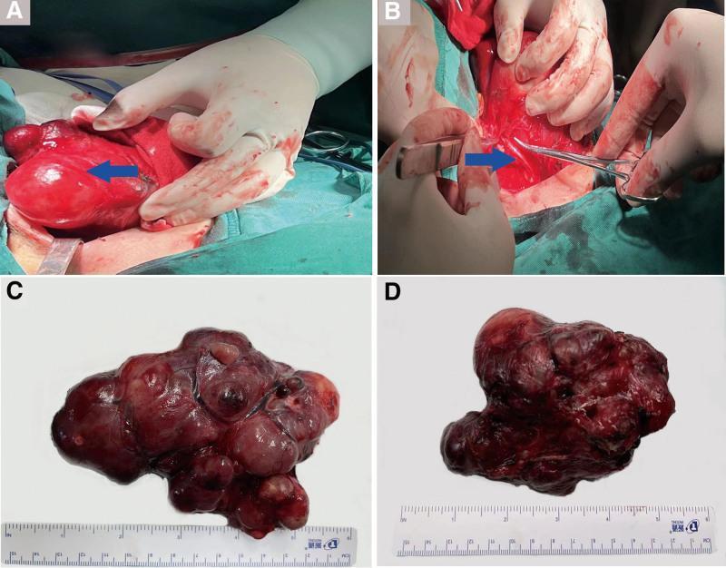
A composite clinical photograph documenting a total thyroidectomy for a giant goiter. Panel A shows an intraoperative view of the lateral dissection of the right thyroid lobe, exhibiting significant glandular hypertrophy and vascular congestion. Panel B displays the surgical field with a blue arrow identifying the recurrent laryngeal nerve (RLN) in the right tracheoesophageal groove, preserved during dissection. Panels C and D present the gross macroscopic appearance of the resected specimens against a white background with a centimeter scale. Panel C shows the resected right lobe and isthmus (12 x 7 x 5 cm), characterized by a multinodular, lobulated surface with prominent venous congestion and varied reddish-brown coloration. Panel D shows the resected left lobe (12 x 9 x 6 cm), which is similarly enlarged with a nodular texture and dense vascular appearance. The image illustrates surgical anatomy and the gross pathology of multinodular goiter, highlighting the importance of nerve identification during radical neck surgery.
parathyroid gland identification preservation thyroidectomy
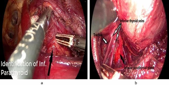
Two-panel endoscopic intraoperative photograph showcasing a thyroidectomy procedure. Panel (a) illustrates the dissection of the lower thyroid pole using laparoscopic instruments, including a blunt-tipped grasper and a vessel sealer. A black arrow indicates the identification of the right inferior parathyroid gland within a field of dissected vascular soft tissue. Panel (b) provides a detailed anatomical view of the left surgical field, featuring the trachea (labeled as a ridged, pale tubular structure), the inferior thyroid veins (thin longitudinal vessels), and the left inferior parathyroid gland (a distinct, rounded, reddish-brown structure). The images emphasize the preservation of the inferior parathyroid glands and their relationship to thyroid vasculature and the trachea during neck surgery. This educational material is suitable for surgical residency training, specifically focusing on endocrine surgery techniques and cervical anatomy preservation.
A clinical photograph of an intraoperative surgical field during a thyroidectomy, demonstrating the use of methylene blue spraying for tissue differentiation. The image illustrates the anatomical relationship between the thyroid gland (labeled 'T'), parathyroid gland ('P'), and the recurrent laryngeal nerve ('R'). The thyroid gland and surrounding perithyroidal connective tissues appear heavily stained with dark blue/black dye. In contrast, the parathyroid gland ('P') is visible as a relatively unstained, lighter-colored nodule, facilitating its identification and preservation. The recurrent laryngeal nerve ('R') is located within the deeply stained operative bed. Surgical retractors and stay sutures are visible, providing exposure of the neck anatomy. This image serves as an educational example of vital staining techniques used in endocrine surgery to distinguish parathyroid tissue from thyroid and adipose tissues, minimizing the risk of accidental excision or nerve injury.
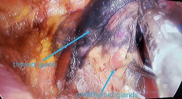
This intraoperative clinical photograph demonstrates the concept of 'negative opacification' during a thyroidectomy and central lymph node dissection. Following the injection of a nanocarbon suspension, the thyroid gland and central compartment lymph nodes exhibit significant black staining as they absorb the lymphatic tracer. In contrast, the parathyroid glands are highlighted by their failure to take up the nanocarbon, appearing as distinct, lighter-colored (yellow-tan) structures against the darkened thyroid tissue. Blue arrows and text labels specifically identify the blackened thyroid gland and the relatively pale parathyroid glands. This technique serves as a surgical aid to facilitate the identification and preservation of parathyroid glands in situ while ensuring the radical clearance of potentially metastatic, stained lymph nodes. The image illustrates a critical step in endocrine surgery for protecting parathyroid function and improving oncological outcomes in thyroid carcinoma cases.
Recurrent Laryngeal Nerve (RLN) and Parathyroid Identification & Preservation During Thyroid Surgery
PART 1: RECURRENT LARYNGEAL NERVE (RLN)
A. Anatomy - Why it "Recurs"
- Loops under the ligamentum arteriosum at the aortic arch (deeper in the chest)
- Travels in a straight, deep course in the tracheoesophageal groove
- More consistent and predictable position
- Loops under the right carotid-subclavian artery junction (higher up)
- Enters the neck at a more oblique, anterolateral angle - more variable
- Lies slightly more anterior and superficial than the left
B. Functional Importance
| Nerve | Function | Effect of Injury |
|---|---|---|
| RLN | Motor to ALL intrinsic laryngeal muscles EXCEPT cricothyroid; sensory to subglottis | Unilateral: hoarseness, aspiration. Bilateral: may need tracheostomy |
| External branch of SLN (EBSLN) | Motor to cricothyroid (pitch/tension) | Loss of high-pitched voice, vocal fatigue |
C. Surgical Landmarks for RLN Identification
- Low in the neck, the RLN is first found in the angle between the trachea and the inferior thyroid artery
- Identify it here first - it is safer, away from the thyroid
- Then follow it cephalad toward the gland
- A posterior projection of thyroid tissue on the posterior surface of the thyroid lobe
- The RLN lies just posteromedial to it
- One of the most reliable landmarks - find the tubercle, find the nerve
- The RLN crosses the ITA near the cricoid cartilage
- In ~70% of cases, the ITA lies anterior to the RLN
- The nerve can be found posterior to, anterior to, or between branches of the ITA - always identify before ligating
- Inferior parathyroid: RLN lies posteromedial to it
- Superior parathyroid: RLN lies inferolateral to it
- So if you find a parathyroid gland, the RLN is nearby
- At the level of the cricoid cartilage, the nerve has an intimate relationship with the Berry ligament (posterior suspensory ligament of thyroid to trachea)
- The RLN typically curves anteriorly toward the ligament before diving posteriorly to enter the larynx
- This is where the nerve is closest to the thyroid - most dangerous zone
D. Step-by-Step RLN Exposure Technique
- Medialize the thyroid lobe - retract it anteromedially with finger, retractor, or clamp
- Excise the investing fascia in a craniocaudal direction to expose the posterior aspect
- Identify ITA and trachea - RLN is at their bisected angle, low in the neck
- Dissect the nerve cephalad - follow it toward the gland, always keeping it in view
- Identify Tubercle of Zuckerkandl - nerve lies just behind it
- Carefully dissect around Berry ligament - most dangerous step; the nerve often curves anteriorly here
- Do NOT apply traction or thermal energy near the nerve at any step
- Dissect bluntly near the nerve
- Do not stretch or pull the nerve
- Don't use monopolar cautery near it (bipolar only, if at all)
E. Non-Recurrent Laryngeal Nerve (NRLN) - An Exam Favourite
- Occurs on the right side in ~1% of people
- Associated with aberrant right subclavian artery (arteria lusoria) arising directly from the aortic arch, crossing behind the esophagus
- Because the normal loop-down never happens embryologically, the nerve takes a direct, lateral, horizontal course from the vagus to the larynx
- If you are looking for the RLN in the TE groove and cannot find it - suspect NRLN
- A CT scan showing aberrant vascular anatomy should raise preoperative suspicion
F. RLN Branching
- The RLN may branch before entering the larynx in 20-30% of cases
- The anterior branches are predominantly motor - most important to preserve
- Always preserve ALL branches
G. Intraoperative Neuromonitoring (IONM)
- EMG-based monitoring: endotracheal tube with surface electrodes on the vocal cords monitors muscle activity when the nerve is stimulated
- A positive signal = nerve is intact and responsive
- Loss of signal = potential nerve injury
- Does not replace visual identification - it supplements it
- Particularly useful in: reoperation, large goiters, thyroid cancer with posterior extension
- Evidence shows routine identification alone is as effective as identification + IONM in experienced hands, but IONM reduces anxiety and aids teaching
PART 2: PARATHYROID GLAND IDENTIFICATION AND PRESERVATION
A. Anatomy Recap - Why It's Hard
| Superior Parathyroid | Inferior Parathyroid | |
|---|---|---|
| Embryologic origin | 4th pharyngeal pouch | 3rd pharyngeal pouch |
| Location | 1 cm above ITA at lateral thyroid edge; near cricoid | More variable - lower pole, thyrothymic ligament |
| Blood supply | Inferior thyroid artery (enters from lateral) | Inferior thyroid artery |
| Size/color | 30-70 mg; caramel/tan color | Same |
- 80% of people have exactly 4 glands; 10%+ have more than 4
- Superior glands are more predictable - at the level of the cricoid, medial to the RLN-ITA intersection
- Inferior glands are highly variable - can be found anywhere from the lower thyroid pole to the mediastinum along the thyrothymic ligament
- A useful rule: the position on one side mirrors the other side - use contralateral gland as a guide
B. How to Identify Parathyroid Glands
- Slightly browner / caramel color compared to the surrounding yellow fat
- With trauma, they become mahogany colored (sign of devascularization)
- They are small oval nodules on the posterior thyroid surface
- Distinguish from fat (yellow, less structured), lymph nodes (paler, firmer), and thyroid tissue (darker red)
- Ideally identify parathyroids before the thyroid is fully medialized
- Certainly identify before ligating the ITA (main blood supply)
- Methylene blue - injected IV; parathyroids take up less dye than thyroid, making them lighter against the stained background (now rarely used due to neurotoxicity)
- Nanocarbon suspension - lymphatic tracer that stains thyroid and lymph nodes black; parathyroids remain unstained (light) = "negative opacification" method
- Near-infrared autofluorescence (NIRAF) - newest technique; parathyroids emit a strong autofluorescent signal under near-infrared light, allowing real-time identification without any dye
C. Technique of Parathyroid Preservation
- Identify early - before clamping or dividing anything on the posterior thyroid
- Do NOT grasp the gland directly - grasp the surrounding fat/connective tissue for retraction; parathyroids are fragile and bruise easily
- Preserve the lateral vascular pedicle - the blood supply enters from the lateral side via a branch of the ITA. Do NOT ligate the ITA trunk proximally - ligate branches close to the thyroid capsule instead ("capsular ligation")
- Sweep the gland laterally - carefully divide the thin attachment between parathyroid and thyroid using bipolar cautery, then mobilize the gland laterally with its pedicle intact
- Leave a broad lateral attachment - this ensures vascularity is maintained
- After mobilization, check viability - a healthy parathyroid is caramel-tan; a dark purple/mahogany gland is devascularized
D. When Parathyroid is Inadvertently Removed or Devascularized - Autotransplantation
- Any parathyroid found on the surgical specimen
- Any gland that appears dark and non-viable after mobilization (confirm it's not just a lymph node - send a sliver for frozen section)
- Confirm it is parathyroid tissue (frozen section if in doubt)
- Mince the gland into small 1-mm³ fragments
- Place into multiple pockets in the ipsilateral sternocleidomastoid (SCM) muscle or brachioradialis (if future ipsilateral surgery is anticipated)
- Mark with a non-absorbable suture and clip so it can be found if the patient later develops hyperparathyroidism
E. Consequences of Parathyroid Loss - Hypoparathyroidism
| Type | Timing | Mechanism | Features |
|---|---|---|---|
| Transient | 24-48 hrs post-op | Devascularization, bruising | Hypocalcemia, tingling, Chvostek's, Trousseau's; recovers within weeks |
| Permanent | >6 months | All glands removed/devascularized | Requires lifelong calcium + vitamin D supplementation |
- Post-thyroidectomy hypocalcemia is the most common complication of total thyroidectomy
- Check serum Ca²⁺ at 6 hours and 24 hours post-op routinely
PART 3: INTEGRATED SURGICAL STEPS SUMMARY
Expose thyroid → Divide middle thyroid vein
        ↓
Ligate SUPERIOR thyroid vessels CLOSE to capsule (protect EBSLN)
        ↓
Medialize thyroid lobe medially
        ↓
Identify PARATHYROIDS early (before ITA ligation)
Sweep them laterally with intact pedicle
        ↓
Identify RLN low in neck (TE groove / angle of ITA & trachea)
Trace cephalad past Tubercle of Zuckerkandl to Berry ligament
        ↓
Ligate inferior thyroid vessels AFTER RLN is in full view
(Ligate branches close to thyroid, NOT trunk)
        ↓
Divide Berry ligament carefully (RLN closest to thyroid here)
        ↓
Remove thyroid - inspect specimen for any parathyroid tissue
Autotransplant if found
PART 4: HIGH-YIELD EXAM POINTS (MNEMONICS)
- T - Tracheoesophageal groove (initial search zone)
- B - Bisected angle of trachea + ITA (first identification point)
- Z - Zuckerkandl's tubercle (RLN just posterior to it)
- B - Berry ligament (danger zone - nerve closest to thyroid)
- S - Spot them early (before ITA ligation)
- L - Leave lateral pedicle intact
- I - Invest surrounding tissue (not the gland itself) for traction
- M - Mobilize laterally with bipolar only
- Right RLN is more oblique than left (due to different anatomical origin)
- Non-RLN = aberrant right subclavian artery = approaches from lateral NOT from below
- RLN branches in 20-30% of cases - anterior branch is predominantly motor
- ITA is anterior to RLN in 70% of cases
- Superior parathyroids = 4th pouch (more constant), Inferior = 3rd pouch (more variable)
- "Treat every parathyroid as the patient's last" = cardinal rule
- IONM supplements but does NOT replace visual identification
- Autotransplant into SCM; mark with clip/suture
- Fischer's Mastery of Surgery, 8th Ed. - p. 1073 (dedicated section on RLN exposure and parathyroid identification)
- Sabiston Textbook of Surgery, 21st Ed. - p. 1488 (RLN anatomy, NRLN, neuromonitoring)
- Cummings Otolaryngology Head and Neck Surgery, 7th Ed. (surgical anatomy, parathyroid management)
- Scott-Brown's Otorhinolaryngology Head & Neck Surgery, 8th Ed. (IONM evidence)
- Mulholland and Greenfield's Surgery, 7th Ed. (parathyroid anatomy during thyroidectomy)