
I have my university exam of 1 st year of subject biochemistry These are the important long answer give me answer of this question "Hexose Monophosphate (HMP) shunt" and remeber it's a university examination
Reading File
Now let me search the medical library for comprehensive HMP shunt content in parallel.
Finding Sources
Finding Sources
Excellent results. Now let me read the most comprehensive biochemistry sources in parallel - Lippincott, Harper's, Basic Medical Biochemistry, and Goldman-Cecil.
Reading File
Reading File
Reading File
Reading File
Excellent content from multiple textbooks. Now let me get more detail from Lippincott on the reactions and G6PD deficiency, plus fetch key pathway images.
Reading File
Reading File
Loading Image
Let me get the correct full image URL and G6PD deficiency content from Lippincott.
Reading File
Loading Image
I now have all the content I need for a comprehensive university-level answer. Let me also get the G6PD deficiency and NADPH uses section quickly.
Reading File
I now have all the necessary information from multiple authoritative textbooks. Let me compose a comprehensive university-level long answer.
Hexose Monophosphate (HMP) Shunt (Pentose Phosphate Pathway)
Introduction
The Hexose Monophosphate (HMP) shunt, also called the Pentose Phosphate Pathway (PPP) or Phosphogluconate Pathway, is an alternative pathway for glucose oxidation that runs parallel to glycolysis. Unlike glycolysis, it does not produce ATP nor consume it. Its two primary products are:
- NADPH - a reducing coenzyme essential for reductive biosynthesis and protection against oxidative stress
- Ribose 5-phosphate - a pentose sugar required for nucleotide and nucleic acid biosynthesis
All reactions occur in the cytosol. The pathway is found in virtually all cells but is especially active in the liver, lactating mammary glands, adipose tissue, adrenal cortex, gonads, and red blood cells.
Location in the Cell
All enzymes of the HMP shunt are cytosolic - just like the enzymes of glycolysis. Oxidation uses NADP+ (not NAD+) as the hydrogen acceptor, which is a key distinction from glycolysis.
Starting Material
Glucose 6-phosphate is the substrate. It is the common entry point for both glycolysis and the HMP shunt - the cell's metabolic needs determine which pathway glucose 6-phosphate enters.
Overview of the Pathway
The HMP shunt is divided into two phases:
| Phase | Type | Reversibility |
|---|---|---|
| Oxidative Phase | Generates NADPH + CO₂ | Irreversible |
| Non-oxidative Phase | Interconverts sugar phosphates | Reversible |
Phase 1: Oxidative Phase (Irreversible)
This phase converts glucose 6-phosphate into ribulose 5-phosphate while generating 2 NADPH and 1 CO₂ per molecule processed.
Reaction 1 - Glucose 6-phosphate Dehydrogenase (G6PD)
- Substrate: Glucose 6-phosphate
- Product: 6-Phosphogluconolactone
- Coenzyme: NADP+ → NADPH
- Enzyme: Glucose 6-phosphate dehydrogenase (G6PD)
- This is the rate-limiting, committed, and regulated step of the entire pathway
- G6PD is competitively inhibited by NADPH (product inhibition) - when NADPH/NADP+ ratio falls, enzyme activity increases
- Insulin upregulates G6PD gene expression
Reaction 2 - 6-Phosphogluconolactone Hydrolase
- Substrate: 6-Phosphogluconolactone
- Product: 6-Phosphogluconate
- Enzyme: 6-Phosphogluconolactone hydrolase (gluconolactone hydrolase)
- This is a hydrolysis reaction (no NADPH generated)
Reaction 3 - 6-Phosphogluconate Dehydrogenase
- Substrate: 6-Phosphogluconate
- Products: Ribulose 5-phosphate + CO₂
- Coenzyme: NADP+ → NADPH (second NADPH generated)
- Enzyme: 6-Phosphogluconate dehydrogenase
- This is an oxidative decarboxylation reaction (carbon 1 of glucose is released as CO₂) - mechanistically similar to isocitrate dehydrogenase in the TCA cycle
Net result of oxidative phase (per glucose 6-phosphate):
Glucose 6-phosphate + 2 NADP⁺ + H₂O → Ribulose 5-phosphate + CO₂ + 2 NADPH + 2 H⁺
Phase 2: Non-oxidative Phase (Reversible)
The ribulose 5-phosphate produced in the oxidative phase is either:
- Converted to ribose 5-phosphate (for nucleotide synthesis), OR
- Converted back to glycolytic intermediates (fructose 6-phosphate and glyceraldehyde 3-phosphate)
The direction depends entirely on the cell's needs.
Key Enzymes
a. Phosphopentose Isomerase
- Ribulose 5-phosphate → Ribose 5-phosphate (needed for nucleotide synthesis)
b. Phosphopentose Epimerase
- Ribulose 5-phosphate → Xylulose 5-phosphate (changes stereochemistry at C-3)
c. Transketolase (coenzyme: Thiamine Pyrophosphate / TPP)
- Transfers 2-carbon fragments (ketol group) from a ketose donor to an aldose acceptor
- Reaction 1: Xylulose 5-P (5C) + Ribose 5-P (5C) → Glyceraldehyde 3-P (3C) + Sedoheptulose 7-P (7C)
- Reaction 2: Xylulose 5-P (5C) + Erythrose 4-P (4C) → Glyceraldehyde 3-P (3C) + Fructose 6-P (6C)
d. Transaldolase
- Transfers 3-carbon fragments (dihydroxyacetone unit) from a ketose to an aldose
- Sedoheptulose 7-P (7C) + Glyceraldehyde 3-P (3C) → Fructose 6-P (6C) + Erythrose 4-P (4C)
The final products - fructose 6-phosphate and glyceraldehyde 3-phosphate - can re-enter the glycolytic pathway.
The Complete Cycle and Overall Equation
If all three phases are run as a complete cycle, 6 molecules of glucose 6-phosphate yield 6 CO₂ and reform 5 molecules of glucose 6-phosphate. This means one molecule of glucose is completely oxidized per cycle.
Overall equation:
C₆H₁₂O₆ + 12 NADP⁺ + 6 H₂O → 6 CO₂ + 12 NADPH + 12 H⁺
The pathway diagram from Lippincott Illustrated Reviews:
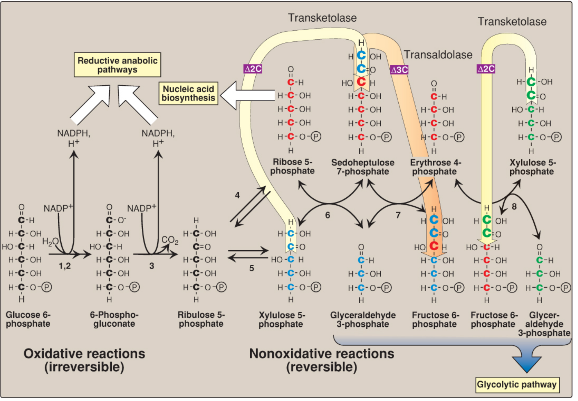
Figure: The pentose phosphate pathway. Left side shows irreversible oxidative reactions generating NADPH; right side shows reversible non-oxidative interconversions via transketolase and transaldolase, feeding back into the glycolytic pathway. (Lippincott Illustrated Reviews: Biochemistry, 8e)
Regulation
| Regulator | Effect on G6PD |
|---|---|
| High NADPH/NADP⁺ ratio | Inhibits G6PD (product inhibition) |
| Low NADPH/NADP⁺ ratio | Activates G6PD - flux increases |
| Insulin | Upregulates G6PD gene expression |
| High glucose-6-phosphate | Promotes flux through pathway |
Functions / Importance of NADPH
NADPH produced by the HMP shunt is used in the following reactions:
- Fatty acid synthesis - NADPH is required by fatty acid synthase for synthesis of long-chain fatty acids (liver, adipose, lactating mammary gland)
- Cholesterol synthesis - NADPH used by HMG-CoA reductase
- Steroid hormone synthesis - in adrenal cortex, gonads, placenta
- Reductive detoxification of H₂O₂ via glutathione - NADPH reduces oxidized glutathione (G-S-S-G) back to reduced glutathione (G-SH) via glutathione reductase. G-SH then reduces H₂O₂ to H₂O via glutathione peroxidase - protecting RBCs and other cells from oxidative damage
- NADPH oxidase in phagocytes - for the "respiratory burst" used to kill microorganisms
- Cytochrome P450 reactions - drug detoxification in the liver
- Regeneration of reduced glutathione and protection against reactive oxygen species (ROS)
Clinical Significance - G6PD Deficiency
Definition
G6PD deficiency is the most common enzyme deficiency in humans, affecting ~400 million people worldwide. It is an X-linked recessive disorder.
Pathophysiology
- Without G6PD, the HMP shunt cannot produce adequate NADPH
- Red blood cells (RBCs) are especially vulnerable because they have no mitochondria and rely entirely on the HMP shunt for NADPH
- Without NADPH, glutathione cannot be regenerated in its reduced form
- H₂O₂ and other ROS accumulate → oxidative damage to RBC membranes and hemoglobin (forming Heinz bodies - denatured hemoglobin precipitates)
- Result: Hemolytic anemia triggered by oxidative stress
Triggers
- Infections (most common trigger)
- Drugs: Primaquine, dapsone, nitrofurantoin, sulfonamides
- Fava beans (favism) - contain vicine and convicine which generate free radicals
- Naphthalene (mothballs)
Clinical Features
- Usually asymptomatic until exposed to oxidant stress
- Acute hemolytic anemia: jaundice, dark urine (hemoglobinuria), pallor
- "Bite cells" and "Heinz bodies" on peripheral blood smear
Diagnosis
- G6PD enzyme assay on RBCs
- Transketolase activity in RBCs is also used to assess thiamine (Vitamin B₁) status - since transketolase requires TPP as a coenzyme, its activity is low in thiamine deficiency
Tissues Where HMP Shunt is Most Active
| Tissue | Reason |
|---|---|
| Liver | Fatty acid and cholesterol synthesis; drug detoxification |
| Adipose tissue | Fatty acid synthesis |
| Lactating mammary gland | Fatty acid synthesis for milk |
| Adrenal cortex | Steroid hormone synthesis |
| RBCs | Protection against oxidative hemolysis |
| Gonads | Steroid synthesis |
Comparison with Glycolysis
| Feature | Glycolysis | HMP Shunt |
|---|---|---|
| ATP production | Yes (net 2 ATP) | None |
| NADPH production | No | Yes (2 per glucose 6-P) |
| CO₂ produced | No (only in PDH step) | Yes (from C-1 of glucose) |
| Hydrogen acceptor | NAD+ | NADP+ |
| Pentose sugars produced | No | Yes (ribose 5-P) |
| Location | Cytosol | Cytosol |
| Reversibility | Mostly irreversible | Oxidative: irreversible; Non-oxidative: reversible |
Summary
The HMP shunt is a metabolically flexible pathway that:
- Provides NADPH for reductive biosynthesis and antioxidant defense
- Provides ribose 5-phosphate for nucleotide synthesis
- Allows interconversion of 3, 4, 5, 6, and 7-carbon sugars
- Links to glycolysis via fructose 6-phosphate and glyceraldehyde 3-phosphate
- Is regulated primarily by the NADPH/NADP⁺ ratio and G6PD activity
- G6PD deficiency leads to hemolytic anemia under oxidative stress due to NADPH insufficiency
Sources:
- Lippincott Illustrated Reviews: Biochemistry, 8th ed, Chapter 13 (Pentose Phosphate Pathway and NADPH)
- Harper's Illustrated Biochemistry, 32nd ed, Chapter 20 (Pentose Phosphate Pathway)
- Basic Medical Biochemistry: A Clinical Approach, 6th ed, Chapter 27 (Pentose Phosphate Pathway)
- Guyton and Hall Textbook of Medical Physiology (Pentose Phosphate Pathway and NADPH)
- Goldman-Cecil Medicine, International Edition (HMP Shunt Disorders)
I have my university exam of 1 st year of subject biochemistry These are the important long answer give me answer of this question"Uronic acid pathway" and remeber it's a university examination
Finding Sources
Finding Sources
Good - found excellent sources in both Harper's and Lippincott. Let me read them in parallel now.
Reading File
Reading File
Now let me get the detailed Harper's uronic acid pathway section and fetch the key diagram images in parallel.
Reading File
Loading Image
Excellent! Now let me get the Harper's disruption section (essential pentosuria) and the full pathway diagram.
Reading File
Finding Sources
Now let me fetch the main uronic acid pathway diagram from Harper's and get the full pathway figure from Lippincott.
Finding Sources
Loading Image
I now have comprehensive content from Harper's and Lippincott, plus two excellent diagrams. Let me write the full university-level long answer.
Uronic Acid Pathway
Introduction
The Uronic Acid Pathway (also called the Glucuronate Pathway) is an alternative metabolic route for glucose oxidation that occurs primarily in the liver. It is of minor quantitative importance as an energy-producing pathway - it does not generate ATP - but is of major physiological and clinical significance for:
- Synthesis of glucuronic acid for conjugation and detoxification
- Synthesis of ascorbic acid (Vitamin C) in most mammals (but NOT in humans)
- Incorporation of glucuronate into proteoglycans and glycosaminoglycans (GAGs)
- Providing a route for dietary D-xylulose to enter central metabolic pathways
Location
The pathway operates in the cytosol of hepatocytes (liver cells). The liver is the primary site, though some activity occurs in other tissues.
Starting Material
Glucose (via Glucose 1-phosphate and UDP-Glucose) is the entry substrate.
Steps of the Uronic Acid Pathway
Step 1 - Glucose → Glucose 6-phosphate
- Glucose is phosphorylated to glucose 6-phosphate by hexokinase/glucokinase
Step 2 - Glucose 6-phosphate → Glucose 1-phosphate
- Isomerization by phosphoglucomutase
Step 3 - Glucose 1-phosphate + UTP → UDP-Glucose + PPi
- Enzyme: UDP-glucose pyrophosphorylase (UDPGlc pyrophosphorylase)
- This is the same reaction as in glycogen synthesis
- UTP provides energy and activates glucose as a nucleotide sugar
Step 4 - UDP-Glucose → UDP-Glucuronate (UDP-Glucuronic Acid)
- Enzyme: UDP-glucose dehydrogenase (NAD-dependent)
- Coenzyme: 2 NAD⁺ → 2 NADH (two-step oxidation)
- Carbon 6 of glucose (-CH₂OH) is oxidized to a carboxyl group (-COOH)
- This is the key committed step that produces UDP-glucuronic acid
Diagram - Oxidation of UDP-Glucose to UDP-Glucuronic acid:
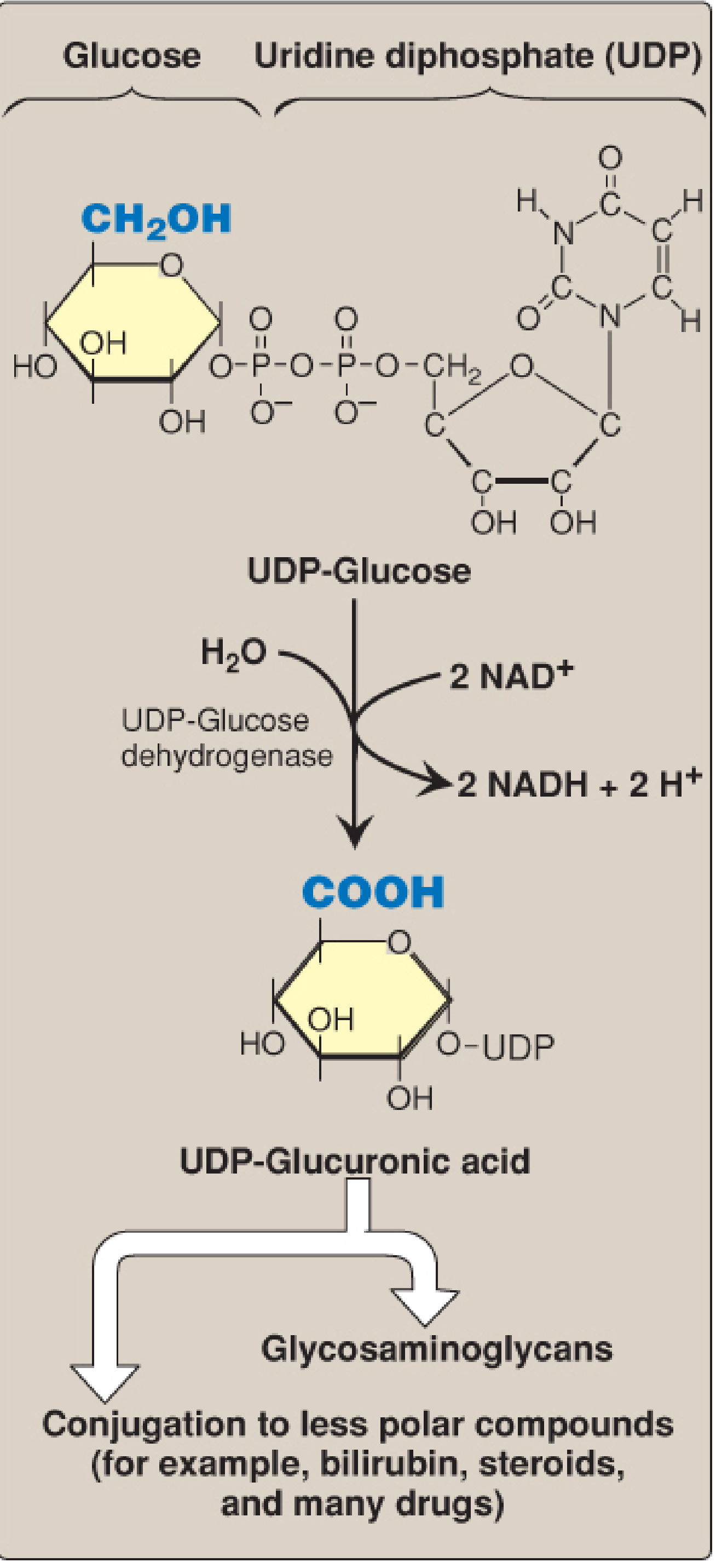
Figure: Formation of UDP-Glucuronic acid from UDP-Glucose. (Lippincott Illustrated Reviews: Biochemistry, 8e)
Step 5 - UDP-Glucuronate → D-Glucuronic Acid
- UDP is cleaved to release free D-glucuronic acid
Step 6 - D-Glucuronic Acid → L-Gulonate
- Enzyme: Glucuronate reductase
- Coenzyme: NADPH (NADPH-dependent reduction)
- Glucuronic acid is reduced at C-1 (carboxyl → aldehyde)
Branch Point: In most mammals (but NOT humans, primates, guinea pigs), L-Gulonate is converted to L-Ascorbic acid (Vitamin C) by L-gulonolactone oxidase. This enzyme is absent in humans due to a gene mutation, making Vitamin C an essential dietary nutrient for us.
Step 7 - L-Gulonate → 3-Keto-L-Gulonate
- Enzyme: L-Gulonate dehydrogenase (L-gulonate oxidase)
- Coenzyme: NAD⁺
Step 8 - 3-Keto-L-Gulonate → L-Xylulose + CO₂
- Enzyme: 3-Keto-L-gulonate decarboxylase
- Decarboxylation releases CO₂
Step 9 - L-Xylulose → Xylitol
- Enzyme: L-Xylulose reductase (NADPH-dependent xylulose reductase)
- Coenzyme: NADPH
- ⚠ This is the enzyme deficient in Essential Pentosuria (see clinical section)
Step 10 - Xylitol → D-Xylulose
- Enzyme: Xylitol dehydrogenase (Xylitol oxidase)
- Coenzyme: NAD⁺
Step 11 - D-Xylulose → D-Xylulose 5-phosphate
- Enzyme: Xylulokinase
Step 12 - D-Xylulose 5-phosphate → Pentose Phosphate Pathway
- D-Xylulose 5-phosphate enters the non-oxidative phase of the HMP shunt
- Ultimately converted to fructose 6-phosphate and glyceraldehyde 3-phosphate (glycolytic intermediates)
Pathway Summary Diagram
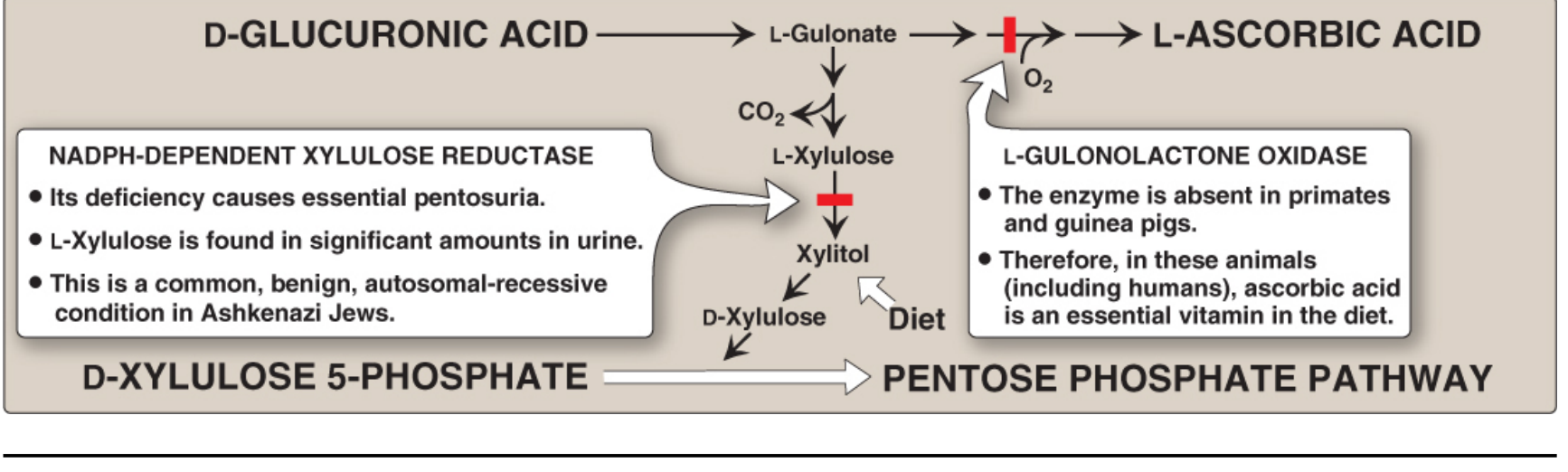
Figure: Metabolism of glucuronic acid (Uronic Acid Pathway). Red bars indicate enzyme deficiencies: (1) L-gulonolactone oxidase - absent in humans, causing Vitamin C dependency; (2) L-xylulose reductase - deficiency causes essential pentosuria. (Lippincott Illustrated Reviews: Biochemistry, 8e)
Key Enzymes and Coenzymes Summary Table
| Step | Enzyme | Coenzyme |
|---|---|---|
| Glucose 1-P → UDP-Glucose | UDPGlc Pyrophosphorylase | UTP |
| UDP-Glucose → UDP-Glucuronate | UDP-Glucose Dehydrogenase | 2 NAD⁺ |
| D-Glucuronate → L-Gulonate | Glucuronate Reductase | NADPH |
| L-Gulonate → L-Ascorbate | L-Gulonolactone Oxidase | O₂ (absent in humans) |
| L-Gulonate → 3-Keto-L-Gulonate | L-Gulonate Dehydrogenase | NAD⁺ |
| 3-Keto-L-Gulonate → L-Xylulose | Decarboxylase | - (CO₂ released) |
| L-Xylulose → Xylitol | L-Xylulose Reductase | NADPH |
| Xylitol → D-Xylulose | Xylitol Dehydrogenase | NAD⁺ |
| D-Xylulose → D-Xylulose 5-P | Xylulokinase | ATP |
Functions / Significance of the Uronic Acid Pathway
1. Glucuronidation (Detoxification and Conjugation)
- UDP-glucuronic acid donates glucuronate to lipophilic, poorly water-soluble compounds
- This process, called glucuronidation, is catalyzed by UDP-glucuronyl transferase in the liver (endoplasmic reticulum)
- Products are glucuronide conjugates which are highly water-soluble and excreted in urine or bile
- Substrates for glucuronidation include:
- Bilirubin (from hemoglobin breakdown) - conjugated bilirubin is water-soluble and excreted in bile
- Steroid hormones (estrogens, androgens, corticosteroids)
- Drugs and xenobiotics - morphine, acetaminophen, statins, sulfonamides
- Thyroid hormones
2. Proteoglycan and Glycosaminoglycan (GAG) Synthesis
- UDP-glucuronic acid is the activated form incorporated into the backbone of GAGs
- GAGs containing glucuronic acid include: hyaluronic acid, chondroitin sulfate, dermatan sulfate, heparan sulfate, heparin
- These are structural components of connective tissue, cartilage, and extracellular matrix
3. Ascorbic Acid (Vitamin C) Synthesis
- In most mammals, L-gulonate is oxidized by L-gulonolactone oxidase to form L-ascorbic acid
- In humans and primates: this enzyme is absent → Vitamin C must be obtained from the diet
- In guinea pigs: same enzyme deficiency - also require dietary Vitamin C
- Ascorbic acid is needed for collagen synthesis (hydroxylation of proline and lysine residues)
4. Entry of Dietary Pentoses into Metabolism
- Dietary D-xylulose can enter this pathway (via xylitol) and eventually be phosphorylated to D-xylulose 5-phosphate, entering the HMP shunt and glycolysis
Clinical Significance
Essential Pentosuria
Definition: A rare, benign, hereditary condition caused by a deficiency of L-xylulose reductase (NADPH-dependent xylulose reductase).
Inheritance: Autosomal recessive - most common in Ashkenazi Jews
Pathophysiology:
- L-Xylulose cannot be reduced to xylitol (enzyme block)
- L-Xylulose accumulates and is excreted in urine (L-xylosuria / pentosuria)
- Urine contains 1-4 g of L-xylulose per day
Clinical features:
- Completely benign - no clinical symptoms
- No metabolic consequences
- Patients have a normal life expectancy
Important diagnostic pitfall:
- L-xylulose is a reducing sugar → gives a false-positive result for glucosuria when urine is tested with alkaline copper reagents (Benedict's test / Fehling's test)
- Specific glucose oxidase strips (Clinistix) are negative - confirming it is NOT glucose
- This distinction is clinically important to avoid misdiagnosis as diabetes mellitus
Alimentary Pentosuria:
- Temporary, non-hereditary pentosuria after ingestion of large amounts of pentose-rich fruits (e.g., pears, cherries)
- Benign and self-limiting
Drugs and the Uronic Acid Pathway
- Several drugs increase glucose entry into the uronic acid pathway:
- Barbital, chlorobutanol - increase conversion of glucose → glucuronate → ascorbate in rats
- Aminopyrine, antipyrine - increase excretion of xylulose in pentosuric patients
Vitamin C (Ascorbic Acid) Deficiency - Scurvy
- Due to absent L-gulonolactone oxidase in humans, Vitamin C is dietary-essential
- Deficiency causes scurvy: impaired collagen synthesis → bleeding gums, poor wound healing, perifollicular hemorrhages
Relationship to Other Pathways
| Connection | How? |
|---|---|
| Glycolysis | Glucose → G6P → G1P enters the pathway; D-Xylulose 5-P exits back to glycolysis via PPP |
| HMP Shunt (PPP) | D-Xylulose 5-phosphate (end product) enters the non-oxidative phase |
| Glycogen Synthesis | Shares the UDP-glucose intermediate and UDPGlc pyrophosphorylase enzyme |
| GAG/Proteoglycan Synthesis | UDP-glucuronate is the building block for GAG chains |
| Drug Metabolism | UDP-glucuronate conjugates xenobiotics in Phase II hepatic drug metabolism |
Summary
The Uronic Acid Pathway is an alternative oxidative pathway for glucose, operating mainly in the liver cytosol. Its central product, UDP-glucuronic acid, serves as:
- A glucuronyl donor for detoxification/conjugation of bilirubin, drugs, and steroids
- A structural precursor for GAGs (hyaluronic acid, heparan sulfate, chondroitin sulfate)
- A precursor for Vitamin C synthesis in most animals (but not humans, who lack L-gulonolactone oxidase)
A deficiency of L-xylulose reductase causes essential pentosuria - a benign autosomal recessive condition in which L-xylulose is excreted in urine and may give false-positive results on copper-based urine glucose tests. The pathway connects back to the pentose phosphate pathway via D-xylulose 5-phosphate, and ultimately to glycolysis.
Sources:
- Harper's Illustrated Biochemistry, 32nd ed, Chapter 20 (Uronic Acid Pathway - Glucuronate, Pentosuria)
- Lippincott Illustrated Reviews: Biochemistry, 8th ed, Chapter 14 (Acidic Sugar Synthesis - Uronic Acid Pathway)
I have my university exam of 1 st year of subject biochemistry These are the important long answer give me answer of this question"Uronic acid pathway" and remeber it's a university examination
I already gave you a full, detailed answer on the Uronic Acid Pathway just a few minutes ago in this conversation. Here is the complete answer again for your convenience:
Uronic Acid Pathway
Introduction
The Uronic Acid Pathway (also called the Glucuronate Pathway) is an alternative metabolic route for glucose oxidation that occurs primarily in the liver. It is of minor quantitative importance as an energy-producing pathway - it does not generate ATP - but is of major physiological and clinical significance for:
- Synthesis of glucuronic acid for conjugation and detoxification
- Synthesis of ascorbic acid (Vitamin C) in most mammals (but NOT in humans)
- Incorporation of glucuronate into proteoglycans and glycosaminoglycans (GAGs)
- Providing a route for dietary D-xylulose to enter central metabolic pathways
Location
The pathway operates in the cytosol of hepatocytes (liver cells). The liver is the primary site.
Starting Material
Glucose (via Glucose 1-phosphate → UDP-Glucose) is the entry substrate.
Steps of the Uronic Acid Pathway
Step 1 - Glucose → Glucose 6-phosphate
- Phosphorylated by hexokinase/glucokinase
Step 2 - Glucose 6-phosphate → Glucose 1-phosphate
- Isomerization by phosphoglucomutase
Step 3 - Glucose 1-phosphate + UTP → UDP-Glucose + PPi
- Enzyme: UDP-glucose pyrophosphorylase
- This is the same reaction as in glycogen synthesis
- UTP activates glucose as a nucleotide sugar
Step 4 - UDP-Glucose → UDP-Glucuronate (UDP-Glucuronic Acid)
- Enzyme: UDP-glucose dehydrogenase (NAD-dependent)
- Coenzyme: 2 NAD⁺ → 2 NADH (two-step oxidation)
- Carbon 6 of glucose (-CH₂OH) is oxidized to a carboxyl group (-COOH)
- This is the key committed step producing UDP-glucuronic acid
Figure: Formation of UDP-Glucuronic acid from UDP-Glucose. (Lippincott Illustrated Reviews: Biochemistry, 8e)
Step 5 - UDP-Glucuronate → D-Glucuronic Acid
- UDP is cleaved to release free D-glucuronic acid
Step 6 - D-Glucuronic Acid → L-Gulonate
- Enzyme: Glucuronate reductase
- Coenzyme: NADPH (reduction at C-1)
Branch Point: In most mammals (but NOT humans, primates, guinea pigs), L-Gulonate is converted to L-Ascorbic acid (Vitamin C) by L-gulonolactone oxidase. This enzyme is absent in humans due to a gene mutation, making Vitamin C an essential dietary nutrient for us.
Step 7 - L-Gulonate → 3-Keto-L-Gulonate
- Enzyme: L-Gulonate dehydrogenase
- Coenzyme: NAD⁺
Step 8 - 3-Keto-L-Gulonate → L-Xylulose + CO₂
- Enzyme: 3-Keto-L-gulonate decarboxylase
- Decarboxylation releases CO₂
Step 9 - L-Xylulose → Xylitol
- Enzyme: L-Xylulose reductase (NADPH-dependent)
- Coenzyme: NADPH
- ⚠ This enzyme is deficient in Essential Pentosuria
Step 10 - Xylitol → D-Xylulose
- Enzyme: Xylitol dehydrogenase
- Coenzyme: NAD⁺
Step 11 - D-Xylulose → D-Xylulose 5-phosphate
- Enzyme: Xylulokinase
- Coenzyme: ATP
Step 12 - D-Xylulose 5-phosphate → Pentose Phosphate Pathway (HMP Shunt)
- Enters the non-oxidative phase of the PPP
- Ultimately yields fructose 6-phosphate and glyceraldehyde 3-phosphate (glycolytic intermediates)
Pathway Diagram
Figure: Metabolism of glucuronic acid (Uronic Acid Pathway). Red bars = enzyme deficiencies: (1) L-gulonolactone oxidase - absent in humans causing Vitamin C dependency; (2) L-xylulose reductase - deficiency causes essential pentosuria. (Lippincott Illustrated Reviews: Biochemistry, 8e)
Key Enzymes and Coenzymes Summary Table
| Step | Enzyme | Coenzyme |
|---|---|---|
| Glucose 1-P → UDP-Glucose | UDPGlc Pyrophosphorylase | UTP |
| UDP-Glucose → UDP-Glucuronate | UDP-Glucose Dehydrogenase | 2 NAD⁺ |
| D-Glucuronate → L-Gulonate | Glucuronate Reductase | NADPH |
| L-Gulonate → L-Ascorbate | L-Gulonolactone Oxidase | O₂ (absent in humans) |
| L-Gulonate → 3-Keto-L-Gulonate | L-Gulonate Dehydrogenase | NAD⁺ |
| 3-Keto-L-Gulonate → L-Xylulose | Decarboxylase | - (CO₂ released) |
| L-Xylulose → Xylitol | L-Xylulose Reductase | NADPH |
| Xylitol → D-Xylulose | Xylitol Dehydrogenase | NAD⁺ |
| D-Xylulose → D-Xylulose 5-P | Xylulokinase | ATP |
Functions / Significance of the Uronic Acid Pathway
1. Glucuronidation (Detoxification and Conjugation)
- UDP-glucuronic acid donates glucuronate to lipophilic, poorly water-soluble compounds
- This process (glucuronidation) is catalyzed by UDP-glucuronyl transferase in the liver (ER)
- Products are water-soluble glucuronide conjugates excreted in urine or bile
- Key substrates for glucuronidation:
- Bilirubin - conjugated bilirubin is water-soluble, excreted in bile
- Steroid hormones - estrogens, androgens, corticosteroids
- Drugs/xenobiotics - morphine, acetaminophen, statins, sulfonamides
- Thyroid hormones
2. Proteoglycan and Glycosaminoglycan (GAG) Synthesis
- UDP-glucuronic acid is incorporated into the backbone of GAGs
- GAGs containing glucuronic acid: hyaluronic acid, chondroitin sulfate, dermatan sulfate, heparan sulfate, heparin
- These are structural components of connective tissue, cartilage, and extracellular matrix
3. Ascorbic Acid (Vitamin C) Synthesis
- In most mammals: L-gulonate → L-ascorbic acid via L-gulonolactone oxidase
- In humans and primates: enzyme is absent → Vitamin C must come from the diet
- In guinea pigs: same gene defect - also require dietary Vitamin C
- Ascorbic acid is required for hydroxylation of proline and lysine in collagen synthesis
4. Entry of Dietary Pentoses into Metabolism
- Dietary D-xylulose enters via xylitol, is phosphorylated to D-xylulose 5-phosphate, and enters the HMP shunt and glycolysis
Clinical Significance
Essential Pentosuria
| Feature | Details |
|---|---|
| Enzyme deficiency | L-Xylulose reductase (NADPH-dependent xylulose reductase) |
| Inheritance | Autosomal recessive |
| Common population | Ashkenazi Jews |
| Metabolite accumulated | L-Xylulose excreted in urine (1-4 g/day) |
| Clinical features | Completely benign - no symptoms, normal life expectancy |
| Important pitfall | L-xylulose is a reducing sugar → false-positive Benedict's/Fehling's test (misdiagnosed as diabetes mellitus) |
| Specific glucose test | Glucose oxidase strips (Clinistix) are negative - confirming NOT glucose |
Alimentary Pentosuria:
- Temporary pentosuria after eating large amounts of pentose-rich fruits (pears, cherries) - benign and self-limiting
Drugs Affecting the Pathway
- Barbital, chlorobutanol - increase glucose entry into the uronic acid pathway in rats
- Aminopyrine, antipyrine - increase xylulose excretion in pentosuric subjects
Scurvy (Vitamin C Deficiency)
- Because humans lack L-gulonolactone oxidase, Vitamin C is dietary-essential
- Deficiency → Scurvy: impaired collagen synthesis, bleeding gums, poor wound healing, perifollicular hemorrhages
Relationship to Other Pathways
| Pathway | Connection |
|---|---|
| Glycolysis | Glucose is the starting material; D-Xylulose 5-P exits back via PPP → fructose 6-P + G3P |
| HMP Shunt (PPP) | D-Xylulose 5-P (final product) enters the non-oxidative phase |
| Glycogen Synthesis | Shares UDP-glucose intermediate and UDPGlc pyrophosphorylase enzyme |
| GAG Synthesis | UDP-glucuronate is the building block for chondroitin sulfate, heparin, hyaluronic acid |
| Phase II Drug Metabolism | UDP-glucuronate conjugates xenobiotics in hepatic detoxification |
Summary
The Uronic Acid Pathway is an alternative oxidative pathway for glucose in the liver cytosol that does not produce ATP. Its central product, UDP-glucuronic acid, is:
- A glucuronyl donor for detoxification/conjugation of bilirubin, drugs, and steroids (glucuronidation)
- A structural precursor for GAGs (hyaluronic acid, heparan sulfate, chondroitin sulfate)
- A precursor for Vitamin C synthesis in most animals (blocked in humans due to absent L-gulonolactone oxidase)
Deficiency of L-xylulose reductase causes essential pentosuria - a benign autosomal recessive condition where L-xylulose is excreted in urine and gives a false-positive result on copper-based urine glucose tests. The pathway ultimately connects back to the HMP shunt via D-xylulose 5-phosphate and then to glycolysis.
Sources:
- Harper's Illustrated Biochemistry, 32nd ed, Chapter 20
- Lippincott Illustrated Reviews: Biochemistry, 8th ed, Chapter 14
This is a shared conversation. Sign in to Orris to start your own chat.