Systemic Lupus Erythematosus (SLE): Complete Notes with Ayurvedic Correlation and Treatment
1. Definition and epidemiology
- SLE is a chronic, relapsing-remitting, multisystem autoimmune disease. Autoantibodies and immune complexes cause inflammation and damage in many organs.
- It is classically a disease of women, with a female:male ratio of about 9:1. Onset is mostly between ages 15 and 45.
- It is more frequent and more severe in people of African, Hispanic and Asian ancestry. Men and children tend to have more severe disease, including more nephritis.
- Main causes of death: infection, lupus nephritis and renal failure, and accelerated atherosclerotic cardiovascular disease. Active CNS disease also contributes.
2. Etiopathogenesis
Multifactorial: genes, hormones and environment
| Factor | Details |
|---|
| Genetic | HLA-DR2 and DR3. Early complement deficiencies (C1q, C2, C4) strongly predispose. Other genes: IRF5, STAT4, TREX1 and FcγR variants. |
| Hormonal | Estrogen promotes autoimmunity. This fits the female predominance, flares in pregnancy and the association with Klinefelter syndrome (47,XXY). |
| Environmental | UV light, EBV, smoking, silica, and drugs. |
| Drug-induced lupus | Hydralazine, procainamide, isoniazid, minocycline, TNF inhibitors. Anti-histone antibodies are typical. |
Core mechanism
- Defective clearance of apoptotic cells exposes nuclear antigens (nucleosomes, dsDNA, Sm, Ro, La).
- Plasmacytoid dendritic cells produce type I interferon (the "interferon signature"), which activates the immune system.
- Self-reactive B cells escape tolerance. BAFF/BLyS survival signals and T-cell help produce autoantibodies.
- Immune complexes deposit in the glomeruli, skin and vessels. This activates the classical complement pathway, with consumption of C3 and C4.
- Neutrophil extracellular traps (NETs) add more nuclear antigen.
- Result: tissue injury, vasculitis, thrombosis (with antiphospholipid antibodies) and organ damage.
Pathology (hallmarks): fibrinoid necrosis, hematoxylin bodies, "onion-skin" periarteriolar fibrosis in the spleen, and wire-loop lesions in the glomeruli. Skin biopsy shows interface dermatitis, with a granular IgG/C3 band at the dermo-epidermal junction on direct immunofluorescence (the lupus band test).
3. Clinical features
General: fatigue, fever, weight loss, lymphadenopathy. Skin is involved in about 80% of cases and 4 of the 11 ACR criteria are mucocutaneous (Andrews' Diseases of the Skin).
| System | Features |
|---|
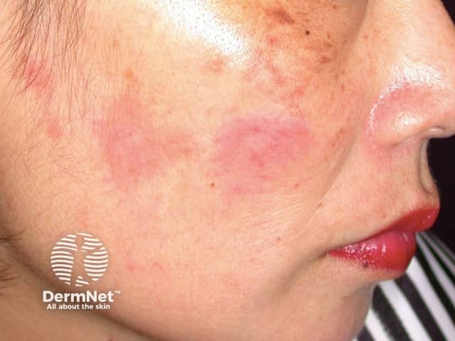
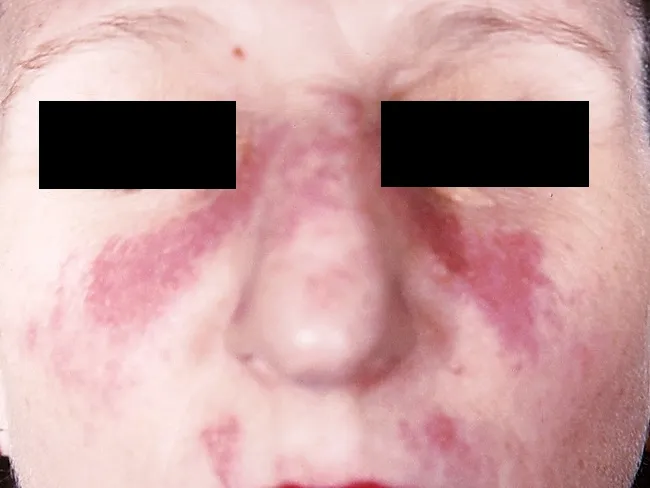
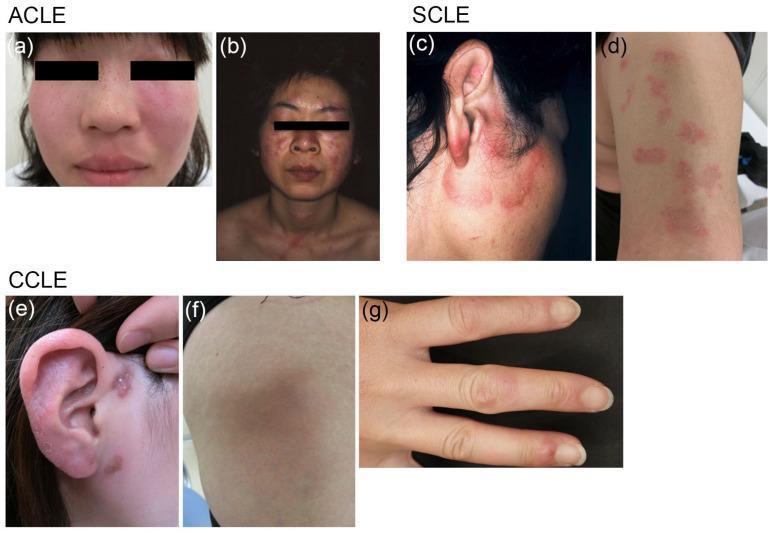
| Mucocutaneous | Acute cutaneous lupus: malar "butterfly" rash that spares the nasolabial folds, and photosensitivity. Subacute cutaneous lupus (SCLE): annular or papulosquamous lesions, associated with anti-Ro. Chronic cutaneous lupus: discoid lupus (scarring, atrophic plaques), lupus profundus, chilblain lupus. Also non-scarring alopecia, painless oral or nasal ulcers, vasculitic lesions, Raynaud phenomenon. |
| Musculoskeletal | Arthralgia or non-erosive arthritis in more than 90%, mainly small joints of the hands, wrists and knees. Jaccoud arthropathy (deformities that can be corrected passively). Myositis. Avascular necrosis, mostly steroid-related. |
| Renal | Lupus nephritis in about 40 to 60%: proteinuria, hematuria, casts, hypertension, falling GFR, nephrotic syndrome. |
| Hematologic | Autoimmune hemolytic anemia (Coombs positive), leukopenia and lymphopenia, thrombocytopenia (ITP-like), anemia of chronic disease. Antiphospholipid syndrome gives venous or arterial thrombosis and pregnancy loss. |
| Neuropsychiatric (NPSLE) | Seizures, psychosis, stroke, aseptic meningitis, myelopathy, chorea, neuropathy, cognitive dysfunction, mood disorders. Anti-ribosomal P is associated with psychosis. |
| Cardiac | Pericarditis (most common), myocarditis, Libman-Sacks (verrucous) endocarditis, accelerated atherosclerosis, and congenital heart block in neonates of anti-Ro mothers. |
| Pulmonary | Pleuritis with or without effusion, acute lupus pneumonitis, diffuse alveolar hemorrhage, ILD, "shrinking lung" syndrome, pulmonary hypertension. |
| GI/hepatic | Peritonitis, mesenteric vasculitis, protein-losing enteropathy, pancreatitis, lupus hepatitis (rare). |
| Ocular | Sicca, retinal vasculitis, cotton-wool spots, optic neuritis. Also hydroxychloroquine retinopathy as a drug effect. |
4. Diagnosis
4.1 Investigations
- CBC: cytopenias. ESR is raised, while CRP is often normal unless there is serositis or infection.
- Urine: protein, RBCs, cellular casts. Do a spot urine protein:creatinine ratio. Serum creatinine and eGFR. Kidney biopsy for any suspected nephritis.
- Direct Coombs, lupus anticoagulant, anticardiolipin, anti-β2 glycoprotein I.
Autoantibodies
| Antibody | Comment |
|---|
| ANA (IIF on HEp-2) | About 95 to 98% sensitive. Poorly specific. A negative ANA makes SLE unlikely. |
| Anti-dsDNA | Highly specific. Titers track activity and nephritis. |
| Anti-Sm | Highly specific, but only in about 20 to 30%. |
| Anti-Ro/SSA, anti-La/SSB | SCLE, neonatal lupus, congenital heart block, sicca. |
| Anti-RNP | Mixed connective tissue disease overlap. |
| Anti-histone | Drug-induced lupus. |
| Antiphospholipid antibodies | Antiphospholipid syndrome, thrombosis, pregnancy loss. |
| Anti-ribosomal P | NPSLE. |
- Complement: low C3, C4 and CH50 indicate activity.
- Activity scores: SLEDAI-2K, BILAG, SELENA-SLEDAI. Damage index: SLICC/ACR.
4.2 Classification criteria
2019 EULAR/ACR criteria
- Entry criterion: ANA titer of at least 1:80 on HEp-2 cells.
- Then add weighted points across 10 domains (a clinical domain's highest-scoring item counts). Classify as SLE if the total is 10 or more and there is at least one clinical criterion.
| Domain | Items (points) |
|---|
| Constitutional | Fever (2) |
| Hematologic | Leukopenia (3), thrombocytopenia (4), autoimmune hemolysis (4) |
| Neuropsychiatric | Delirium (2), psychosis (3), seizure (5) |
| Mucocutaneous | Non-scarring alopecia (2), oral ulcers (2), SCLE or discoid (4), acute cutaneous lupus (6) |
| Serosal | Pleural or pericardial effusion (5), acute pericarditis (6) |
| Musculoskeletal | Joint involvement (6) |
| Renal | Proteinuria over 0.5 g/24 h (4), class II or V nephritis (8), class III or IV nephritis (10) |
| Antiphospholipid antibodies | (2) |
| Complement | Low C3 or low C4 (3), both low (4) |
| SLE-specific antibodies | Anti-dsDNA or anti-Sm (6) |
SLICC 2012: 4 or more criteria with at least 1 clinical and 1 immunologic, or biopsy-proven lupus nephritis with ANA or anti-dsDNA (Andrews' Diseases of the Skin, p. 188).
1997 ACR (11 criteria): malar rash, discoid rash, photosensitivity, oral ulcers, arthritis, serositis, renal disorder, neurologic disorder, hematologic disorder, immunologic disorder, ANA. Four or more are needed (Andrews' Diseases of the Skin, p. 188).
4.3 Differential diagnosis
Rheumatoid arthritis, dermatomyositis, mixed connective tissue disease, Sjogren syndrome, vasculitis, ITP, TTP, viral infections (parvovirus, HIV, hepatitis C), and drug-induced lupus.
5. Lupus nephritis (ISN/RPS classification)
| Class | Lesion | Notes |
|---|
| I | Minimal mesangial | Normal on light microscopy, immune deposits on IF |
| II | Mesangial proliferative | Mild proteinuria or hematuria |
| III | Focal (under 50% of glomeruli) | Active (A), chronic (C) or both |
| IV | Diffuse (50% or more of glomeruli) | Most common and most severe. Hypocomplementemia, high anti-dsDNA, nephritic-nephrotic picture |
| V | Membranous | Nephrotic syndrome, thrombosis risk. Can coexist with III or IV |
| VI | Advanced sclerosing (90% or more globally sclerosed) | Irreversible |
Treatment (EULAR 2023 and the 2025 kidney update)
- Biopsy before treatment where possible. Add renoprotection: ACE inhibitor or ARB, blood pressure control, SGLT2 inhibitor where appropriate.
- Induction, class III/IV: glucocorticoids (pulse methylprednisolone, then a low-dose taper) plus mycophenolate or low-dose IV cyclophosphamide (Euro-Lupus: 500 mg every 2 weeks for 6 doses). For high-risk disease, add belimumab or a calcineurin inhibitor (voclosporin or tacrolimus).
- Maintenance: mycophenolate or azathioprine for at least 3 years. Taper steroids to 5 mg/day or less, or stop.
- Pure class V with nephrotic-range proteinuria: mycophenolate-based therapy.
- Refractory disease: rituximab, obinutuzumab (2025 update).
- Class VI and ESRD: dialysis or transplant. Lupus often quiets once on dialysis.
6. Modern management of SLE overall
6.1 Principles (EULAR 2023 update: 5 principles, 13 recommendations)
- The target is remission or low disease activity (LLDAS), with prevention of flares and organ damage.
- Use an SLE activity score at each visit.
- Hydroxychloroquine (HCQ) for all patients, at 5 mg/kg/day or less of real body weight. HCQ lowers flares, damage, thrombosis and mortality. Do baseline eye screening and annual screening after 5 years (earlier with renal impairment or higher doses). It is the only drug that can be continued in pregnancy without concern.
- Glucocorticoids as bridging only. Pulse methylprednisolone for severe flares. Long-term maintenance should be 5 mg/day prednisone equivalent or less, and withdrawn when possible.
- Immunosuppressants (methotrexate, azathioprine, mycophenolate) should be started early to control disease and spare steroids.
- Biologics: anifrolumab (type I interferon receptor antibody) and belimumab (anti-BLyS) in non-renal SLE that persists despite standard therapy. Rituximab for refractory disease. Cyclophosphamide for organ-threatening disease.
6.2 Organ-specific approach
| Manifestation | Treatment |
|---|
| Mild disease (skin, joints, fatigue) | HCQ, short low-dose steroid course, NSAIDs (avoid with renal disease). Add methotrexate for arthritis or skin if needed. |
| Cutaneous | Sun protection, smoking cessation, topical steroids or calcineurin inhibitors, HCQ, then methotrexate or mycophenolate. Anifrolumab for refractory disease. |
| Hematologic (ITP, hemolysis) | Steroids, then azathioprine, mycophenolate, rituximab or IVIG. |
| NPSLE | Treat on the cause: immunosuppression (steroids and cyclophosphamide or rituximab) for inflammatory causes. Anticoagulation for thrombotic causes. Symptomatic drugs as needed. |
| Serositis | NSAIDs or steroids. |
| Antiphospholipid syndrome | Warfarin for thrombosis. Avoid DOACs in high-risk (triple-positive) APS. |
6.3 Supportive care and general measures
- Sun protection (SPF 50 or higher), smoking cessation, exercise, vitamin D and calcium, and a bone-health plan if on steroids.
- Cardiovascular risk management: lipids, blood pressure, aspirin in selected patients.
- Vaccinations: inactivated vaccines (influenza, pneumococcal, HPV, recombinant zoster, COVID-19). Avoid live vaccines when on high-dose immunosuppression.
- Infection screening before immunosuppression (TB, hepatitis B and C).
6.4 Pregnancy
- Plan conception during quiescent disease (at least 6 months of remission).
- Continue HCQ. Add low-dose aspirin from about 12 weeks to lower pre-eclampsia risk.
- Safe options: azathioprine, tacrolimus, cyclosporine, low-dose prednisolone.
- Stop mycophenolate, cyclophosphamide, methotrexate and ACE inhibitors before conception.
- Anti-Ro/La positive mothers: fetal echocardiography surveillance and HCQ for prevention of congenital heart block.
6.5 Flares
Flares are common. Reassess activity, check adherence (especially HCQ), and look for infection before escalating therapy.
6.6 Recent evidence check
Recent reviews are consistent with the library and the guidance above:
- The 2023 EULAR update (PMID 37827694) and its systematic review (PMID 38777375). They support anifrolumab and belimumab in extrarenal SLE, and belimumab and voclosporin add-on in lupus nephritis.
- The 2025 EULAR kidney update (PMID 41107121). It adds obinutuzumab as an option.
- A 2026 network meta-analysis of interventions for SLE arthritis (PMID 42055851).
Newer drugs may postdate the textbooks in the library, so check current labeling and local availability before prescribing.
7. Ayurvedic correlation
Important framing: Ayurveda has no single named disease that equals SLE. The correlations below are interpretive and come from reviews and case reports, not from Samhita text that describes SLE as such. They are a framework for understanding and treating, not an established equivalence.
7.1 Disease correlations
| Ayurvedic concept | Why it fits |
|---|
| Vatarakta (especially Vata-pradhana, and Raktavrita Vata) | Joint pain, swelling and stiffness, skin discoloration, Raynaud-like changes, vasculitic ulcers, and a relapsing course. This is the most commonly used correlation in published case reports (Ayurveda360 case report on Vatapradhana Vatarakta; Gururaja 2019, PMC6938929). |
| Amavata | Polyarthritis with fever, fatigue and heaviness, aggravated by ama and Vata. It fits the early inflammatory, joint-predominant phase. |
| Rakta-pradoshaja vikara / Pitta-rakta dushti | Malar rash, photosensitivity, oral ulcers, fever, and bleeding tendencies. This fits the Pitta-Rakta heat component of the rash and mucosal lesions. |
| Ojas vyapat / Ojakshaya (Vishramsa, Vyapad) | Immune dysregulation and loss of self-tolerance. Ojas is the substrate of bala (immunity) and vyadhikshamatva. This is the conceptual correlate of autoimmunity. |
| Dushi visha / Gara visha | The chronic, relapsing, toxin-like course with residual toxins (visha) lodged in the tissues. This is an interpretive concept. |
Feature-wise mapping (interpretive)
| Feature | Ayurvedic concept |
|---|
| Fever | Jwara (often Vishama) |
| Malar rash and photosensitivity | Pitta-Rakta vitiation, Visarpa or Mandala-type kushtha |
| Facial pigmentation | Vyanga |
| Arthritis | Vatarakta, Amavata |
| Oral ulcers | Mukhapaka |
| Alopecia | Khalitya, Indralupta |
| Nephritis and edema | Shotha (Mutravaha srotas and Medovaha srotas involvement) |
| Cytopenias, pallor | Pandu, Rakta kshaya |
| Neuropsychiatric | Vata-Pitta unmada, apasmara |
| Pleural or cardiac involvement | Parshvashula, Hridroga |
7.2 Nidana (causes)
Excess amla, lavana, katu, vidahi and viruddha ahara, curd and fermented foods, excess sun and heat exposure (atapa), day sleep with night wakefulness, anger and chronic stress (manasika nidana), suppression of natural urges, and a Sukumara (delicate) constitution. Hormonal and genetic susceptibility corresponds to bija dosha.
7.3 Samprapti (pathogenesis)
- Agnimandya leads to ama formation.
- Nidana vitiates Pitta and Rakta. Vata is aggravated and obstructed (avarana) by vitiated Rakta (Raktavrita Vata) and ama.
- This produces srotorodha, with Vata-Pitta-Rakta moving through the srotas and depositing in sites of khavaigunya (weakness). Sites: twak (skin), sandhi (joints), vrikka (kidneys), hridaya, majja.
- Dhatu involvement: Rasa and Rakta first, then Mamsa, Asthi and Majja. Ojakshaya follows, which weakens vyadhikshamatva and perpetuates the process.
- The chronic phase has a tridoshaja character with relapses and remissions.
Samprapti ghataka summary
| Ghataka | Features |
|---|
| Dosha | Vata-Pitta (Pitta predominant in rash and fever, Vata in joint pain) |
| Dushya | Rasa, Rakta, Mamsa, Asthi, Majja, Ojas |
| Agni | Jatharagni and dhatvagni mandya |
| Ama | Present |
| Srotas | Rasavaha, Raktavaha, Mamsavaha, Asthivaha, Mutravaha |
| Srotodushti | Sanga, vimargagamana |
| Udbhava sthana | Amashaya and pakvashaya |
| Vyakta sthana | Twak, sandhi, vrikka |
| Rogamarga | Madhyama (internal) |
8. Ayurvedic treatment
Evidence note: The evidence for the regimens below is mostly classical principles and published case reports or small series (Gururaja 2019; the Ayurveda360 2025 Vatarakta report). I did not find randomized trials. Ayurveda should be used as an adjunct to HCQ, steroids and immunosuppressants. Never stop or withhold them, especially in nephritis, NPSLE or other organ-threatening disease.
8.1 Principles
- Treatment follows Vatarakta and Amavata chikitsa, adjusted for dosha predominance and the patient's strength (bala).
- Nidana parivarjana: remove the causes (diet, sun exposure, stress, sleep).
- Langhana and Pachana for ama, then Snehana and Shodhana when the patient is strong enough, then Shamana and Rasayana (Ojas support).
- Shodhana should be done only in a stable phase, avoiding active flares, severe cytopenia, active nephritis with renal failure, or high-dose steroid cover without physician coordination.
8.2 Stage-wise plan
A. Acute or ama stage (active flare, fever, arthritis)
- Deepana-pachana: Trikatu, Panchakola, Chitrakadi vati, Shunthi (dry ginger) and Guduchi decoctions.
- Light diet (laghu ahara): mudga yusha, rice gruel, and warm water (kept lukewarm, not hot).
- Fever: Sudarshana ghana vati or churna, Guduchi, Amritottara kashaya.
- Rasna-based or Amruta-based kashayas (Rasnasaptaka, Amrutottara) for joints in the Amavata phase.
B. Shodhana (once ama has settled and the patient is stable)
- Mridu virechana (mild purgation) is the main shodhana for Pitta-Rakta predominance. Example: Aragvadhadi avaleha or Avipattikara churna, Trivrit lehya, or Gandharvahastadi eranda taila, after snehana (internal Tiktaka ghrita or Mahatiktaka ghrita).
- Basti: for Vata predominance and joint disease. Ksheera basti with Dashamoola, Punarnavashtaka or Guduchi (as in the Ayurveda360 case report), or Tiktaka ksheera basti, Madhutailika basti, or Erandamuladi niruha. The Gururaja case combined Guduchi, Kaishora guggulu and basti.
- Raktamokshana (bloodletting, leech) is used in Vatarakta, but it is generally avoided in SLE because of anemia, thrombocytopenia and bleeding or thrombotic risk.
- External therapy: Pariseka or dhara with Pitta-Vata-shamaka dravyas (cold medicated milk or Kshirabala taila), Takradhara or Shirodhara for neuropsychiatric symptoms, Lepa with Chandana, Usheera or Yashtimadhu for rash, and Abhyanga with Chandanadi taila, Kshirabala taila or Pinda taila (avoid heat in a flare).
C. Shamana (oral medicines)
| Purpose | Single drugs and formulations (typical use) |
|---|
| Immunomodulation, Rakta-Pitta shamana | Guduchi (Tinospora cordifolia) is the main drug in case reports. Also Amalaki, Haridra, Manjishtha, Sariva, Chandana, Usheera, Yashtimadhu, Neem/Nimba, Patola, Kirata-tikta |
| Vatarakta | Kaishora guggulu, Amrita guggulu, Guduchyadi kashaya, Punarnavadi kashaya, Manjishthadi kashaya, Mahatiktaka ghrita, Panchatikta guggulu ghrita (arthritis or skin-heavy disease) |
| Pitta-Rakta heat | Praval pishti, Mukta pishti, Kamadudha rasa, Chandrakala rasa, Giloy satva, Sarivadyasava, Chandanasava, Amritarishta |
| Skin | Mahamanjishtadi kashaya, Gandhaka rasayana, Arogyavardhini vati, Nimba-patola-based preparations |
| Renal or edema (physician-supervised) | Punarnavadi kashaya, Gokshuradi guggulu, Varunadi kashaya. Check kidney function and avoid nephrotoxic metal preparations. |
| Anemia | Punarnavadi mandura, Navayasa lauha. Use only after checking iron indices (lauha bhasmas can overload in hemolysis). |
| Neuropsychiatric | Brahmi, Shankhapushpi, Jatamansi, Ashwagandha (caution: may stimulate immunity, so use physician judgement), Saraswatarishta |
D. Rasayana (maintenance, remission phase)
Guduchi, Amalaki (Chyawanprash is a classic base), Shatavari, Yashtimadhu, Brahmi, Gandhaka rasayana, Ashwagandha, Pippali rasayana. These aim to restore Ojas and reduce relapse. Avoid hot, penetrating rasayanas (such as Bhallataka) in Pitta-Rakta disease.
8.3 Pathya (wholesome) and apathya (unwholesome)
| Items |
|---|
| Pathya (favour) | Old rice (shashtika), barley, wheat in moderation, mudga (green gram), masoor, bitter vegetables (patola, karela, parwal), ash gourd, pomegranate, amla, coconut water, cow's ghee, cow's milk, buttermilk with caution, adequate sleep, yoga, pranayama, meditation, and stress management. Cooling and light foods in Pitta phases. |
| Apathya (avoid) | Curd, fermented foods, sour, salty, spicy, fried and heavy foods, viruddha ahara (such as milk with fish or sour fruits), alcohol, tobacco, excess sesame, black gram (masha), day sleep, night wakefulness, direct sun exposure, excess heat and intense exercise, anger, grief, and suppression of urges. |
Yoga and manasika: gentle asanas (Sukhasana, Vajrasana, Setu bandhasana), Shitali and Sheetkari pranayama, Anuloma-viloma, and meditation to reduce stress, which is a recognized flare trigger.
8.4 Reported outcomes and limits
- Case reports describe symptom relief (pain, swelling, rash, fatigue), ulcer healing, steroid dose reduction and no relapse over follow-up with Guduchi, Kaishora guggulu, mild virechana and basti-based regimens (Gururaja 2019, PMC6938929; Ayurveda360 2025 report; an IJDDT review also compiles several). These are uncontrolled single-patient or small observations, so they cannot show efficacy or safety.
- No randomized controlled trial of an Ayurvedic regimen in SLE has been found in my search.
8.5 Safety cautions (important)
- Do not replace HCQ, steroids or immunosuppressants. Taper steroids only under rheumatology supervision.
- Immunostimulant herbs (Guduchi, Ashwagandha) have a theoretical risk of aggravating autoimmunity. Reports of herb-induced liver injury, including autoimmune-hepatitis-like injury, have been described with Tinospora cordifolia. Monitor liver function.
- Guggulu preparations may affect the liver and interact with other drugs.
- Rasaushadhi and bhasma (metal-mineral preparations) carry a risk of heavy-metal contamination, kidney and liver toxicity. Use only well-tested, standardized products, especially in lupus nephritis.
- Turmeric (Haridra) and similar herbs may increase bleeding risk with anticoagulants or antiplatelet drugs.
- Pregnancy: avoid purgation and strongly acting herbs.
- Monitor CBC, LFT, RFT and urine regularly while combining therapies. Keep the rheumatologist and the Ayurvedic physician informed of everything the patient takes.
9. Integrative summary
| Phase | Conventional | Ayurvedic adjunct |
|---|
| Active flare | Steroids, HCQ, immunosuppressants, biologics, organ-directed treatment | Ama pachana, light diet, Sudarshana, Guduchi, Pitta-shamana. No shodhana. |
| Stable or low activity | HCQ, steroid-sparing agents, monitoring | Mridu virechana, basti (selected patients), shamana (Kaishora guggulu, Mahatiktaka ghrita) |
| Remission | HCQ maintenance, taper, vaccination, cardiovascular and bone care | Rasayana, pathya, yoga, stress control, sun protection |
Key points to remember
- SLE is diagnosed by the 2019 EULAR/ACR criteria (ANA at least 1:80 entry, 10 or more points). Anti-dsDNA and anti-Sm are the most specific antibodies. Low C3/C4 indicates activity.
- HCQ for everyone, steroids as bridging only, and early steroid-sparing drugs. Belimumab and anifrolumab are used for persistent disease. Lupus nephritis needs a biopsy and mycophenolate or Euro-Lupus cyclophosphamide, with belimumab or voclosporin added in high-risk cases.
- Ayurvedic correlation is mainly Vatarakta (Raktavrita Vata) with Amavata, Pitta-Rakta dushti and Ojakshaya. Treatment is staged: ama pachana, then mridu virechana and basti, then shamana and rasayana, with strict pathya. It supports but does not replace modern therapy.
Sources: Andrews' Diseases of the Skin (SLE section, p. 188); Henry's Clinical Diagnosis and Management by Laboratory Methods and Tietz Textbook of Laboratory Medicine (classification criteria); EULAR 2023 recommendations (
PMID 37827694); published Ayurvedic case reports and reviews as cited above.