
Hydronephrosis give laq of the topic from standard textbook of surgery
Reading File
Now let me search the library for hydronephrosis content in surgical textbooks.
Finding Sources
Finding Sources
Excellent! I found rich content in S Das (Manual on Clinical Surgery), Bailey & Love, Campbell-Walsh Urology, and Robbins Pathology. Let me read all the key surgical sources in parallel.
Reading File
Reading File
Reading File
Reading File
Good. Now let me read more of the S Das content (which has the most comprehensive LAQ content) and Bailey & Love surgical content:
Reading File
Reading File
Finding Sources
Now let me read the S Das content for treatment/surgery details and Bailey & Love for the full urinary tract obstruction section:
Reading File
Reading File
Now let me read the treatment section from S Das more carefully - I need to find the surgical treatment/pyeloplasty section:
Reading File
Finding Sources
Now let me get the treatment section from S Das (the main surgical LAQ content) and the Robbins pathology morphology:
Reading File
Reading File
Now let me get the treatment section for hydronephrosis from S Das (the block just before renal calculus section, which should have treatment):
Reading File
Finding Sources
Let me get the Smith & Tanagho General Urology content on hydronephrosis which is likely the most complete for a surgical LAQ:
Finding Sources
Reading File
Let me now get the dedicated hydronephrosis chapter content from Smith & Tanagho General Urology and also the Campbell Walsh content for treatment:
Finding Sources
Reading File
I now have comprehensive information from multiple standard textbooks. Let me compile the complete LAQ answer.
Loading Image
I now have all the information needed to compile a comprehensive LAQ on hydronephrosis. Here is the complete answer:
HYDRONEPHROSIS
Long Answer Question (LAQ) - Standard Surgical Textbook
DEFINITION
Hydronephrosis is the distension of the calyces and pelvis of the kidney caused by obstruction to the outflow of urine, resulting in progressive dilation of the renal collecting system with accompanying atrophy of the renal parenchyma.
- S Das, Manual on Clinical Surgery, 13th Ed.
- Robbins & Kumar Basic Pathology
ETIOLOGY / CAUSES
A. UNILATERAL HYDRONEPHROSIS
1. Pelviureteric (PUJ) obstruction:
- Idiopathic pelviureteric junction (PUJ) stenosis - commonest cause; results from an aperistaltic/hypoplastic muscular segment
- Calculus at PUJ
- Tumour of the renal pelvis
- Pressure from an aberrant (crossing) renal artery at PUJ
- High insertion of ureter into the pelvis
2. Ureteric obstruction:
- Ureteric calculus
- Tumour of the ureter
- Tumour infiltrating from adjacent organs (bladder, cervix, rectum, colon)
- Ureterocele
- Schistosomiasis
- Bladder tumour (involving one ureteric orifice)
- Retroperitoneal fibrosis
B. BILATERAL HYDRONEPHROSIS (obstruction below the level of ureters)
- Benign prostatic hyperplasia (BPH)
- Carcinoma of the prostate
- Carcinoma of the bladder
- Urethral stricture or posterior urethral valve (PUV)
- Phimosis
- Neurogenic bladder (spinal cord damage)
- Schistosomiasis
- Retroperitoneal lymphoma
- Carcinoma of the cervix or uterus
C. CONGENITAL CAUSES
- Atresia of the urethra
- Valve formations in ureter or urethra
- Aberrant renal artery compressing ureter
- Abnormal position of kidney with torsion/kinking of ureter
D. MISCELLANEOUS
- Pregnancy (mild, physiological - due to progesterone effect and uterine compression on right ureter)
- Inflammatory lesions: prostatitis, ureteritis, urethritis, retroperitoneal fibrosis
Note: Bilateral hydronephrosis occurs only when obstruction is below the level of the ureters. If blockage is at the ureters or above, the lesion is unilateral.
PATHOPHYSIOLOGY
Even with complete obstruction, glomerular filtration persists for some time. The filtrate subsequently diffuses back into the renal interstitium and perirenal spaces, returning to lymphatic and venous systems via backflow.
Due to continued filtration, the calyces and pelvis become progressively dilated. The high pelvic pressure transmits back through collecting ducts and compresses the renal vasculature, producing:
- Arterial insufficiency and venous stasis
- Most severe effects at the papillae (greatest pressure concentration)
- Early: tubular dysfunction - impaired concentrating ability
- Late: glomerular filtration decreases
- Interstitial inflammatory reaction leading to fibrosis
With sudden and complete obstruction, glomerular filtration is compromised early, and renal function may cease while dilation remains mild.
- Robbins & Kumar Basic Pathology
PATHOLOGICAL ANATOMY (MORPHOLOGY)
Gross pathology:
- With subtotal/intermittent obstruction: kidney massively enlarged (up to 20 cm length)
- Organ consists almost entirely of greatly distended pelvicalyceal system
- Renal parenchyma compressed and atrophied
- Obliteration of papillae and flattening of pyramids
- Depending on level of obstruction, ureter also dilated (hydroureter)
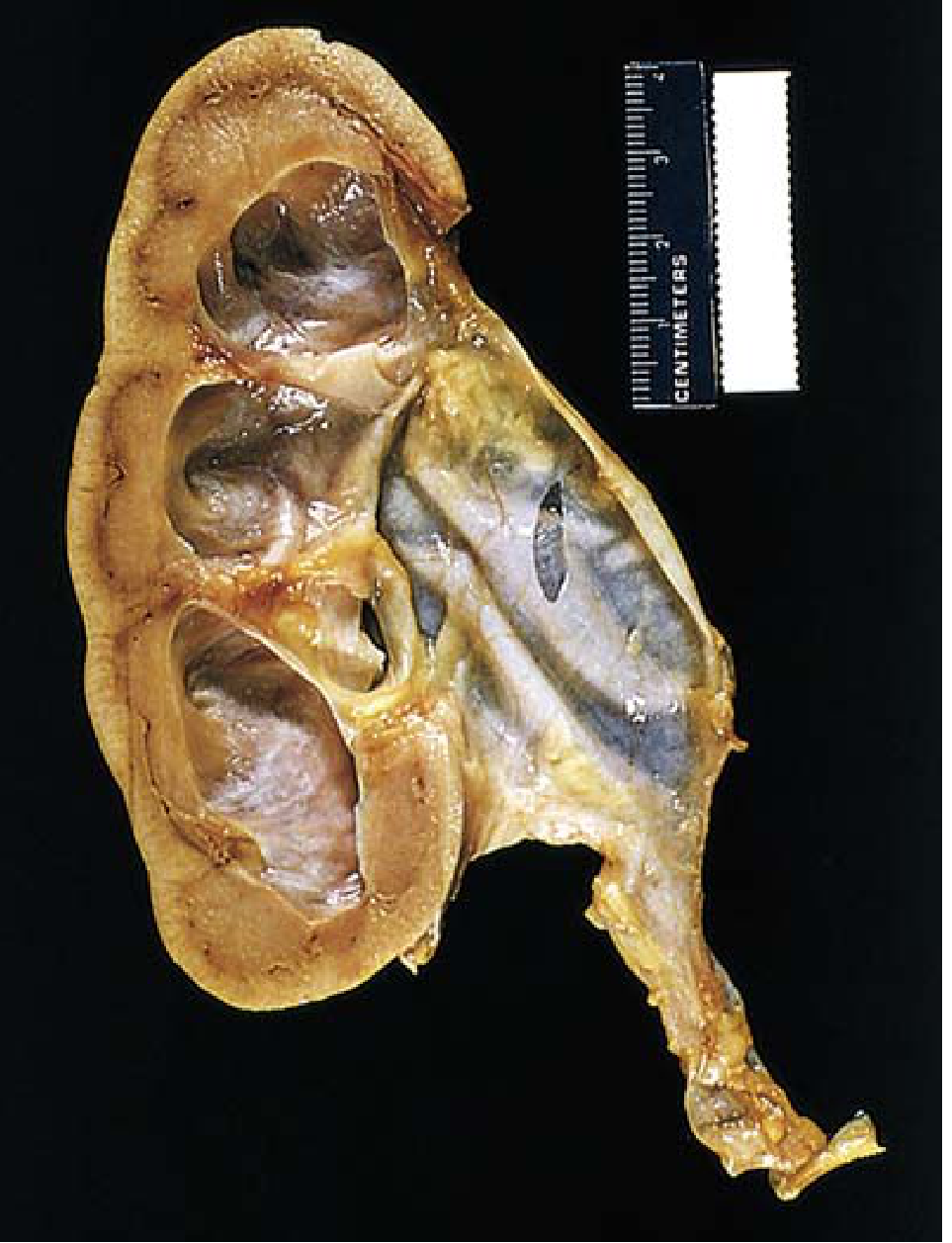
Microscopic pathology:
-
Early: tubular dilation and atrophy
-
Progressive loss of glomeruli
-
Replacement of renal parenchyma by fibrous tissue
-
Minimal inflammatory reaction in uncomplicated cases
-
Superimposed pyelonephritis is common
-
Robbins & Kumar Basic Pathology
CLINICAL FEATURES
Symptoms
- Onset is insidious; females more commonly affected than males
- Dull ache or sense of weight/dragging in the loin
- May be dismissed as mild backache
- If hydronephrosis develops quickly: severe colicky pain
- Pain exacerbated by drinking excess water, alcohol, or taking diuretics
- Referred pain to epigastrium - may mimic duodenal ulcer
- Dietl's crisis: After severe pain, large quantity of urine is passed and swelling reduces in size - due to intermittent obstruction at PUJ being transiently overcome
- Haematuria (if calculus or tumour present)
- Recurrent UTI / pyelonephritis
- In bilateral cases: features of the underlying cause dominate
Signs
- Cystic renal swelling in the loin/flank - smooth, fluctuant
- Ballottable (due to retroperitoneal position)
- Better palpable bimanually (one hand in loin, one anteriorly)
- Moves with respiration
- In advanced bilateral disease: features of chronic renal failure (anuria, uraemia, hypertension)
- Paradox: incomplete bilateral obstruction may cause polyuria (not oliguria) due to tubular concentrating defects
INVESTIGATIONS
1. Ultrasound (USG)
- First-line investigation - least invasive
- Shows dilated pelvicalyceal system
- Confirms cystic nature of renal swelling
- Can detect cause (e.g. calculi)
- Can detect hydronephrosis in utero (antenatal detection of PUJ obstruction)
2. Intravenous Urography (IVP / Excretory Urography)
Confirmatory investigation. Sequential changes:
- Early films may fail to visualize pelvis and calyces (poor contrast excretion)
- Better delineation after 6-hour delayed films
- Earliest change depends on type of renal pelvis:
- Extrarenal pelvis (majority): pelvis itself dilates first
- Intrarenal pelvis: decreasing concavity then flattening of minor calyces
- Progressive dilation of major calyces
- Clubbing (convexity) of minor calyces
- Pelvis becomes markedly distended; its convex lower margin forms an acute angle with the ureter
- If pelvis and calyces not seen properly: Retrograde urography needed
- Sometimes the cause (stone) visible on plain X-ray (KUB)
3. Retrograde Ureteropyelography
- When IVP fails to show pelvis/calyces clearly
- Defines the site and cause of obstruction
4. Diuretic Renography (MAG-3 / DTPA scan)
- Isotope renography detects dilatation of the renal collecting system
- MAG-3 preferred in children and patients with impaired function
- Obstructive pattern: delayed transit on time-activity curve, hold-up of tracer
- Provides differential renal function - critical for management decisions
- Helps distinguish obstructed from non-obstructed dilated systems
5. CT Scan / CT Urography
- Best for defining the cause of obstruction
- Shows level of obstruction
- Useful for retroperitoneal pathology (fibrosis, lymphoma)
6. Whitaker Test
- Used in specialised units
- Percutaneous puncture of kidney to monitor intrapelvic pressure
- Perfusion pressure > 22 cm H2O = significant obstruction
7. VCUG (Voiding Cystourethrogram)
- To detect vesicoureteral reflux (VUR)
- Assess posterior urethral valves in bilateral cases
8. Blood Tests
- Serum creatinine, urea - assess renal function
- FBC - infection markers
- PSA in elderly males
TREATMENT
GENERAL PRINCIPLES
Treatment is directed at:
- Relieving the obstruction
- Preserving/restoring renal function
- Treating infection
- Removing the cause
A. CONSERVATIVE (Watchful Waiting)
- Asymptomatic antenatal/mild hydronephrosis without functional deterioration
- Prenatal fetal hydronephrosis: most mild-to-moderate cases resolve spontaneously after birth
- Postnatal ultrasound at least 48 hours after birth; MAG-3 at 4-6 weeks
- High-risk infants: VCUG, antibiotic prophylaxis, functional scan, monthly follow-up
- Low-risk infants: 1-3 monthly ultrasound follow-up
B. SURGICAL - BASED ON CAUSE
1. PUJ Obstruction (Pyeloplasty)
Indications for surgery:
- Symptomatic patients (pain, UTI, haematuria)
- Obstructive pattern on diuretic renography
- Deterioration of differential renal function
- Progressive hydronephrosis on serial ultrasound
- Renal function < 40% on MAG-3
Anderson-Hynes Dismembered Pyeloplasty (gold standard):
- Transection at the PUJ obstruction
- Excision of the aperistaltic/stenotic segment
- Fashioning a funnel-like, tension-free, dependent anastomosis
- Temporary ureteral stent and drain placed
- Can handle crossing vessels (vessels are transposed behind the anastomosis)
- Success rate ~95%
Foley Y-V Plasty:
- For a high ureteral insertion (not crossing vessel)
- Success rate ~86%
Minimally Invasive Options:
- Laparoscopic pyeloplasty: now the standard in many centres; equal success to open
- Robotic-assisted pyeloplasty: increasing use
- Endopyelotomy (incision of the PUJ endoscopically): less invasive; results inferior to pyeloplasty; best for short segment stricture without a crossing vessel
2. For Ureteric Obstruction
- Ureteric calculus: ESWL, ureteroscopy, PCNL, or open ureterolithotomy
- Ureteric stricture: Balloon dilation, endoscopic incision, ureteric reimplantation, or substitution (ileal ureter)
- Ureterovesical junction (UVJ) obstruction / Megaureter: Ureteric reimplantation
- Retroperitoneal fibrosis: Ureterolysis ± corticosteroids; wrap ureter in omentum
- External compression by tumour: Treat primary tumour; stenting
3. For Bladder Outlet / Lower Urinary Tract Obstruction
- BPH: TURP, open prostatectomy, or medical (alpha-blockers, 5-alpha reductase inhibitors)
- Carcinoma prostate: Hormonal/radiotherapy; TURP for palliation
- Urethral stricture: Urethrotomy, urethroplasty
- Posterior urethral valves (PUV): Transurethral valve ablation
4. Temporary Drainage Procedures
- Nephrostomy (percutaneous): Emergency drainage for infected obstructed kidney, impaired function awaiting definitive surgery, anuria
- Ureteral stenting (JJ stent): Bypasses obstruction; maintains ureteric patency
- Urethral catheter: Initial drainage for bladder outlet obstruction
5. Nephrectomy
Indications:
- Grossly hydronephrotic kidney with <15% differential function (poorly functioning/non-functioning kidney)
- Chronic pyonephrosis
- Recurrent calculi with destroyed parenchyma
- Irreversible renal damage with no salvageable function
- Note: After removal of obstruction, function may return if obstruction duration was short
COMPLICATIONS
- Pyonephrosis - infected hydronephrosis (pus in collecting system) - surgical emergency
- Pyelonephritis - recurrent infection
- Hypertension - due to renal ischaemia (renin-angiotensin activation)
- Renal failure - bilateral hydronephrosis or single kidney involvement
- Rupture - rare; periurinoma / urinoma
- Renal calculi - secondary to stasis and infection
- Irreversible renal damage if obstruction unrelieved
KEY SURGICAL POINTS (Summary Box)
| Feature | Detail |
|---|---|
| Definition | Dilation of calyces + pelvis due to obstruction |
| Commonest cause (unilateral) | Idiopathic PUJ stenosis |
| Commonest cause (bilateral) | BPH / PUV |
| First-line investigation | Ultrasound |
| Confirmatory investigation | IVP (excretory urography) |
| Functional assessment | MAG-3 / DTPA diuretic renography |
| Gold standard surgery for PUJ | Anderson-Hynes dismembered pyeloplasty |
| Dietl's crisis | Intermittent colicky pain + large urine output relieving swelling |
| Emergency drainage | Percutaneous nephrostomy |
| Nephrectomy indication | Differential function <15% |
Sources:
- S Das, A Manual on Clinical Surgery, 13th Edition (primary surgical reference)
- Bailey and Love's Short Practice of Surgery, 28th Edition
- Robbins & Kumar Basic Pathology
- Campbell-Walsh-Wein Urology, 3-Volume Set
- Smith & Tanagho's General Urology, 19th Edition
This is a shared conversation. Sign in to Orris to start your own chat.