I now have everything I need. Let me compile the comprehensive MCQ set with all the material from Dr. Abdelhamid's lectures and textbook cross-references.
🫁 BMC RESPIRATION & CIRCULATION BLOCK
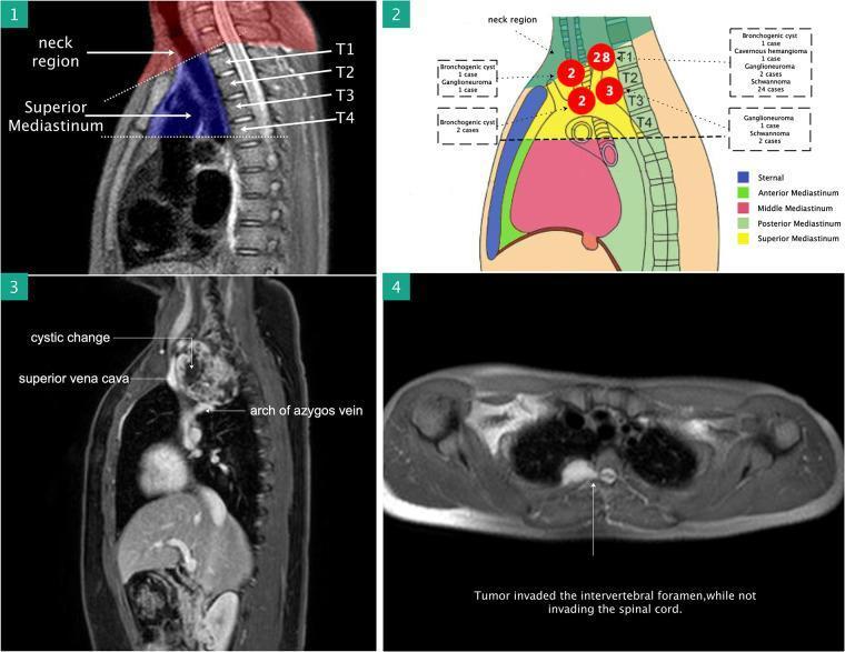
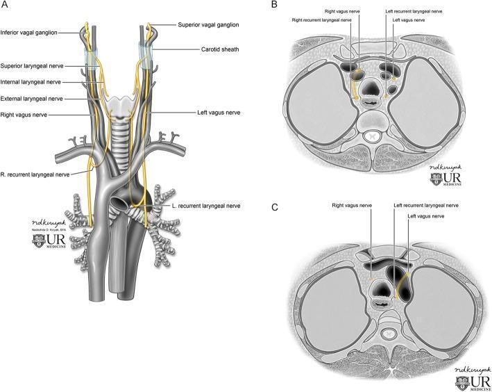
High-Yield Anatomy MCQs — Dr. Abdelhamid Elsify's Objectives
Mediastinum (Parts I & II) + Lung Development
━━━━━━━━━━━━━━━━━━━━━━━━━━━━━━━
🔴 SECTION 1 — MEDIASTINUM BOUNDARIES & DIVISIONS
━━━━━━━━━━━━━━━━━━━━━━━━━━━━━━━
MCQ 1 ⭐⭐⭐ (HIGH FREQUENCY)
The mediastinum is divided into superior and inferior parts by an imaginary plane that passes through which two landmarks?
- A) Manubriosternal joint and lower border of T5
- B) Sternal angle and the disc between T4 and T5 ✅
- C) Jugular notch and the body of T3
- D) Xiphisternal joint and T6 vertebra
- E) Sternal angle and the disc between T3 and T4
Answer: B
Explanation: The plane of the sternal angle (angle of Louis) passes horizontally at the level of the T4–T5 intervertebral disc. This is one of the most tested anatomy landmarks at BMC — it divides the entire mediastinum. Everything ABOVE this line = superior mediastinum. Everything BELOW = inferior mediastinum.
Gray's Anatomy for Students — The horizontal plane passes through the sternal angle and the intervertebral disc between vertebrae TIV and TV.
🩺 Clinical Correlation at BMC:
When you examine a chest X-ray in your clinical years, you'll use the sternal angle as a landmark to identify where the aortic arch begins, where the trachea bifurcates (carina sits near T4–T5), and to localize mediastinal masses. A mediastinal mass ABOVE this line is in the superior mediastinum — common causes include thymoma, retrosternal goiter, and teratoma. On ward rounds, your consultant may ask you to classify a mass based on its location relative to this landmark.
MCQ 2 ⭐⭐⭐ (HIGH FREQUENCY)
Which of the following structures forms the posterior boundary of the superior mediastinum?
- A) Body of sternum
- B) Pericardium
- C) Upper 4 thoracic vertebrae ✅
- D) All 12 thoracic vertebrae
- E) Lower 8 thoracic vertebrae
Answer: C
Explanation: The superior mediastinum is bounded posteriorly by the upper 4 thoracic vertebrae (T1–T4). Contrast this with the posterior mediastinum, which is bounded posteriorly by the lower 8 thoracic vertebrae (T5–T12).
📚 BMC Tip: Dr. Abdelhamid frequently asks "which vertebrae bound which mediastinum." A quick memory trick: Superior = upper 4, Posterior = lower 8.
🩺 Clinical Correlation:
In your internal medicine rotation, superior mediastinal masses (e.g., lymphoma, aortic aneurysm) cause compression of structures at T1–T4 level. You'll see classic "superior mediastinal syndrome" presentations — facial and arm swelling from SVC compression, hoarse voice from compression of the left recurrent laryngeal nerve, and dysphagia from tracheal/esophageal compression.
MCQ 3 ⭐⭐⭐
The anterior mediastinum contains which of the following structures?
- A) Heart and pericardium
- B) Esophagus and thoracic duct
- C) Sternopericardial ligaments, thymic remnants, and a few lymph nodes ✅
- D) Descending thoracic aorta and azygos vein
- E) Phrenic nerves and deep cardiac plexus
Answer: C
Explanation: The anterior mediastinum is the smallest compartment, bounded anteriorly by the sternum body and posteriorly by the pericardium. It contains only sternopericardial ligaments, remnants of the thymus, and a few lymph nodes — essentially an almost empty space.
🩺 Clinical Correlation:
The anterior mediastinum is the most common site for the "4 T's" of anterior mediastinal masses: Thymoma, Teratoma/Germ cell tumor, Thyroid (retrosternal), and "Terrible lymphoma." As a clinical student during your surgery/radiology rotation, whenever you see an anterior mediastinal mass on CT, you'll run through this differential. Thymoma is associated with myasthenia gravis — so if your patient has ptosis + diplopia, check for a mediastinal mass!
MCQ 4 ⭐⭐⭐ (FREQUENTLY EXAMINED)
Which of the following structures is found in the MIDDLE mediastinum?
- A) Esophagus
- B) Thoracic duct
- C) Azygos vein
- D) Phrenic nerve ✅
- E) Thoracic sympathetic chain
Answer: D
Explanation: The middle mediastinum contains the heart within its pericardium, plus: ascending aorta, pulmonary trunk and its branches, SVC (lower ½), IVC (thoracic part), four pulmonary veins, azygos vein termination, right and left main bronchi, phrenic nerves on the sides of the pericardium, and the deep cardiac plexus.
📚 BMC Tip: Remember: phrenic nerves run ON THE SIDES of the pericardium — they are in the middle mediastinum, even though students often place them elsewhere.
🩺 Clinical Correlation:
The phrenic nerve (C3, C4, C5) runs alongside the pericardium — so any pericarditis or cardiac surgery that involves the pericardium can cause phrenic nerve injury, leading to ipsilateral diaphragm paralysis. In your surgery rotation, after cardiac surgery, watch for elevated hemidiaphragm on post-op CXR — this is phrenic nerve palsy. The mnemonic: "C3, 4, 5 keeps the diaphragm alive."
━━━━━━━━━━━━━━━━━━━━━━━━━━━━━━━
🔴 SECTION 2 — SUPERIOR MEDIASTINUM CONTENTS
━━━━━━━━━━━━━━━━━━━━━━━━━━━━━━━
MCQ 5 ⭐⭐⭐ (VERY HIGH FREQUENCY)
Which nerve is unique to the LEFT side of the superior mediastinum (i.e., present on the left but NOT the right)?
- A) Phrenic nerve
- B) Vagus nerve
- C) Left recurrent laryngeal nerve ✅
- D) Greater splanchnic nerve
- E) Cardiac sympathetic nerve
Answer: C
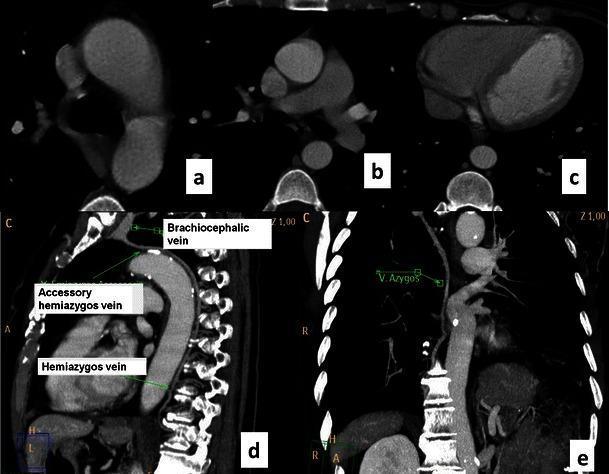
Explanation: The LEFT recurrent laryngeal nerve hooks around the arch of the aorta (specifically around the ligamentum arteriosum) in the superior mediastinum before ascending in the tracheoesophageal groove. The RIGHT recurrent laryngeal nerve hooks around the right subclavian artery in the NECK — it never enters the mediastinum. This asymmetry is extremely high-yield.
🩺 Clinical Correlation:
In your ENT and internal medicine rotations, you'll encounter Ortner's syndrome (cardiovocal syndrome) — hoarseness caused by left RLN compression from an enlarged left atrium (mitral stenosis) or aortic aneurysm. When a patient with mitral stenosis develops a hoarse voice, the enlarged left atrium in the posterior mediastinum stretches the left RLN. Lung cancer in the left upper lobe (near the aortopulmonary window) is the #1 clinical cause of left RLN palsy.
MCQ 6 ⭐⭐⭐
The arch of the aorta gives rise to three branches in the following order (from right to left):
- A) Left subclavian, left common carotid, brachiocephalic artery
- B) Brachiocephalic, left common carotid, left subclavian ✅
- C) Right common carotid, left common carotid, left subclavian
- D) Brachiocephalic, left subclavian, left common carotid
- E) Left common carotid, brachiocephalic, left subclavian
Answer: B
Explanation: The arch of aorta gives off three branches in this sequence: (1) Brachiocephalic artery (divides into right common carotid + right subclavian), (2) Left common carotid artery, (3) Left subclavian artery.
📚 BMC Memory Trick: "Big Lazy Scotsman" — Brachiocephalic, Left carotid, Left Subclavian.
🩺 Clinical Correlation:
Atherosclerosis can affect any of these branches. Subclavian steal syndrome occurs when the left subclavian artery is blocked proximal to the vertebral artery — blood "steals" from the vertebral artery, causing vertebrobasilar insufficiency (dizziness, visual changes) when the left arm is exercised. You'll diagnose this by checking bilateral blood pressures — a >15 mmHg difference between arms is significant.
MCQ 7 ⭐⭐
The superior vena cava (SVC) is formed by union of the right and left brachiocephalic veins. Which part of the SVC is found in the superior mediastinum?
- A) Entire SVC
- B) Lower ½ only
- C) Upper ½ only ✅
- D) The SVC is entirely in the middle mediastinum
- E) Just the very origin
Answer: C
Explanation: The SVC is about 7 cm long. The upper ½ is in the superior mediastinum; the lower ½ (including where the azygos vein drains into it) is in the middle mediastinum. This split is frequently tested.
🩺 Clinical Correlation:
SVC syndrome (obstruction causing facial and arm swelling, venous distension, dyspnea) is commonly caused by lung cancer or lymphoma compressing the SVC in the superior mediastinum. In your clinical years, you'll see patients with puffy face, dilated neck veins, and dilated subcutaneous veins on the chest wall. Treatment is oncological (radiation/chemotherapy) or stenting. Time-sensitive!
MCQ 8 ⭐⭐ (CLINICAL CASE)
A 25-year-old male is found to have a mediastinal mass on CXR. The mass is found to be in the superior mediastinum and is located anterior to the large vessels. What is the most likely structure enlarged?
- A) Esophagus
- B) Thoracic duct
- C) Thymus gland ✅
- D) Trachea
- E) Azygos vein
Answer: C
Explanation: Dr. Abdelhamid's lecture specifies that the thymus gland is located ANTERIOR to the large vessels (arch of aorta and its branches) in the superior mediastinum. It extends from the 4th costal cartilage to the lower poles of the thyroid gland and also occupies the anterior mediastinum. In young adults, thymoma is the #1 anterior/superior mediastinal mass.
🩺 Clinical Correlation:
Thymoma is associated with myasthenia gravis, pure red cell aplasia, and hypogammaglobulinemia. On your neurology rotation, a young patient with ptosis, diplopia, fatigable weakness → order a CT chest to look for thymoma. Thymectomy improves myasthenia gravis in many cases.
━━━━━━━━━━━━━━━━━━━━━━━━━━━━━━━
🔴 SECTION 3 — POSTERIOR MEDIASTINUM CONTENTS
━━━━━━━━━━━━━━━━━━━━━━━━━━━━━━━
MCQ 9 ⭐⭐⭐ (HIGH FREQUENCY)
Which of the following structures is NOT a content of the posterior mediastinum?
- A) Descending thoracic aorta
- B) Esophagus
- C) Thoracic duct
- D) Azygos, hemiazygos, and accessory hemiazygos veins
- E) Pulmonary trunk ✅
Answer: E
Explanation: The pulmonary trunk is in the MIDDLE mediastinum (not posterior). The posterior mediastinum contains: descending thoracic aorta, esophagus, thoracic duct, azygos venous system, posterior mediastinal lymph nodes, and autonomic nerves (vagi and sympathetic chains).
📚 BMC Tip: "DELTA" for posterior mediastinum contents: Descending aorta, Esophagus, Lymphatics (thoracic duct), Thorax azygos veins, Autonomic nerves.
🩺 Clinical Correlation:
Posterior mediastinal masses are most commonly neurogenic tumors (schwannoma, neurofibroma — arise from sympathetic chain). On imaging, they sit in the "paravertebral" location. In neurofibromatosis (café-au-lait spots + neurofibromas), always check for posterior mediastinal masses.
MCQ 10 ⭐⭐⭐ (VERY HIGH FREQUENCY)
The azygos vein arches over which structure to drain into the SVC?
- A) Right main bronchus ✅
- B) Left main bronchus
- C) Right pulmonary artery
- D) Trachea
- E) Aortic arch
Answer: A
Explanation: The azygos vein arches anteriorly over the right main bronchus (and right pulmonary artery root) to drain into the posterior aspect of the SVC at the level of T4.
🩺 Clinical Correlation:
On a chest X-ray, the azygos vein knob is visible at the right tracheobronchial angle (right paratracheal area). It should be <1 cm in normal patients. An enlarged azygos vein (>1 cm) indicates elevated central venous pressure — seen in right heart failure, SVC obstruction, or portal hypertension with collateral drainage. This is a simple CXR finding you'll interpret daily on the wards.
━━━━━━━━━━━━━━━━━━━━━━━━━━━━━━━
🔴 SECTION 4 — ESOPHAGUS (Part II Objectives)
━━━━━━━━━━━━━━━━━━━━━━━━━━━━━━━
MCQ 11 ⭐⭐⭐ (EXTREMELY HIGH FREQUENCY)
The esophagus has 4 normal constrictions. Which is located at 9 inches from the incisor teeth?
- A) Pharyngo-esophageal junction
- B) Where crossed by the aortic arch ✅
- C) Where crossed by the left main bronchus
- D) Where it pierces the diaphragm
- E) At the cardiac end of the stomach
Answer: B
Explanation: The 4 esophageal constrictions from the incisors:
- Pharyngo-esophageal junction → 6 inches
- Crossed by arch of aorta → 9 inches
- Crossed by left main bronchus → 11 inches
- Diaphragmatic hiatus (T10) → 16 inches
📚 Memory Trick: "6 – 9 – 11 – 16" or remember: "People Always Leave During Finals" (Pharyngo, Aortic arch, Left bronchus, Diaphragm).
🩺 Clinical Correlation:
These constrictions are clinically critical! Foreign bodies (in children), food bolus impactions, and carcinomas most commonly occur at these sites because swallowing slows here. During upper GI endoscopy (scope), you report distances from the incisors. Barrett's esophagus and adenocarcinoma occur at the diaphragmatic constriction (gastroesophageal junction). Swallowed batteries or coins most often lodge at 6 inches (cricopharyngeus level) or 9 inches (aortic level).
MCQ 12 ⭐⭐⭐
The esophagus pierces the diaphragm at which vertebral level?
- A) T8
- B) T9
- C) T10 ✅
- D) T11
- E) T12
Answer: C
Explanation: The esophagus pierces the diaphragm at T10, 1 inch to the left of the midline. It ends by joining the stomach at T11. The aorta passes through the diaphragm at T12; the IVC at T8.
📚 BMC Mnemonic: "I 8 (ate) 10 of the 12 eggs" → IVC=T8, Esophagus=T10, Aorta=T12.
🩺 Clinical Correlation:
Hiatus hernia occurs at the esophageal opening (T10). In a sliding hiatus hernia (95%), the gastroesophageal junction slides up into the thorax — causing GERD. In a rolling (paraesophageal) hernia, the fundus rolls up but the GE junction stays below — risk of strangulation. You'll see this on barium swallow studies in your gastroenterology rotation.
MCQ 13 ⭐⭐⭐ (CLINICAL CASE — HIGH YIELD)
A 55-year-old man with known liver cirrhosis presents with sudden massive hematemesis. What is the likely source of bleeding and its anatomical basis?
- A) Duodenal ulcer — erosion of the gastroduodenal artery
- B) Esophageal varices — porto-systemic anastomosis at the lower esophagus ✅
- C) Mallory-Weiss tear — mucosal laceration at GEJ
- D) Gastric cancer — tumor erosion
- E) Aortoenteric fistula — aorta communicating with esophagus
Answer: B
Explanation: The lower esophagus is a site of PORTO-SYSTEMIC ANASTOMOSIS. The left gastric vein (portal system) drains the abdominal esophagus and connects with the azygos/hemiazygos veins (systemic system). In portal hypertension, this anastomosis dilates forming esophageal varices. Rupture causes life-threatening hematemesis.
📚 Sites of porto-systemic anastomosis (BMC High Yield):
- Lower esophagus (left gastric ↔ azygos)
- Umbilicus (paraumbilical ↔ epigastric)
- Anal canal (superior rectal ↔ inferior/middle rectal)
- Bare area of liver (portal ↔ phrenic/intercostal)
🩺 Clinical Correlation:
In your GI/surgery rotation, you'll manage variceal bleeds with terlipressin, Sengstaken-Blakemore tube, band ligation, or TIPS procedure. The anatomy of the anastomosis is essential for understanding WHY portal hypertension causes upper GI bleeding and NOT lower GI bleeding (varices are at the esophagus and upper rectum, not throughout the bowel).
MCQ 14 ⭐⭐
Enlargement of the LEFT ATRIUM (e.g., in mitral stenosis) would most likely compress which anterior relation of the esophagus?
- A) Trachea
- B) Left recurrent laryngeal nerve
- C) Left main bronchus
- D) Pericardium and left atrium — shown on barium swallow ✅
- E) Right pulmonary artery
Answer: D
Explanation: The anterior relations of the thoracic esophagus include (from above down): trachea → left RLN → left main bronchus → pericardium + left atrium. An enlarged left atrium pushes anteriorly on the pericardium and POSTERIORLY on the esophagus, creating a posterior indentation visible on barium swallow.
🩺 Clinical Correlation:
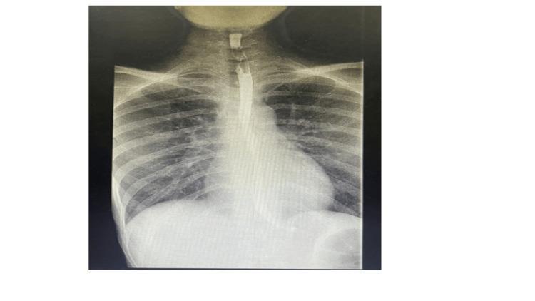
In cardiology rotation, a barium swallow is an OLD clinical sign for mitral stenosis. You'll see a posterior deviation/indentation of the esophagus on the lateral view of a barium swallow when the left atrium is massively enlarged. This is also why mitral stenosis causes dysphagia (difficulty swallowing).
MCQ 15 ⭐⭐
The thoracic duct crosses from the right side to the left side of the posterior mediastinum at which vertebral level?
- A) T4
- B) T5 ✅
- C) T7
- D) T8
- E) T10
Answer: B
Explanation: The thoracic duct enters the thorax at T12, ascends on the RIGHT side through the posterior mediastinum, then at the level of T5, crosses to the LEFT side. It continues up through the superior mediastinum on the left of the esophagus and eventually drains into the junction of the left subclavian and left internal jugular veins.
📚 BMC Tip: Right from T12 to T5 → crosses → Left from T5 to neck. Drains into LEFT venous angle.
🩺 Clinical Correlation:
Chylothorax (milky pleural fluid rich in chylomicrons) results from thoracic duct injury — most commonly from surgery (esophagectomy, cardiac surgery) or trauma. Because the duct crosses from right to left at T5, injury BELOW T5 causes a right chylothorax; injury ABOVE T5 causes a left chylothorax. On pleural fluid analysis, triglycerides >110 mg/dL confirms chylothorax.
━━━━━━━━━━━━━━━━━━━━━━━━━━━━━━━
🔴 SECTION 5 — THYMUS
━━━━━━━━━━━━━━━━━━━━━━━━━━━━━━━
MCQ 16 ⭐⭐
Which of the following best describes the blood supply of the thymus gland?
- A) Subclavian and aortic arch arteries
- B) Inferior thyroid artery and internal thoracic artery ✅
- C) Brachiocephalic and internal carotid arteries
- D) Superior thyroid and common carotid arteries
- E) Intercostal and phrenic arteries
Answer: B
Explanation: The thymus receives blood from (1) the inferior thyroid artery and (2) the internal thoracic (mammary) artery. The thymus gland does NOT receive afferent lymphatics (unique among lymphoid organs — antigen-naive T cells enter via blood, not lymphatics). Efferent lymphatics drain to parasternal, brachiocephalic, and tracheobronchial lymph nodes.
🩺 Clinical Correlation:
In cardiothoracic surgery, the internal thoracic (mammary) artery is harvested for coronary artery bypass grafting (CABG). It's the most durable graft for the LAD artery. Knowing this artery supplies the thymus (and the anterior chest wall) is important — patients who've had CABG may have altered blood supply to these structures.
━━━━━━━━━━━━━━━━━━━━━━━━━━━━━━━
🔴 SECTION 6 — LUNG DEVELOPMENT (HIGH FREQUENCY)
━━━━━━━━━━━━━━━━━━━━━━━━━━━━━━━
MCQ 17 ⭐⭐⭐ (EXTREMELY HIGH FREQUENCY)
The respiratory diverticulum appears as an outgrowth from the ventral wall of the foregut at approximately which week of embryonic development?
- A) 3 weeks
- B) 4 weeks ✅
- C) 5 weeks
- D) 6 weeks
- E) 8 weeks
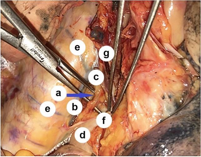
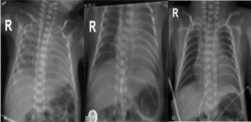
Answer: B
Explanation: At approximately week 4, the respiratory diverticulum appears as a ventral outgrowth from the foregut. The tracheoesophageal ridges then fuse in a caudo-cranial direction to form the tracheoesophageal septum, dividing the foregut into:
- Dorsal: Esophagus
- Ventral: Trachea + lung buds
🩺 Clinical Correlation:
This developmental step is the basis for TEF (tracheoesophageal fistula). If the ridges fail to fuse properly, abnormal connections form between the trachea and esophagus. You'll encounter TEF in your first on-call night as a neonatology intern — baby with excessive frothy saliva, cyanosis during feeds, and failure to pass an NG tube.
MCQ 18 ⭐⭐⭐ (CASE PROBLEM FROM DR. ABDELHAMID'S SLIDES)
A 5-year-old girl comes for a routine well-child examination and is normal. Which of the following structures would continue to INCREASE IN NUMBER in this child?
- A) Skeletal myocytes
- B) Nephrons
- C) Brown adipocytes
- D) Alveoli ✅
- E) Oocytes
Answer: D
Explanation (this case is directly from Dr. Abdelhamid's slides!): Alveoli continue to multiply from birth until approximately 8–10 years of age. At birth, only 1/6 of adult alveoli are present (~50 million). The remaining 5/6 (~300 million) develop postnatally. No other structure listed undergoes postnatal numerical increase:
- Nephrons: formed by week 36, no new ones after birth
- Oocytes: maximum at 20 weeks gestation, decline afterward
- Skeletal myocytes: enlarge (hypertrophy) but don't increase in number significantly
- Brown fat: decreases after birth
🩺 Clinical Correlation:
This is why premature babies who survive may have long-term lung problems — their alveoli haven't fully formed. In your pediatrics rotation, bronchopulmonary dysplasia (BPD) in ex-premature infants means abnormal alveolar development. Giving supplemental oxygen can impair alveolar development (oxygen toxicity). Pediatric pulmonologists track alveolar development; exercise capacity in these children may be reduced.
MCQ 19 ⭐⭐⭐ (VERY HIGH FREQUENCY)
Surfactant is produced by which cell type, and adequate levels are reached at which month of gestation?
- A) Type I pneumocytes; 5th month
- B) Type II pneumocytes; 7th month ✅
- C) Clara cells; 6th month
- D) Type I pneumocytes; 7th month
- E) Type II pneumocytes; 5th month
Answer: B
Explanation: Type II alveolar epithelial cells (pneumocytes) develop by the end of the 6th month and produce surfactant (a phospholipid-rich fluid). Adequate levels are reached by the 7th month (28 weeks). Surfactant lowers alveolar surface tension, preventing collapse (atelectasis) during expiration.
📚 Important facts:
- Type I pneumocytes → line the alveolus (thin, flat, gas exchange)
- Type II pneumocytes → produce surfactant, can regenerate Type I cells
- Surfactant composition: mainly dipalmitoylphosphatidylcholine (DPPC)
🩺 Clinical Correlation:
Babies born before 28 weeks are at high risk for RDS (formerly called hyaline membrane disease). Treatment:
- Maternal corticosteroids (betamethasone/dexamethasone) → given to mother if preterm labor expected → stimulates fetal surfactant production
- Exogenous surfactant (beractant, poractant) → given directly into trachea of premature newborn
In your neonatology rotation, RDS presents as increasing respiratory effort within hours of birth, with classic "ground glass" appearance on CXR.
MCQ 20 ⭐⭐⭐
A premature infant born at 26 weeks develops progressive respiratory distress within hours of birth. Chest X-ray shows diffuse bilateral ground-glass opacities. What is the immediate pathophysiological mechanism?
- A) Meconium aspiration obstructing airways
- B) Insufficient surfactant causing alveolar collapse during expiration ✅
- C) Cardiac defect causing pulmonary edema
- D) Group B Streptococcus pneumonia
- E) Diaphragmatic hernia with bowel in thorax
Answer: B
Explanation: RDS (respiratory distress syndrome) is caused by surfactant deficiency in premature infants. Without surfactant, surface tension in alveoli cannot be overcome during expiration → alveolar collapse → atelectasis → V/Q mismatch → hypoxia → acidosis. The collapsed alveoli filled with proteinaceous fluid form hyaline membranes (seen on histology).
🩺 Clinical Correlation:
Management of RDS:
- CPAP (continuous positive airway pressure) → keeps alveoli open
- Surfactant replacement therapy
- Oxygen supplementation (carefully — too much O₂ causes retinopathy of prematurity, BPD)
- Prevent preterm birth with maternal steroids
MCQ 21 ⭐⭐⭐ (STAGES — VERY HIGH FREQUENCY)
During which stage of lung maturation does a fetus first become viable (can survive with intensive care support)?
- A) Pseudoglandular period (up to 4th month)
- B) Canalicular period (4–6 months)
- C) Terminal sac (saccular) period (6 months–birth) ✅
- D) Alveolar period (late prenatal–8 years)
- E) At birth, all stages are complete
Answer: C
Explanation: The 4 stages of lung maturation:
| Stage | Timing | Key Feature | Viability |
|---|
| Pseudoglandular | Up to 4th month | Terminal bronchioles only, no alveoli | NOT viable |
| Canalicular | 4–6 months | Respiratory bronchioles + alveolar ducts form | NOT viable (dies even in ICU) |
| Terminal sac | 6 months–birth | Primitive alveoli form; surfactant starts at 7 months | VIABLE with ICU |
| Alveolar | Late prenatal–8 years | Mature alveoli | Viable |
🩺 Clinical Correlation:
The limit of viability is generally 23–24 weeks in modern NICUs. Before the terminal sac stage, there is no gas exchange surface, so survival is impossible regardless of intervention. After 28 weeks (adequate surfactant), survival rates exceed 90% in well-equipped NICUs. This is important in obstetrics and neonatology for counseling parents with preterm labor.
MCQ 22 ⭐⭐⭐ (VERY HIGH FREQUENCY — FROM DR. ABDELHAMID'S SLIDES)
Most common type of tracheoesophageal fistula (TEF), accounting for approximately 90% of cases, is:
- A) Isolated esophageal atresia (type B)
- B) H-type fistula without esophageal atresia (type C)
- C) Upper esophageal blind pouch + lower segment fistula with trachea (type A) ✅
- D) Double fistula above and below atresia
- E) Fistula with normal esophagus
Answer: C
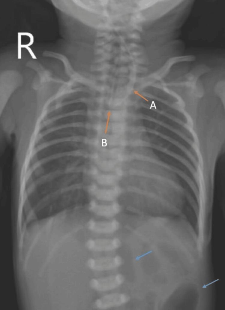
Explanation: Type A TEF (90%) = Upper esophageal segment ends in a BLIND POUCH + lower segment forms a FISTULA with the trachea. This means air enters the stomach from the trachea via the fistula (seen as bowel gas on X-ray), but food cannot reach the stomach (proximal pouch is blind).
Incidence: ~1 in 3,000 births.
Other types:
- Type B (4%): isolated esophageal atresia, NO fistula → no bowel gas on X-ray
- Type C (4%): H-type (no atresia) — presents late with recurrent chest infections
🩺 Clinical Correlation:
Classic presentation: neonate with excessive frothy saliva (can't swallow), choking/cyanosis during first feed, NG tube cannot be passed (coils in proximal pouch on X-ray). Diagnosis: NG tube coiled on CXR + abdominal gas (confirms fistula). Treatment: SURGICAL — primary esophageal anastomosis + fistula ligation. Associated with VACTERL syndrome (Vertebral, Anorectal, Cardiac, Tracheoesophageal, Renal, Limb anomalies).
MCQ 23 ⭐⭐
Fetal breathing movements begin before birth and are important for:
- A) Delivering oxygen to the fetus
- B) Stimulating lung development and conditioning respiratory muscles ✅
- C) Pumping amniotic fluid away from the lungs
- D) Establishing the surfactant gradient
- E) Preventing polyhydramnios
Answer: B
Explanation: Fetal breathing movements (detectable on ultrasound) cause aspiration of amniotic fluid into the airways. These movements are important for:
- Stimulating lung growth and development
- Conditioning (training) respiratory muscles for birth
- Distributing surfactant on alveolar membranes as fluid is reabsorbed
🩺 Clinical Correlation:
On biophysical profile (BPP) in obstetrics, fetal breathing movements are one of the 5 parameters scored. Absent breathing movements for >30 minutes in a 30-minute observation = score of 0 (abnormal) → indicator of fetal compromise. Polyhydramnios occurs in TEF type B (no fistula) or other swallowing disorders because the fetus CANNOT swallow amniotic fluid.
MCQ 24 ⭐⭐ (EMBRYOLOGY — TISSUE DERIVATIVES)
The epithelial lining of the larynx, trachea, bronchi, and alveoli is derived from:
- A) Ectoderm
- B) Somatic mesoderm
- C) Splanchnic mesoderm
- D) Neural crest cells
- E) Endoderm ✅
Answer: E
Explanation: The EPITHELIUM of the entire respiratory tract (from larynx to alveoli) is endodermal origin (from the foregut). The cartilage, smooth muscle, and connective tissue of the tracheobronchial tree come from SPLANCHNIC MESODERM surrounding the foregut. The visceral pleura comes from splanchnic mesoderm; the parietal pleura from somatic mesoderm.
🩺 Clinical Correlation:
Understanding germ layer origins helps with tumor histology. Respiratory tract carcinomas are carcinomas (epithelial tumors) because the mucosa is endodermal. Mesothelioma arises from the mesothelium (mesodermal) of the pleura — caused by asbestos. Distinguishing these on histology (adenocarcinoma vs. squamous cell vs. mesothelioma) is crucial in oncology.
MCQ 25 ⭐⭐⭐ (CASE PROBLEM — FROM DR. ABDELHAMID'S SLIDES)
A 61-year-old man presents with a 5-month history of shooting pain in both legs. Twenty years ago, he had a painless penile ulcer that resolved on its own. Examination shows pupils that do NOT react to light but DO constrict with accommodation (Argyll Robertson pupils). Reflexes are absent bilaterally with broad-based gait. This patient is at increased risk for which complication?
- A) Atrioventricular block
- B) Mitral valve regurgitation
- C) Penile squamous cell carcinoma
- D) Cerebral artery septic embolism
- E) Thoracic aortic aneurysm ✅
Answer: F (E in original) — Thoracic aortic aneurysm
Explanation: This patient has tertiary syphilis (tabes dorsalis):
- Painless penile ulcer 20 years ago = primary syphilis (chancre)
- Now: Argyll Robertson pupils (light-near dissociation), absent patellar reflexes, broad-based gait, lightning pains → tabes dorsalis
- Tertiary syphilis also causes syphilitic aortitis → endarteritis obliterans of the vasa vasorum of the ascending/transverse aorta → weakening of aortic wall → aneurysm
- Location: predominantly ascending aorta and aortic arch
🩺 Clinical Correlation:
Syphilitic aortic aneurysm (unlike atherosclerotic which is typically infrarenal abdominal) affects the ascending aorta and arch. This gives the classic "egg-shell calcification" of the ascending aorta on CXR and can cause aortic regurgitation. Always test for syphilis (RPR/VDRL + TPHA) in patients with unexplained ascending aortic aneurysm.
━━━━━━━━━━━━━━━━━━━━━━━━━━━━━━━
🔴 SECTION 7 — PLEURA & CLINICAL CORRELATES
━━━━━━━━━━━━━━━━━━━━━━━━━━━━━━━
MCQ 26 ⭐⭐⭐
The visceral and parietal pleura are derived from which embryological layer respectively?
- A) Ectoderm and endoderm
- B) Splanchnic mesoderm and somatic mesoderm ✅
- C) Somatic mesoderm and splanchnic mesoderm
- D) Endoderm and splanchnic mesoderm
- E) Neural crest and somatic mesoderm
Answer: B
Explanation: The intraembryonic coelom (pericardioperitoneal canals) becomes the pleural cavities. The mesoderm covering the outside of the lung (splanchnic/visceral mesoderm) = visceral pleura. The somatic mesoderm lining the body wall = parietal pleura.
🩺 Clinical Correlation:
The parietal pleura has pain receptors (somatic sensory) — so parietal pleuritis is PAINFUL and localised. The visceral pleura has NO pain fibers — so visceral pleuritis or pneumothorax from visceral pleura rupture is NOT immediately painful. This distinction matters when a spontaneous pneumothorax patient describes sudden chest pain — the pain comes from parietal pleural irritation by air.
MCQ 27 ⭐⭐
The costodiaphragmatic recess is clinically important because:
- A) It is the entry point for the thoracic duct
- B) It is a potential space that accommodates fluid accumulation and lung expansion ✅
- C) It is where the phrenic nerve enters the diaphragm
- D) It is the narrowest part of the pleural cavity
- E) It forms the posterior mediastinal boundary
Answer: B
Explanation: The costodiaphragmatic recess is the largest pleural recess, found inferiorly between the thoracic wall and diaphragm. It does not normally contain lung — it accommodates lung expansion during deep inspiration and is the first place pleural effusions (fluid) accumulate due to gravity.
🩺 Clinical Correlation:
On a chest X-ray, the first visible sign of pleural effusion is blunting of the costophrenic angle (costodiaphragmatic recess). Approximately 200–300 mL of fluid must accumulate before blunting appears on PA CXR. On lateral decubitus views, as little as 50 mL is detectable. Thoracocentesis is performed in the costodiaphragmatic recess (usually at the 9th intercostal space, mid-scapular line).
━━━━━━━━━━━━━━━━━━━━━━━━━━━━━━━━━━━━━━━━━━━━━━
🏆 BMC HIGH-YIELD CHEATSHEET
Respiration & Circulation Block — Anatomy of Mediastinum + Lung Development
━━━━━━━━━━━━━━━━━━━━━━━━━━━━━━━━━━━━━━━━━━━━━━
📌 MEDIASTINUM DIVISIONS AT A GLANCE
STERNAL ANGLE (T4–T5 disc) = THE DIVIDING LINE

SUPERIOR MEDIASTINUM (above the line):
┌─────────────────────────────────────────────────────┐
│ Tubes: Trachea | Esophagus | Thoracic duct          │
│ Arteries: Arch of Aorta + 3 branches                │
│ Veins: Brachiocephalic R&L | SVC upper ½            │
│ Nerves: Vagus (R&L) | Left RLN | Phrenic (R&L)     │
│         Superficial cardiac plexus                   │
│ Lymphoid: Thymus (anterior to vessels)              │
└─────────────────────────────────────────────────────┘

INFERIOR MEDIASTINUM (below the line):
┌──────────────────────────────────────────────────────────┐
│ ANTERIOR: sternopericardial ligs, thymic remnants, LNs  │
│ MIDDLE: Heart+pericardium, Ascending aorta, Pulmonary   │
│         trunk, SVC lower½, IVC, 4 pulmonary veins,      │
│         Main bronchi, Phrenic nerves, Deep cardiac plexus│
│ POSTERIOR: Descending aorta, Esophagus, Thoracic duct,  │
│            Azygos system, Sympathetic chains, Vagi      │
└──────────────────────────────────────────────────────────┘
📌 THE "MAGIC NUMBERS" — MOST TESTED FACTS
| Landmark | Level / Distance |
|---|
| Esophagus BEGINS | C6 (lower border cricoid) |
| Esophagus CONSTRICTION 1 | 6 inches from incisors (pharyngo-esophageal) |
| Esophagus CONSTRICTION 2 | 9 inches (aortic arch) |
| Esophagus CONSTRICTION 3 | 11 inches (left main bronchus) |
| Esophagus CONSTRICTION 4 / DIAPHRAGM | 16 inches / T10 |
| Esophagus ENDS | T11 (cardiac end of stomach) |
| Thoracic duct BEGINS | T12 (cisterna chyli) |
| Thoracic duct CROSSES midline | T5 (right → left) |
| Thoracic duct DRAINS | Left venous angle (subclavian + IJV) |
| Azygos vein ARCHES over | Right main bronchus → SVC |
| IVC pierces diaphragm | T8 |
| Esophagus pierces diaphragm | T10 |
| Aorta passes through diaphragm | T12 |
| Mediastinum dividing plane | Sternal angle + T4/T5 disc |
📌 LUNG DEVELOPMENT STAGES — EXAM TABLE
| Stage | Timing | Key Events | Viability |
|---|
| Pseudoglandular | Wk 5 – Month 4 | Terminal bronchioles only; looks like gland | ❌ None |
| Canalicular | Month 4–6 | Respiratory bronchioles + alveolar ducts; vascularisation begins | ❌ None (dies in ICU) |
| Terminal sac | Month 6 – birth | Primitive alveoli; Type II cells appear; surfactant starts ~28wks | ✅ With ICU |
| Alveolar | Late fetal – 8 yrs | Mature alveoli; only 1/6 present at birth | ✅ |
📌 CRITICAL EMBRYOLOGY FACTS
- Week 4: Respiratory diverticulum appears from ventral foregut
- Endoderm → epithelium of ALL respiratory tract (larynx to alveoli)
- Splanchnic mesoderm → cartilage, smooth muscle, CT of tracheobronchial tree; visceral pleura
- Somatic mesoderm → parietal pleura
- TEF most common type: Proximal blind pouch + distal fistula with trachea = 90% of cases
- TEF H-type: No atresia, just fistula = presents LATE with recurrent chest infections
- Polyhydramnios in TEF type B (isolated atresia, NO fistula) → fetus can't swallow
- Surfactant: Type II pneumocytes, adequate by 28 weeks (7th month)
- RDS treatment: maternal corticosteroids + exogenous surfactant
- Alveoli at birth: Only 1/6 of adult number; rest develop by age 8–10 years
📌 LEFT vs RIGHT ASYMMETRY — MOST TESTED
| Feature | LEFT | RIGHT |
|---|
| Recurrent laryngeal nerve hooks under | Aortic arch (in mediastinum) | Subclavian artery (in neck) |
| Lung lobes | 2 lobes | 3 lobes |
| Secondary bronchi | 2 | 3 |
| Main bronchus angle | More horizontal, longer | More vertical, shorter, wider |
| Pulmonary emboli more common in | — | Right (more vertical) |
| Thoracic duct side (above T5) | LEFT side | — |
📌 CLINICAL SYNDROME CHEATSHEET
| Syndrome | Cause | Anatomy Basis |
|---|
| Mediastinal syndrome | Mass compressing structures | SVC (engorgement), trachea (dyspnea), esophagus (dysphagia), L-RLN (dysphonia), phrenic nerve (diaphragm paralysis) |
| SVC syndrome | Lung cancer/lymphoma | SVC in superior mediastinum |
| Ortner's syndrome | Mitral stenosis enlarging L. atrium | L. atrium compresses L-RLN |
| Esophageal varices | Portal hypertension | Porto-systemic anastomosis at lower esophagus |
| RDS | Surfactant deficiency | Premature birth before 28 weeks |
| Chylothorax | Thoracic duct injury | Right-sided if below T5; left-sided if above T5 |
| Hiatus hernia | GE junction moves up | Esophageal opening T10 |
| TEF | Failed TE septum formation | Week 4 of development |
📌 NERVE SUPPLY — QUICK REFERENCE
| Nerve | Location | Clinical Consequence if Damaged |
|---|
| Left RLN | Loops under aortic arch | Hoarseness, bovine cough |
| Right RLN | Loops under R. subclavian (neck) | Hoarseness |
| Phrenic nerve (C3,4,5) | Sides of pericardium (middle mediastinum) | Diaphragm paralysis |
| Vagus (both) | Posterior mediastinum → esophageal plexus | Affects esophageal motility |
| Sympathetic chain | Posterior mediastinum | Horner syndrome if T1 affected |
━━━━━━━━━━━━━━━━━━━━━━━━━━━━━━━━━━
🎯 TOP TIPS TO ACE THE BMC EXAM
━━━━━━━━━━━━━━━━━━━━━━━━━━━━━━━━━━
1. Master the "3 Diaphragm Holes" (T8-T10-T12)
Every MCQ about anatomical levels tests this. Write it 10 times: IVC=T8, Esophagus=T10, Aorta=T12.
2. Own the Sternal Angle
The sternal angle = T4/T5 disc = carina level = aortic arch level = SVC begins. One landmark, 4 facts.
3. The 4 Esophageal Constrictions
"6-9-11-16" — every year this comes up in some form, whether as direct distance questions or clinical foreign body/endoscopy scenarios.
4. Left vs Right Asymmetry in Everything
RLN hooks: Left=aorta, Right=subclavian. Bronchi angle: Right more vertical. Thoracic duct: Right below T5, Left above T5. These generate multiple MCQs per year.
5. Lung Development Stages — Know ALL 4 with timings and viability
The pseudoglandular, canalicular, terminal sac, and alveolar stages appear constantly. The "1/6 alveoli at birth" and "8 years for full adult number" facts are repeated across every paper Dr. Abdelhamid releases.
6. TEF = 90% Type A
Blind pouch + distal fistula = 90%. Know this alongside VACTERL association.
7. Surfactant = Type II, adequate at 28 weeks
Treatment = maternal steroids + exogenous surfactant. The case problem in the slides IS in the exam.
8. Both Case Problems in Dr. Abdelhamid's slides ARE exam questions
The 5-year-old alveoli case (Answer: D) and the 61-year-old syphilis case (Answer: thoracic aortic aneurysm) have appeared directly in previous BMC assessments. Memorize both completely.
9. Clinical Correlates Slides = Bonus marks
Dr. Abdelhamid consistently takes MCQ scenarios from his "Clinical Correlates" slides: mediastinal syndrome, esophageal varices, TEF, left atrial enlargement on barium swallow, RDS. Read them carefully.
10. Study the Contents of Each Mediastinum Using Tables
Make a table with 4 columns: Superior, Anterior, Middle, Posterior. Fill in all contents. Dr. Abdelhamid tests which structure belongs where — especially phrenic nerve (middle!), thoracic duct (posterior + superior), thymus (superior + anterior).
Sources: Dr. Abdelhamid Elsify (BMC 2025-2026 lecture PDFs) | Gray's Anatomy for Students, 4th Ed. | Clinically Oriented Anatomy, Moore & Dalley | Langman's Medical Embryology, 14th Ed.Here's everything you need for your BMC Respiration & Circulation block. Here's a summary of what was generated:
What's Included
27 High-Yield MCQs organized into 7 sections, pulled directly from Dr. Abdelhamid Elsify's 2025–2026 lecture objectives:
| Section | MCQs | Topics |
|---|
| 1 | MCQ 1–4 | Mediastinum boundaries & divisions |
| 2 | MCQ 5–8 | Superior mediastinum contents |
| 3 | MCQ 9–10 | Posterior mediastinum |
| 4 | MCQ 11–15 | Esophagus (all 4 constrictions, relations, varices) |
| 5 | MCQ 16 | Thymus |
| 6 | MCQ 17–25 | Lung development (all stages, TEF, RDS, surfactant) |
| 7 | MCQ 26–27 | Pleura & costodiaphragmatic recess |
Each MCQ includes:
- ✅ Correct answer with full explanation
- 🔬 Medical textbook reference (Gray's Anatomy for Students)
- 🩺 Plain-English clinical correlation for your clinical years
- 📸 Relevant medical image where applicable
High-Yield Cheatsheet covers:
- All mediastinum contents in a structured table
- The "magic numbers" (distances, vertebral levels)
- Lung development stage table with viability
- Left vs Right asymmetry comparison table
- Clinical syndrome cheatsheet
- 10 targeted exam tips including the two exact case problems from Dr. Abdelhamid's slides that have appeared in past BMC exams
Highest frequency topics (appears every year based on slide emphasis): Sternal angle landmark, 4 esophageal constrictions, TEF 90% type, surfactant at 28 weeks, lung stages viability, both case problems from the slides.