
Swelling over right lower back differential diagnosis?
Reading File
Swelling over the right lower back (lumbar/loin region) can arise from several structures - skin/subcutaneous tissue, musculoskeletal, retroperitoneal organs (especially the kidney), or referred pathology. Here's a broad overview while I search the textbooks.
Searching sources...
Finding Sources
Finding Sources
Searching the Web
differential diagnosis swelling right lower back loin region surgery
Finding Sources
Finding Sources
Finding Sources
Finding Sources
Finding Sources
Finding Sources
Reading File
Finding Sources
Here is a systematic differential diagnosis for swelling over the right lower back (lumbar/loin region), organized by anatomical layer and category:
Differential Diagnosis: Swelling Over the Right Lower Back (Loin/Lumbar Region)
The right lower back / loin region contains skin, subcutaneous fat, muscles, the lumbar vertebrae and paraspinal structures, and is overlying the retroperitoneal space where the right kidney, ureter, and major vessels reside. Differentials are best classified by layer/origin.
1. Skin & Subcutaneous Tissue
| Condition | Key Features |
|---|---|
| Sebaceous / epidermal cyst | Superficial, fluctuant, punctum visible, non-transilluminant |
| Lipoma | Soft, lobulated, non-tender, slips under fingers ("slip sign") |
| Abscess (pyogenic) | Hot, tender, fluctuant, erythematous; associated fever |
| Carbuncle / furuncle | Skin infection, multiple pustules, very tender |
| Fibroma / other benign soft tissue tumour | Firm, discrete, non-tender |
2. Musculoskeletal / Paraspinal
| Condition | Key Features |
|---|---|
| Lumbar hernia (Petit's / Grynfeltt triangle) | Reducible, expansile cough impulse, through lumbar triangles; rare |
| Haematoma of paraspinal muscle | Post-trauma, tender, fluctuant; ecchymosis may be present |
| Muscle tear / torn ligament | Tender, no discrete swelling; acute onset with activity |
| Cold abscess (tuberculous) | Soft, fluctuant, non-tender (hence "cold"), no erythema, impulse on coughing; arises from caries spine (Pott's disease) and tracks along psoas or directly to lumbar region - S Das Manual of Clinical Surgery, p.298 |
| Meningocele / spina bifida | Midline, soft, transilluminant; impulse on crying/coughing; usually lumbosacral |
| Spinal tumour / osteosarcoma | Hard, fixed, may have neurological signs |
| Aneurysmal bone cyst / osteomyelitis | Usually presents in younger patients, tender, bony swelling |
3. Retroperitoneal / Renal Origin
These present as a deep loin mass, bimanually palpable (ballotable), moving with respiration.
| Condition | Key Features |
|---|---|
| Renal cell carcinoma (RCC) | Classic triad: haematuria, loin pain, loin mass; weight loss, fever; commonest cause of solid renal mass |
| Hydronephrosis | Large, ballotable, smooth; resonant on percussion (bowel anterior); may have flank pain |
| Polycystic kidney disease | Bilateral, irregular masses; family history; associated with hypertension, CRF |
| Wilms' tumour (Nephroblastoma) | Children < 5 years; large, smooth, does not cross midline |
| Perinephric / renal abscess | Tender, fever, rigors; often secondary to pyelonephritis or haematogenous spread |
| Angiomyolipoma | Benign, associated with tuberous sclerosis; may present with flank mass |
| Renal cyst (simple or complex) | Smooth, fluctuant; often incidental; confirmed by ultrasound |
| Renal vein thrombosis | Acute: loin/flank pain, swelling; associated nephrotic syndrome - Comprehensive Clinical Nephrology, 7th Ed |
| Adrenal tumour (phaeochromocytoma, adrenocortical carcinoma) | Upper pole; hypertension (paroxysmal in phaeochromocytoma) |
| Retroperitoneal sarcoma / lymphoma | Large, deep, ill-defined; systemic B symptoms in lymphoma |
4. Vascular
| Condition | Key Features |
|---|---|
| Aortic aneurysm (right extension) | Pulsatile, expansile mass; older male; bruit on auscultation |
| Psoas haematoma | Post-anticoagulation or trauma; tender, may track to groin; associated with hip flexion deformity |
5. Referred / Miscellaneous
| Condition | Key Features |
|---|---|
| Psoas abscess (secondary) | Originates from vertebral TB or Crohn's; presents as loin/iliac fossa mass; no local heat ("cold") |
| Ruptured Baker's cyst (unlikely at this site) | - |
| Ectopic ureterocele | Rare; paediatric presentation |
Key Distinguishing Clinical Points
- Impulse on coughing - present in lumbar hernia and cold abscess (meningocele also)
- Ballotability - bimanually palpable, ballotable mass = renal origin
- Transillumination - positive in cysts and meningocele
- Temperature - hot abscess = pyogenic; cold, non-tender = TB cold abscess
- Reducibility - reducible = hernia
- Haematuria - renal tumour, polycystic kidneys, renal vein thrombosis
- Age - Wilms' tumour (children); RCC (adults > 50); lipoma/hernia (any age)
- Associated spinal signs (kyphosis, gibbus deformity, neurological deficit) - Pott's disease with cold abscess
High-Priority Diagnoses Not to Miss
- Renal cell carcinoma (especially if > 40 years, haematuria, weight loss)
- Pott's disease with cold abscess (especially in TB-endemic regions)
- Perinephric abscess (if febrile, unwell)
- Aortic aneurysm (if pulsatile, elderly male)
- Retroperitoneal sarcoma (if large, deep, progressive)
Sources: S Das Manual on Clinical Surgery 13th Ed, p.298 | ROSEN's Emergency Medicine | Comprehensive Clinical Nephrology 7th Ed | Campbell Walsh Wein Urology
Psoas muscle abnormalities
The psoas muscle can be affected by infections (abscess), haematomas, tumours, and inflammatory conditions. It plays a key role as a clinical sign in many retroperitoneal and spinal pathologies. Let me search the textbooks for detailed coverage.
Searching sources...
Finding Sources
Finding Sources
Searching the Web
psoas muscle abnormalities pathology clinical features diagnosis
Reading File
Reading File
Finding Sources
Reading File
Reading File
Finding Sources
Finding Sources
Finding Sources
Finding Sources
Finding Sources
Reading File
Loading Image
Here is a comprehensive overview of psoas muscle abnormalities, covering anatomy, pathology, clinical signs, and management:
Psoas Muscle - Anatomy Recap
The psoas major originates from the transverse processes and vertebral bodies of T12-L5, runs through the retroperitoneal space, passes under the inguinal ligament, and inserts on the lesser trochanter of the femur. It lies enclosed in the psoas fascial sheath, which is a closed compartment superiorly but opens inferiorly into the thigh. This anatomical arrangement explains why infections, haematomas, and tumours can track from the lumbar spine all the way down to the groin or thigh.
Classification of Psoas Muscle Abnormalities
1. PSOAS ABSCESS
This is the most clinically significant psoas pathology.
Types
| Type | Cause |
|---|---|
| Primary | Haematogenous spread - most commonly Staphylococcus aureus (80%); seen in IV drug users, immunocompromised, diabetics |
| Secondary | Contiguous spread from adjacent structures (far more common) |
Secondary Causes (by source)
- Spinal / vertebral - Pott's disease (TB spondylitis) - the classic historical cause; vertebral osteomyelitis (S. aureus)
- Gastrointestinal - Crohn's disease (most common today in resource-rich countries), appendicitis, diverticulitis, colorectal cancer, perforated bowel
- Genitourinary - perinephric abscess, pyelonephritis, vaginal delivery complications, post-caesarean section
- Vascular - infected aortic graft, infected retroperitoneal haematoma
- Iatrogenic - post-lumbar puncture, post-spinal surgery
Other pathogens: E. coli, Pseudomonas aeruginosa, Serratia, Proteus, Mycobacterium tuberculosis (in TB-endemic regions and immunocompromised hosts)
Clinical Features
- Fever (41%), hip/flank pain (43%), abdominal pain (14%), limp (22%)
- Fixed flexion deformity of the hip - patient lies with hip flexed to relieve psoas spasm; passive hip extension causes pain
- Swelling may track distally under the inguinal ligament and appear as a groin mass (fluctuant, non-tender if TB origin = "cold abscess")
- Symptoms may be insidious and non-specific (weight loss, malaise, nausea)
Provocation test: Place hand just proximal to patient's knee and ask the patient to raise the thigh against resistance - this contracts the psoas and reproduces pain. - Tintinalli's Emergency Medicine
The Cold Abscess (Tuberculous Psoas Abscess)
- Arises when TB of the lumbar vertebrae (Pott's disease) causes caseous necrotic material to track into the psoas sheath
- No heat, no redness, non-tender (hence "cold")
- Tracks: lumbar region → psoas sheath → below inguinal ligament → groin swelling (in front of femoral vessels) or into the medial thigh
- Has an impulse on coughing
- Illustrated below - Gray's Anatomy for Students
Investigations
- CT scan with contrast - gold standard; sensitivity >90% (reduced to 33% if symptoms <6 days)
- MRI - better for soft tissue detail and spinal involvement
- USS - can guide drainage
- Blood cultures, CRP, ESR, WBC (leucocytosis in pyogenic; ESR markedly elevated in TB)
- Mantoux/IGRA if TB suspected
Treatment
- Antibiotics covering S. aureus and enteric organisms (e.g. co-amoxiclav + flucloxacillin; or piperacillin-tazobactam)
- Percutaneous CT-guided drainage - first-line
- Open surgical drainage - if percutaneous fails or if bowel resection required (e.g. Crohn's)
- TB psoas abscess: anti-TB therapy (HRZE regimen) for 6-9 months + drainage
- Treat the underlying cause (e.g. spinal stabilisation for Pott's disease)
CT image of a right-sided psoas abscess (arrow, coronal view):
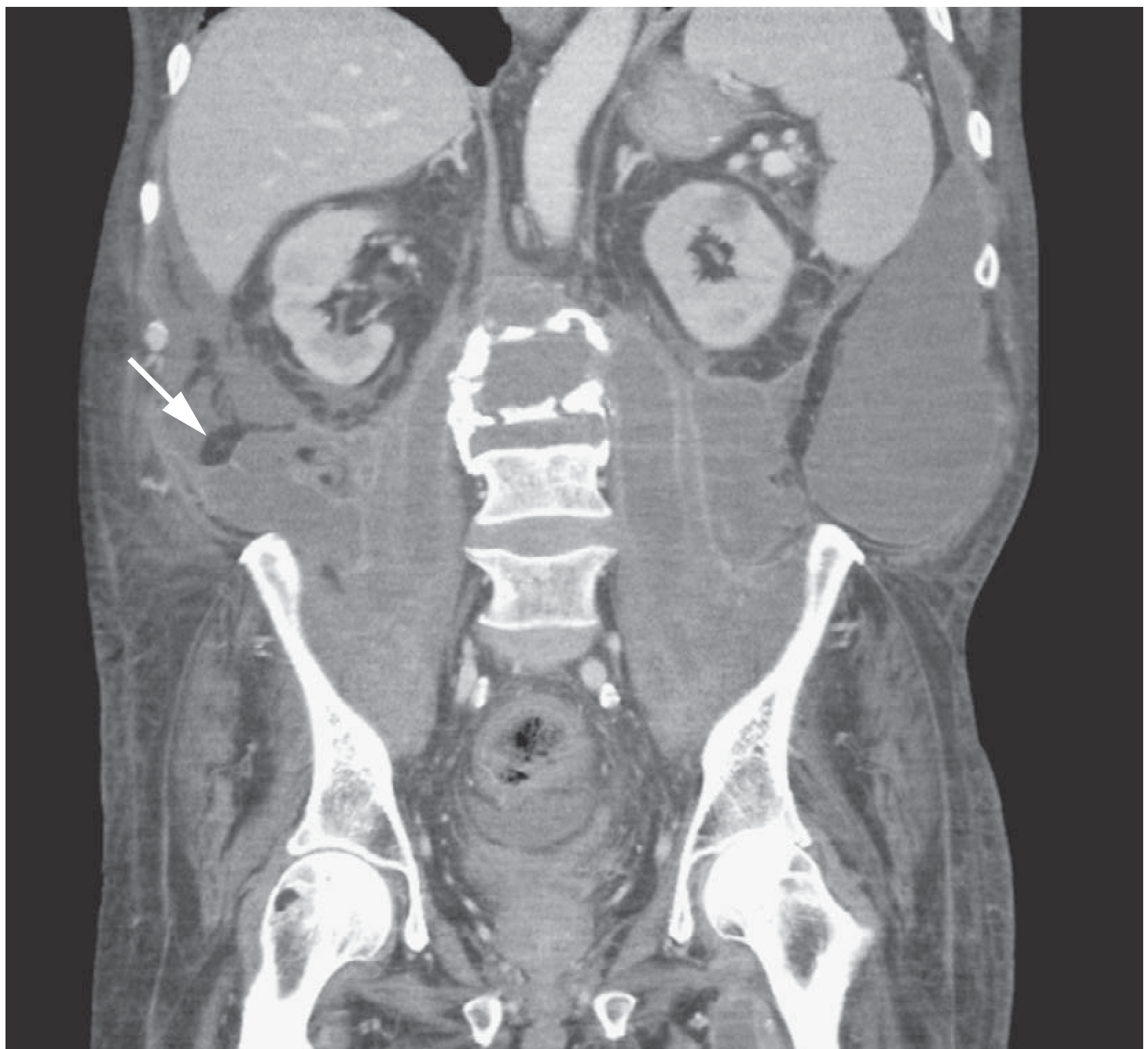
Right-sided psoas abscess (arrow) - coronal CT reconstruction. Bailey and Love's Short Practice of Surgery, 28th Ed.
2. PSOAS HAEMATOMA
| Feature | Detail |
|---|---|
| Cause | Anticoagulation therapy (warfarin, heparin, DOACs), haemophilia, trauma, post-lumbar puncture, aortic aneurysm rupture, retroperitoneal surgery |
| Clinical | Sudden onset groin/hip/flank pain; hip held in flexion; tender iliac fossa mass; may have femoral nerve compression → anterior thigh numbness, quadriceps weakness |
| Complications | Femoral nerve compression (most feared); compartment syndrome within psoas sheath; hypovolaemic shock if large |
| Diagnosis | CT scan (hyperdense collection); USS |
| Treatment | Reverse anticoagulation; transfusion; conservative (most resolve); CT-guided drainage if large; surgical decompression if femoral nerve compression |
3. PSOAS SIGN (Clinical Sign)
The psoas sign is a clinical examination finding indicating retroperitoneal or pelvic inflammation irritating the psoas muscle:
- Elicitation: Patient lies on left side; right hip is passively hyperextended - pain = positive sign
- Indicates: Retroperitoneal appendicitis (classic), psoas abscess, pyelonephritis, retroperitoneal mass, Crohn's disease
- Yamada's Textbook of Gastroenterology: "When elicited on the right side, the psoas sign is suggestive of acute appendicitis, although it may also be elicited in other retroperitoneal inflammatory processes including psoas abscess or pyelonephritis."
4. PSOAS OBLITERATION ON X-RAY
- Normally the psoas shadow is clearly visible on plain AXR as a triangular soft-tissue density on each side of the lumbar spine
- Obliteration of the psoas shadow = important radiological sign of:
- Perinephric haematoma
- Retroperitoneal abscess (including psoas abscess)
- Cold abscess (TB)
- Retroperitoneal haemorrhage (ruptured AAA, trauma)
- Retroperitoneal tumour or lymphadenopathy
5. ILIOPSOAS BURSITIS
- The iliopsoas bursa lies between the iliopsoas tendon and the hip joint capsule - the largest bursa in the body
- Causes: Osteoarthritis of hip, rheumatoid arthritis, overuse (runners, dancers), trauma
- Clinical: Anterior groin/hip pain, worsened by hip extension and resisted flexion; may cause a palpable mass in the groin
- Diagnosis: USS or MRI
- Treatment: Rest, NSAIDs, USS-guided corticosteroid injection, surgical bursectomy (rare)
6. SNAPPING HIP / PSOAS TENDINOPATHY (Coxa Saltans Interna)
- Movement of the iliopsoas tendon snapping over the iliopectineal eminence or femoral head
- Produces an audible/palpable click/snap at the anterior hip with flexion/extension
- Common in dancers, gymnasts, runners
- May coexist with iliopsoas tendinitis or bursitis
- Treat with physiotherapy, stretching, US-guided injection; rarely surgery
7. PSOAS MUSCLE AS A ROUTE OF SPREAD FOR TUMOURS
The psoas muscle or its sheath may be involved by:
- Direct invasion: Renal cell carcinoma, retroperitoneal sarcoma, colorectal cancer, lymphoma
- Metastatic deposits: Rare; lung, breast, prostate
- Lymphoma: Retroperitoneal lymphadenopathy may displace or invade the psoas
- Liposarcoma: Can arise in the retroperitoneum adjacent to or within the psoas
8. PSOAS MINOR ABNORMALITIES
- Psoas minor (absent in ~40% of people) can rarely develop calcification or enthesopathy
- Of little clinical significance
Summary Table
| Abnormality | Key Feature | Key Investigation | Treatment |
|---|---|---|---|
| Primary psoas abscess | S. aureus, immunocompromised | CT scan | Antibiotics + percutaneous drainage |
| Secondary psoas abscess (TB) | Cold, non-tender; tracks to groin; Pott's disease | CT/MRI + IGRA | Anti-TB drugs + drainage |
| Secondary psoas abscess (Crohn's) | Crohn's history; mixed flora | CT + colonoscopy | Antibiotics + drainage + Crohn's treatment |
| Psoas haematoma | Anticoagulants/trauma; femoral nerve palsy | CT | Reverse anticoagulation; drainage if large |
| Iliopsoas bursitis | Anterior groin pain; hip OA/RA | USS/MRI | NSAIDs, injection |
| Snapping hip | Audible snap with hip flexion | Clinical/USS | Physiotherapy |
| Psoas sign positive | Retroperitoneal inflammation | Clinical sign only | Treat underlying cause |
| Psoas shadow obliteration | Retroperitoneal pathology on X-ray | AXR → CT | Treat underlying cause |
Sources: Bailey and Love's Short Practice of Surgery 28th Ed | Harrison's Principles of Internal Medicine 22nd Ed | Tintinalli's Emergency Medicine | Rheumatology 2-Volume Set (Elsevier 2022) | Gray's Anatomy for Students | Yamada's Textbook of Gastroenterology
Transverse colon myenteric plexus degeneration diseases
The myenteric (Auerbach's) plexus governs colonic motility. Degeneration of this plexus leads to conditions like Hirschsprung's disease, Chagas disease, achalasia-related dysmotility, and chronic intestinal pseudo-obstruction. Let me search the textbooks thoroughly.
Searching sources...
Finding Sources
Finding Sources
Searching the Web
myenteric plexus degeneration diseases colon clinical conditions
Finding Sources
Finding Sources
Finding Sources
Finding Sources
Finding Sources
Reading File
Reading File
Finding Sources
Reading File
Finding Sources
Finding Sources
Reading File
Searching the Web
diseases associated with myenteric plexus degeneration colon Hirschsprung Chagas paraneoplastic scleroderma diabetic autonomic neuropathy
Here is a thorough, structured review of diseases associated with transverse colon myenteric (Auerbach's) plexus degeneration:
The Myenteric (Auerbach's) Plexus - Background
The myenteric plexus lies between the inner circular and outer longitudinal layers of the muscularis propria along the entire GI tract. It governs:
- Peristaltic coordination (propulsive contractions)
- Inhibitory relaxation (via NO and VIP-releasing inhibitory neurons)
- Interstitial cells of Cajal (ICC) act as the pacemaker and relay signals
Degeneration, absence, or dysfunction of the myenteric plexus leads to a spectrum of enteric neuropathies producing dilatation, obstruction, dysmotility, and constipation.
Classification of Myenteric Plexus Degeneration Diseases
1. HIRSCHSPRUNG DISEASE (Congenital Aganglionosis)
Mechanism: Failure of neural crest cell migration during embryogenesis, causing complete absence of ganglion cells in both the myenteric (Auerbach) and submucosal (Meissner) plexuses of the distal bowel.
Genetics: RET proto-oncogene mutation (chromosome 10); autosomal dominant with variable penetrance; associated with Down syndrome (3-5% of cases), MEN2B
Extent of aganglionosis (relevant to the transverse colon):
| Segment | Frequency |
|---|---|
| Rectosigmoid only | ~80% |
| Splenic flexure / transverse colon | ~17% ("long segment") |
| Total colonic aganglionosis (entire colon + small bowel) | ~8% |
The transverse colon is therefore specifically involved in long-segment Hirschsprung disease (~17% of cases). - Sabiston Textbook of Surgery
Pathophysiology: Absent ganglia → persistent tonic contraction of the aganglionic segment (no inhibitory relaxation) → functional obstruction → proximal dilation (the dilated proximal bowel is the normal, ganglionated segment)
Clinical features:
- Newborns: failure to pass meconium within 48 hours, abdominal distension, bilious vomiting
- Older children/adults (missed cases): chronic constipation, abdominal distension
- Complications: Hirschsprung-associated enterocolitis (HAEC) - most common cause of death; presents with alternating diarrhoea/obstipation, fever, haematochezia, toxic megacolon, shock
Histopathology:
- Absent ganglion cells in myenteric and submucosal plexus
- Hypertrophied nerve trunks (acetylcholinesterase-positive)
- Calretinin immunostaining - now standard adjunct (negative in aganglionic segment)
Diagnosis:
- Contrast enema: narrow calibre distal segment, transition zone, dilated proximal bowel; failure to evacuate barium after 24 hours
- Anorectal manometry: failure of internal sphincter to relax on balloon distension
- Rectal biopsy - gold standard (suction biopsy in neonates, full-thickness in older children; must be >1 cm above dentate line)
Treatment: Surgical pull-through procedures (Swenson, Soave, Duhamel operations) - resect aganglionic segment and restore continuity with normal ganglionated bowel
2. CHAGAS DISEASE (Acquired Aganglionosis)
Cause: Trypanosoma cruzi (protozoan), transmitted by the reduviid (kissing) bug; endemic in Central Brazil, Venezuela, Northern Argentina; ~20 million infected in South America; ~500,000 in the USA
Mechanism: Chronic phase (developing up to 20 years after acute infection) → autoimmune-mediated destruction of autonomic ganglion cells throughout the body (heart, gut, urinary tract, respiratory tract)
GI involvement:
- Oesophagus, duodenum, and colon most affected
- Progressive ganglion cell destruction → loss of peristaltic coordination
- Abnormal peristalsis begins after 50% ganglion cell loss
- Dilatation occurs after 90% loss
- Results in: megaoesophagus, megaduodenum, megacolon, megarectum, megaureter
Megacolon in Chagas: Diffuse degeneration of myenteric plexus ganglion cells throughout the colon including the transverse colon → massive colonic dilatation, severe constipation, faecaloma formation, volvulus risk
Systemic features: Chronic cardiomyopathy with conduction disturbances (most common cause of death), arrhythmias
Diagnosis:
- Acute phase: parasites visible on blood smear
- Chronic phase: serological tests (complement fixation, PCR)
Treatment: Treatment of the infection is of limited efficacy in chronic phase; treat GI complications symptomatically; surgery for megacolon (colectomy)
- Sleisenger and Fordtran's GI & Liver Disease
3. CHRONIC INTESTINAL PSEUDO-OBSTRUCTION (CIPO) / Enteric Neuropathy
Definition: Severe impairment of intestinal motility causing bowel dilatation in the absence of mechanical obstruction - Bailey and Love's Surgery, 28th Ed
Mechanism relevant to myenteric plexus:
- Neuropathic CIPO = degenerative or inflammatory changes in the myenteric plexus (neuronal inflammation = myenteric ganglionitis, neuronal loss, neuronal inclusion bodies)
- Myopathic CIPO = degeneration of intestinal smooth muscle (separate entity)
Primary causes (rare, often genetic/familial):
- Familial visceral neuropathy (autosomal dominant or recessive)
- Mitochondrial neurogastrointestinal encephalomyopathy (MNGIE - TYMP gene mutation; neuropathic degeneration of the myenteric plexus + leukoencephalopathy)
- Intestinal neuronal dysplasia type B (IND-B) - colonic submucosal hyperganglionosis
Secondary causes (more common in adults):
| Cause | Mechanism |
|---|---|
| Chagas disease | Ganglion cell destruction (as above) |
| Paraneoplastic neuropathy | Anti-Hu (ANNA-1) antibodies; small cell lung cancer most common; autoimmune destruction of ENS neurons |
| Scleroderma / connective tissue disease | Smooth muscle atrophy + secondary neurodegeneration |
| Diabetes mellitus | Autonomic neuropathy → vagal and myenteric degeneration; also loss of ICC |
| Amyloidosis | Amyloid deposits in myenteric plexus → neuronal destruction |
| Radiation injury | Direct toxicity to myenteric neurons |
| Parkinson's disease | Alpha-synuclein (Lewy body) deposition in myenteric plexus - the gut may be an early site of PD pathology |
| Idiopathic | Unknown; may be degenerative |
Clinical presentation: Abdominal distension, vomiting, pain, constipation; mimics mechanical obstruction
Diagnosis: CT (exclude mechanical obstruction); MRI brain + skeletal muscle biopsy (for mitochondrial causes); full-thickness bowel biopsy for neuropathological analysis
4. PARANEOPLASTIC ENTERIC NEUROPATHY
- Most commonly associated with small cell lung carcinoma, also thymoma, carcinoid
- Anti-Hu antibodies (ANNA-1) target RNA-binding proteins in ENS neurons
- Leads to degeneration of myenteric plexus neurons throughout the GI tract, including the colon
- Presents with severe constipation, intestinal pseudo-obstruction, autonomic neuropathy
- May precede the cancer diagnosis
5. DIABETIC AUTONOMIC NEUROPATHY
- Long-standing diabetes damages autonomic nerves supplying the gut, including the vagal input and the myenteric plexus itself
- Loss of interstitial cells of Cajal (ICC) - the pacemaker cells - contributes
- Colonic involvement: diabetic colonic dysmotility → slow transit constipation, megacolon in severe cases
- Associated with diabetic gastroparesis (same mechanism in the stomach)
6. ACHALASIA (Oesophageal Analogue - Same Plexus, Different Level)
While achalasia classically affects the oesophageal myenteric plexus (loss of inhibitory neurons containing NO/VIP), Chagas disease can cause the same picture at the oesophageal level and simultaneously in the colon - both being myenteric plexus degeneration disorders on a spectrum.
7. INTESTINAL NEURONAL DYSPLASIA TYPE B (IND-B)
- Colonic submucosal hyperganglionosis (giant ganglia, >8 neurons per ganglion)
- May mimic Hirschsprung or cause chronic constipation
- Diagnosis: rectal/colonic biopsy
- Often resolves with age; treatment conservative
8. SLOW TRANSIT CONSTIPATION (Colonic Inertia)
- A subset of patients with intractable constipation show reduced myenteric plexus neurons and ICC loss on full-thickness colonic biopsy (particularly in the right and transverse colon)
- Not a distinct neuropathy but a spectrum of degenerative change
- Simultaneous degeneration of myenteric plexus + pelvic parasympathetic colonic nerve fibres has been documented in slow transit constipation
Summary Table
| Disease | Mechanism | Colon Involved | Key Feature |
|---|---|---|---|
| Hirschsprung disease | Absent neural crest migration; congenital aganglionosis | Distal >> transverse (long segment, 17%) | Failure to pass meconium; transition zone on enema |
| Chagas disease | T. cruzi autoimmune ganglion cell destruction | Entire colon including transverse | Megacolon; cardiomyopathy; Latin American endemic |
| CIPO - neuropathic | Myenteric ganglionitis / neuronal degeneration | Small bowel + colon | Pseudo-obstruction; no mechanical cause |
| MNGIE | Mitochondrial TYMP mutation; myenteric plexus degeneration | Small bowel > colon | Leukoencephalopathy, cachexia, peripheral neuropathy |
| Paraneoplastic | Anti-Hu antibodies; SCLC | Entire ENS | Precedes cancer; pan-GI dysmotility |
| Diabetic neuropathy | Autonomic + ICC loss | Colon, stomach | Slow transit constipation; gastroparesis |
| IND-B | Hyperganglionosis | Colon | Chronic constipation in children |
| Slow transit constipation | Reduced myenteric neurons + ICC | Transverse + right colon | Intractable constipation; female > male |
| Parkinson's disease | Alpha-synuclein in ENS | Entire ENS | Constipation precedes motor symptoms |
| Amyloidosis | Amyloid in plexus | Entire GI | Systemic amyloid features |
Sources: Sabiston Textbook of Surgery | Quick Compendium of Clinical Pathology 5th Ed | Sleisenger and Fordtran's GI & Liver Disease | Bailey and Love's Short Practice of Surgery 28th Ed | Pediatric enteric neuropathies review (PMC5475271)
This is a shared conversation. Sign in to Orris to start your own chat.