
Paronychia treatment
paronychia treatment management
paronychia nail fold infection clinical
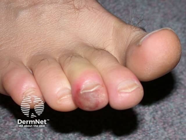
Clinical photography of a toenail unit demonstrating acute paronychia. The image shows a dorsal close‑up of the great toe with surrounding periungual soft tissue erythema and marginal swelling at the proximal nail fold. The nail plate remains present and intact, but the adjacent tissue is inflamed, with a reddish-purple hue and mild edema extending from the cuticle onto the lateral nail folds. No well‑formed abscess is clearly visible in this view, though localized tenderness and warmth are suggested by the tissue characteristics. This dermatologic finding is most consistent with acute paronychia or periungual cellulitis, often secondary to trauma, nail biting, moisture exposure, or minor infection. Differential diagnoses include chronic paronychia, fungal nail infection (onychomycosis), irritant/contact dermatitis, or less likely herpetic infection. Clinically, the image supports decision‑making regarding initial management: warm soaks, analgesia, antiseptic care, and consideration of oral antibiotics directed at common pathogens such as Staphylococcus aureus; incision and drainage if an abscess forms. This photograph serves educational and search-retrieval purposes for dermatology, infectious disease, and primary care, enabling keyword‑rich indexing: paronychia, periungual, nail fold inflammation, toe infection, dorsal toe view, nail unit, soft tissue edema, erythema, swelling, acute, bacterial. Images like this guide clinicians in triaging therapy and patient education.
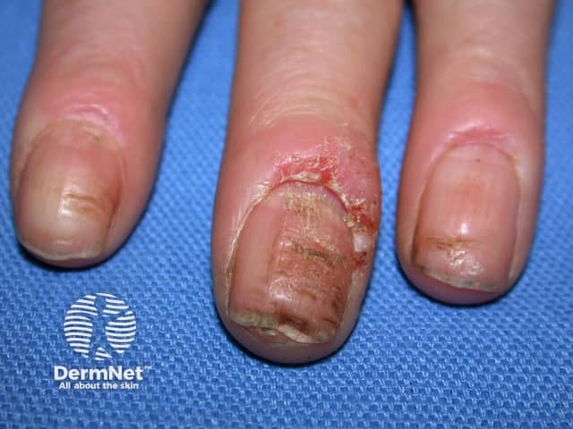
Clinical photography of the periungual digits demonstrating acute paronychia with inflamed, erythematous nail folds. The central and ring fingers show pronounced edema surrounding the proximal nail folds, with fissuring of the cuticle and crusted debris along the eponychial region. The adjacent distal nail plates appear slightly deformed and tender, consistent with early nail-fold infection. No penetrating foreign body is evident. Skin over the dorsal fingertip is uninjured beyond the periungual zones, while surrounding epidermis shows mild desquamation. The left-handed orientation presents a close-up, magnified view of three digits; the middle finger bears the most conspicuous swelling, whereas the index and ring digits display milder involvement. The overall picture is suggestive of acute bacterial paronychia, most commonly caused by Staphylococcus aureus, with potential moisture-related colonization or trauma-induced microfissures providing entry for pathogens. Clinically, this finding warrants supportive care, topical antisepsis, possible incision and drainage if abscess forms, and consideration of oral antibiotics in extensive edema or systemic symptoms. Diagnosis should be distinguished from herpetic whitlow and chronic paronychia by history, distribution, and presence of vesicles or chronic changes. Documentation of nail dystrophy, secondary infection, and risk factors such as diabetes or immunosuppression is essential for management and prognosis. Clinical relevance acknowledged.
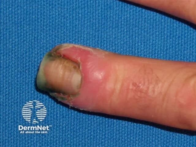
Comprehensive Description: A high-magnification clinical photograph of a single fingernail demonstrates chronic paronychia affecting the periungual region. The image captures the dorsal-lateral aspect of the distal finger, highlighting erythema and edema of the lateral nail fold with fissuring and crusting at the cuticle. The nail plate is partially displaced by surrounding inflammation, and there may be mild hypertrophy of the proximal nail fold. These findings reflect a protracted inflammatory process often linked to repeated moisture exposure, trauma from manicure practices, irritant contact, or candidal colonization. The visual features include localized redness, tenderness, soft tissue swelling, and occasional crusted debris at the cuticle; the adjacent skin appears thickened in places. Clinically, this presentation suggests chronic paronychia rather than an acute infection, and a clinician would consider secondary fungal or bacterial involvement, eczema, or psoriasis of the nail unit in the differential. The diagnostic significance lies in guiding management toward topical or systemic anti-inflammatory therapy, barrier protection, meticulous nail hygiene, and targeted antimicrobial or antifungal treatment when indicated. This image is valuable for dermatology education, clinical case assessment, and comparative studies of nail-fold inflammation or chronic paronychia progression. Early recognition improves outcomes through targeted therapy and preventive hand care for patients daily.
Paronychia - Treatment
Acute Paronychia
Causes
- Most commonly Staphylococcus aureus (including CA-MRSA)
- Anaerobes and gram-negative organisms can also be isolated
- In children: often anaerobes from finger-sucking/nail-biting; rarely Group A beta-hemolytic streptococcus in thumb-suckers with strep pharyngitis
- Mixed aerobic/anaerobic infections are common (~50% of cases)
- Associated with: HIV antiretroviral therapy, EGFR inhibitor therapy, nail cosmetics, occupations like haircutting and meat handling
Stages and Treatment
Stage 1 - Early Cellulitis (no abscess)
- Warm soaks 3x/day (water or chlorhexidine soaks)
- Short course of oral antibiotics (antistaphylococcal):
- Cephalosporin or semisynthetic penicillin (e.g., dicloxacillin, cefalexin)
- If MRSA suspected: TMP-SMX or doxycycline
- Topical antibiotics +/- topical corticosteroids can also be used
- Incision has little value at this stage
Stage 2 - Abscess Formed
- Drainage is the definitive treatment - usually quickly curative
- Technique: lift the eponychial fold away from the nail matrix to allow pus to drain - this is often sufficient without skin incision
- One-sided abscess: incise at the corner, angling the knife away from the nail bed (to avoid creating a nail ridge later)
- Abscess under one corner of the nail root: remove that corner of the nail
- If migrated to the opposite side: second incision, fold skin back proximally, excise the proximal one-third of the nail; pack loosely with iodoform gauze for 48 hours
- A 21- or 23-gauge needle tip can be used to lift the nail fold and drain the abscess - effective in most cases with no anesthesia required
- Actual skin incision or nail removal is rarely required as the initial approach
- Post-drainage: antibiotics are used judiciously (likely unnecessary in most uncomplicated cases)
Stage 3 - Subungual Abscess (pus under the nail)
- More extensive drainage procedure required
- If the nail bed is mobile, infection has extended under the nail
- May require partial or complete nail removal
Chronic Paronychia
Cause
- Prolonged mechanical or environmental trauma damages the cuticle
- Moisture/irritant exposure allows penetration of bacteria, Candida, and irritants under the proximal nail fold
- Results in inflammatory reaction of the nail matrix
Clinical Features
- Edema and redness of the proximal nail fold
- Absence of cuticles (pathognomonic)
- Beau's lines and nail plate surface abnormalities
- Repeated acute flares on a chronic background
Treatment
- Protective measures (first-line):
- Cotton inner gloves + rubber outer gloves to avoid contact with water and irritants
- Avoiding wet work, frequent handwashing, and irritant chemicals
- Topical corticosteroids - reduce the inflammatory component
- Topical antimicrobials - target secondary bacterial or candidal colonization
- Topical antifungals (e.g., clotrimazole, miconazole) if candidal
- Topical antibiotics if bacterial superinfection
- Systemic antifungals (e.g., fluconazole, itraconazole) for refractory candidal cases
- Surgical option for refractory cases: eponychial marsupialization (Swiss roll technique) - excision of a crescent of skin proximal to the nail fold
Special Considerations
| Situation | Notes |
|---|---|
| Herpetic Whitlow | Can mimic paronychia - look for vesicles; do NOT incise (antiviral therapy, not I&D) |
| EGFR inhibitors | Common side effect of cetuximab, erlotinib, etc.; manage with topical antibiotics, steroids |
| Immunocompromised | Broader spectrum coverage; consider atypical organisms |
| Recurrence | Check for inadequate drainage; consider MRSA culture |
| Children | Anaerobes are common; consider strep if thumb-sucking + pharyngitis |
Key Principles Summary
- No abscess → warm soaks + oral antibiotics
- Abscess present → drain first, antibiotics second
- Chronic → protect from moisture + topical steroids/antifungals
- Never incise suspected herpetic whitlow