
Define shocks classify and pathpgysiology of each type with their forensic relevance
classification of shock types pathophysiology diagram
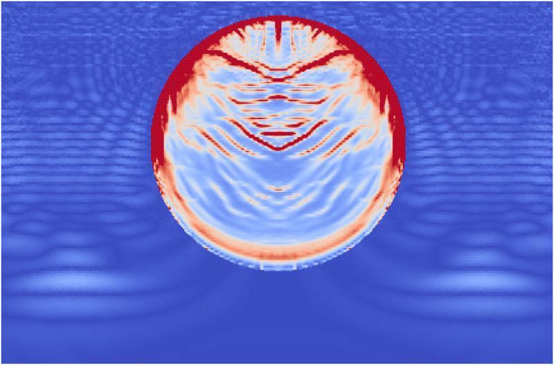
This pathophysiology diagram is a computational visualization using the Material Point Method (MPM) to simulate the effects of Extracorporeal Shock Wave Lithotripsy (ESWL) on a kidney stone. The image depicts a circular, single-phase kidney stone immersed in a fluid medium (modeled as water) at the moment a high-pressure shock wave has reached the end of the stone. The fluid medium displays stress waves as concentric, blue-toned undulating ripples propagating through the domain. Within the stone, damage accumulation is color-coded: red indicates areas of complete failure, while lighter blue and white areas represent internal stress and emerging fractures. Significant damage is concentrated at the proximal side (top) where the wavefront first impacted, showing a thick layer of failure. Distinct angled failure lines extend from the stone's edges toward the center, representing shear stress interactions. Minor damage and tensile fractures are also visible at the distal end (bottom) of the stone, resulting from internal wave reflections and interference patterns. This simulation serves as an educational model for understanding fragmentation mechanisms in urolithiasis treatment.
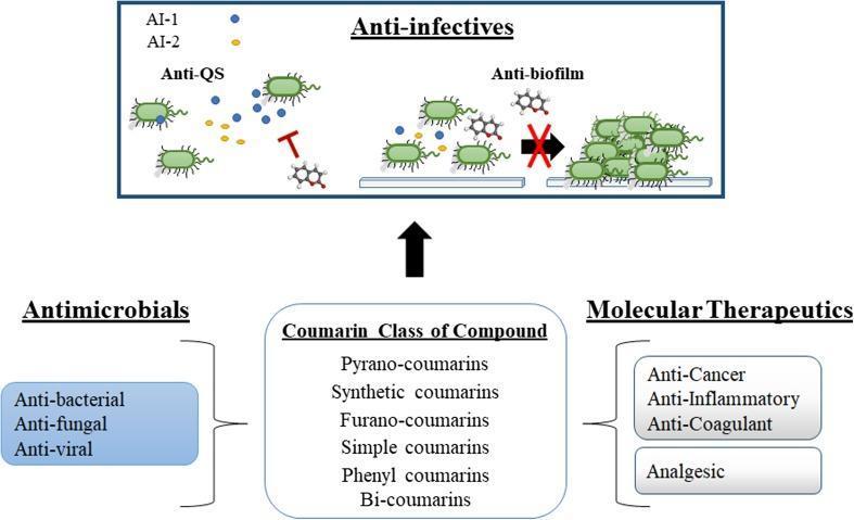
This medical pathophysiology diagram illustrates the multi-functional pharmacological properties of the coumarin class of compounds, focusing on their role as anti-infectives. The schematic is divided into two primary sections: a broad classification of coumarin types and their therapeutic applications, and a detailed mechanistic view of their anti-infective properties. The lower half categorizes coumarins into various structural types (pyrano-coumarins, synthetic, furano-coumarins, etc.) and links them to 'Antimicrobials' (anti-bacterial, anti-fungal, anti-viral) and 'Molecular Therapeutics' (anti-cancer, anti-inflammatory, anti-coagulant, analgesic). The upper section details the 'Anti-infective' mechanism, specifically highlighting 'Anti-QS' (quorum sensing) and 'Anti-biofilm' activities. In the 'Anti-QS' portion, coumarin structures are shown inhibiting signaling molecules AI-1 (blue) and AI-2 (yellow), represented by T-bar inhibition icons between bacteria. In the 'Anti-biofilm' portion, a red cross over a transition arrow signifies the inhibition of individual bacterial cells aggregating into a multicellular biofilm structure. This educational infographic summarizes how coumarins act as non-toxic modulators of microbial behavior by disrupting cell-to-cell communication and structural development in pathogens.
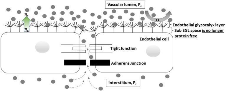
A pathophysiology diagram illustrating the Revised Starling Principle at the capillary-endothelial interface during profound shock. The illustration shows a cross-section of the vascular wall, featuring the vascular lumen (Pc), the endothelial glycocalyx layer (EGL), the sub-EGL space, and the interstitium (Pi). Key structural components include endothelial cells connected by tight and adherens junctions. The diagram highlights the loss of the protein-free status in the sub-EGL space, where proteins (represented by gray circles) diffuse from the interstitium through the inter-endothelial cleft back into the sub-EGL space. This movement indicates a transient reversal from filtration to reabsorption (autotransfusion). Pressures are annotated with arrows: πc represents capillary oncotic pressure and πg represents sub-glycocalyx oncotic pressure. The visual demonstrates how reduced hydrostatic pressure gradients (Pc ≈ Pi) in shock allow for protein accumulation in the sub-EGL, altering the effective oncotic pressure gradient that typically opposes filtration. This educational material is relevant for medical students and clinicians studying microcirculation, fluid dynamics, and critical care physiology.
SHOCK: Definition, Classification, Pathophysiology, and Forensic Relevance
DEFINITION
"Shock is a systemic state of low tissue perfusion that is inadequate for normal cellular respiration. With insufficient delivery of oxygen and glucose, cells switch from aerobic to anaerobic metabolism. If perfusion is not restored in a timely fashion, cell death ensues."
- Bailey and Love's Short Practice of Surgery, 28th Edition
CLASSIFICATION
| Type | Major Subtypes | Core Mechanism |
|---|---|---|
| 1. Hypovolemic | Hemorrhagic, non-hemorrhagic | Reduced circulating volume |
| 2. Cardiogenic | Intrinsic, obstructive/extracardiac | Pump failure |
| 3. Distributive | Septic, neurogenic, anaphylactic | Peripheral vasodilation / maldistribution |
| 4. Obstructive | PE, tamponade, tension pneumothorax | Mechanical obstruction to cardiac output |
| 5. Endocrine | Adrenal crisis, hypothyroid, thyrotoxic | Hormonal dysregulation (from Bailey & Love) |
PATHOPHYSIOLOGY OF EACH TYPE
1. Hypovolemic Shock
- Loss of circulating blood/plasma volume reduces venous return (preload)
- Decreased preload -> decreased stroke volume -> decreased cardiac output
- Baroreceptors detect fall in BP -> sympathetic activation -> catecholamine release
- Result: tachycardia, vasoconstriction (high SVR), oliguria (RAAS activation), ADH release (water retention)
- Cells switch to anaerobic glycolysis -> lactic acid accumulation -> metabolic acidosis
- Persistent hypoperfusion -> endothelial injury -> capillary leak -> DIC -> multiorgan failure (MOF)
- Non-progressive (compensated): Baroreceptor reflexes, catecholamines, ADH, RAAS maintain vital organ perfusion
- Progressive: Widespread tissue hypoxia, lactic acidosis, arteriolar dilation, microcirculatory pooling, DIC begins; vital organs fail
- Irreversible: Lysosomal enzyme leakage, myocardial depression (NO-mediated), bowel flora translocation, renal failure, death despite intervention
2. Cardiogenic Shock
- Primary failure of the heart as a pump -> reduced stroke volume and cardiac output despite adequate volume
- Decreased CO -> reflex sympathetic activation -> tachycardia and vasoconstriction
- High SVR increases afterload, further straining a failing heart (vicious cycle)
- Increased ventricular end-diastolic pressure -> pulmonary venous congestion -> pulmonary edema
- Tissue hypoxia, lactic acidosis, progressive MOF
Goldman-Cecil: "Cardiogenic shock is due to pump failure caused most commonly by acute myocardial ischemia but also by acute valvular dysfunction, arrhythmias, myocarditis, or ventricular wall rupture."
3. Distributive Shock
A. Septic Shock
- Microbial PAMPs (LPS/endotoxin, peptidoglycan, fungal antigens) bind TLRs, G-protein-coupled receptors, and C-type lectin receptors on macrophages, neutrophils, dendritic cells
- Massive cytokine storm: TNF, IL-1, IL-12, IL-18, IFN-gamma, HMGB1
- Complement activation: C3a, C5a (anaphylatoxins) -> further inflammation
- Reactive oxygen species, prostaglandins, PAF released
- Endothelial activation -> upregulation of adhesion molecules, vascular permeability -> leukocyte margination, tissue edema
- Widespread coagulation activation -> DIC
- Arteriovenous shunting at microvascular level -> maldistribution (tissues cannot extract O2 properly)
- Later phases: hypovolemia from capillary leak + myocardial depression (combined shock pattern)
- Counter-regulatory immunosuppression (IL-10, TGF-beta, lymphocyte apoptosis) may supervene, leading to secondary infections
B. Neurogenic Shock
- Loss of sympathetic outflow from spinal cord injury -> unopposed parasympathetic tone
- Peripheral vasodilation and venous pooling -> profound hypotension
- Bradycardia (unlike most other shock states where tachycardia is expected) due to loss of cardiac sympathetic innervation
- No catecholamine-mediated compensatory vasoconstriction
C. Anaphylactic Shock
- Antigen crosslinks IgE on mast cells and basophils -> massive degranulation
- Histamine release + other mediators (leukotrienes, PAF, prostaglandins) -> systemic vasodilation, increased vascular permeability
- Bronchoconstriction (may cause airway obstruction and superimposed obstructive/hypoxic component)
- Rapid fluid shift from intravascular to extravascular space -> functional hypovolemia
4. Obstructive Shock
- Mechanical obstruction of cardiac filling (reduced preload) or outflow
- In tamponade: pericardial fluid compresses cardiac chambers -> reduced diastolic filling -> reduced stroke volume
- In tension pneumothorax: mediastinal shift compresses the great veins and the right heart
- In massive PE: right ventricular outflow obstruction + right heart failure -> left ventricular underfilling -> low CO
- Result: low CO despite intact myocardium; SVR is high (reflex vasoconstriction)
5. Endocrine Shock (Bailey & Love)
- Adrenal insufficiency (Addisonian crisis): Cortisol deficiency -> failure of vascular responsiveness to catecholamines + hypovolemia (mineralocorticoid deficiency) -> combined distributive/hypovolemic picture
- Hypothyroid shock (myxedema coma): Disordered catecholamine responsiveness -> low inotropy, bradycardia, low CO; resembles neurogenic shock hemodynamically
- Thyrotoxic crisis: High-output cardiac failure
MORPHOLOGICAL CHANGES IN SHOCK (Autopsy Findings)
| Organ | Morphological Changes |
|---|---|
| Brain | Ischemic encephalopathy; neuronal loss (irreversible) |
| Heart | Subendocardial coagulative necrosis; "contraction band necrosis"; cardiomyocyte loss (irreversible) |
| Kidney | Acute tubular necrosis (ATN) - proximal tubules + loop of Henle; fibrin thrombi in glomeruli |
| Adrenals | Cortical lipid depletion (lipid used for increased steroid synthesis - stress response) |
| Lungs | Diffuse alveolar damage ("shock lung" / ARDS) - especially in septic and traumatic shock; resistant in pure hemorrhagic shock |
| GI Tract | Hemorrhagic mucosal necrosis (stress ulcers); bacterial translocation in irreversible stage |
| Liver | Centrilobular necrosis (most susceptible zone) |
"Except for neuronal and cardiomyocyte loss, affected tissues can recover completely if the patient survives." - Robbins, Cotran & Kumar
FORENSIC RELEVANCE OF SHOCK
1. Shock as a Mechanism of Death
- Cause of death: The injury, disease, or poisoning that started the chain of events (e.g., "multiple fractures due to run over by truck")
- Mechanism of death: The physiological/biochemical disturbance incompatible with life - shock is one of the most common mechanisms listed (along with sepsis, metabolic acidosis, ventricular fibrillation, respiratory arrest)
- Manner of death: Natural / Homicide / Suicide / Accident / Undetermined
2. Hypovolemic/Hemorrhagic Shock - Forensic Significance
- Most common forensic shock type - seen in gunshot wounds, stab wounds, blunt force trauma, vehicular accidents, explosive injuries
- Forensic pathologist must document:
- Site and nature of injury causing hemorrhage
- Estimated blood loss (external + internal hemorrhage)
- Signs of shock at autopsy: pale organs, empty heart chambers, pallor of mucosae
- Absence of clotted blood (blood may remain fluid in rapid death from hemorrhage - forensically significant)
- Wound ballistics: High-velocity missile injuries create shock waves in tissues causing injury to organs remote from the actual wound track (Essentials of Forensic Medicine and Toxicology, 36th edition)
3. Cardiogenic Shock and Trauma - Forensic Significance
- Traumatic myocardial contusion (e.g., steering wheel injury) can cause cardiogenic shock; if victim dies, forensic pathologist must establish cause-and-effect relationship
- Blunt chest trauma may cause coronary artery thrombosis -> MI -> cardiogenic shock -> death
- From Dikshit's Textbook of Forensic Medicine: Injury sufficiently severe to cause hypovolemic shock can precipitate MI in a person with pre-existing atherosclerotic coronary disease - a forensically important scenario in medicolegal cases
- "Even threatening the person can cause cardiovascular changes, sudden rise in blood pressure that can rupture an atheromatous plaque" - relevant in cases of sudden death following altercation
- Two basic situations in court: (a) Direct cardiac injury (laceration, perforation - easier to prove); (b) Indirect injury (arrhythmia, exacerbation of pre-existing disease - harder to prove)
4. Septic Shock - Forensic Significance
- May follow neglected/infected wounds, illegal abortions, hospital-acquired infections in trauma victims
- Relevant in criminal negligence cases (e.g., delayed treatment of injuries leading to septic shock and death)
- The mechanism - sepsis -> MOF -> death - may be the critical link in proving that an assault caused death even though the original injury was not immediately fatal
5. Neurogenic Shock - Forensic Significance
- Seen in hanging, neck injuries, spinal cord injuries from assault or accidents
- The characteristic bradycardia + hypotension without tachycardia can be a clue at autopsy
- Relevant in road traffic accident (RTA) deaths with cervical spine injuries
6. Anaphylactic Shock - Forensic Significance
- Sudden, unexpected death after drug injection, insect sting, or food ingestion
- Must be considered in unexplained sudden deaths
- Autopsy may show: laryngeal edema, mucosal congestion, pulmonary hyperinflation
- Histology: mast cell degranulation; elevated serum tryptase levels (forensic biochemistry)
- Forensically relevant in medical negligence cases (e.g., failure to screen for allergies before drug administration)
7. Electrocution Shock - Forensic Significance
- Death from electrocution may result from "instant shock" causing ventricular fibrillation
- The term "shock" in electrocution overlaps with the physiological concept of neurogenic/distributive shock
- Blast-related "shock waves" from high-velocity projectiles or explosions cause secondary injuries to remote organs (forensically documented in gunshot wound analysis)
8. Shock at Autopsy - Key Forensic Findings
| Finding | Significance |
|---|---|
| Empty, contracted heart | Severe hemorrhage |
| Pale, ischemic organs | Prolonged hypoperfusion |
| Acute tubular necrosis | Survived the initial insult; died later from MOF |
| Adrenal lipid depletion | Stress response - confirms ante-mortem shock state |
| DIC changes (fibrin thrombi) | Septic shock, massive trauma |
| "Shock lung" (ARDS) | Trauma, sepsis, aspiration - may indicate survivability window |
| Stress (Curling's) ulcers | Survived period of shock (burn patients) |
| Dusky/congested bowel | Mesenteric ischemia from shock |
9. Shock in Relation to Survivability and Manner of Death
- The length of the agonal period (time between lethal insult and death) can be estimated by the degree of organ changes
- If victim survived hours to days after injury, autopsy will show more developed organ changes (ATN, stress ulcers, DIC)
- This is relevant in establishing whether an assault "caused" death or whether the death was from a supervening complication
SUMMARY TABLE: Hemodynamic Profile
| Parameter | Hypovolemic | Cardiogenic | Distributive (Septic) | Obstructive |
|---|---|---|---|---|
| Cardiac Output | Low | Low | High (early) / Low (late) | Low |
| SVR | High | High | Low | High |
| CVP/Venous Pressure | Low | High | Low | High |
| Mixed Venous O2 | Low | Low | Low (impaired extraction) | Low |
| Skin | Cool, pale, clammy | Cool, pale, clammy | Warm, flushed (early) | Cool, clammy |
Neurogenic shock
neurogenic shock spinal cord injury sympathetic pathway diagram
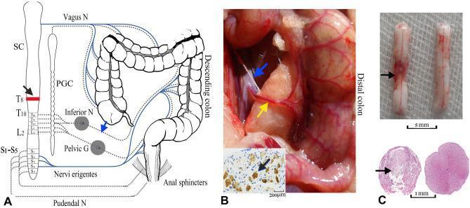
This composite educational graphic illustrates the neuroanatomical basis for peri-arterial sympathectomy of the inferior mesenteric artery (PSIMA) in a spinal cord injury (SCI) model. Panel A presents a human anatomical diagram showing the autonomic innervation of the colon; sympathetic outflow (T10-L2) is indicated by dashed lines via the paravertebral ganglia chain (PGC) and inferior mesenteric ganglia, while parasympathetic innervation (S2-S4) via the pelvic nerves is shown in blue. A black arrow indicates the T8 SCI level. Panel B provides an intraoperative photograph of a rat model, detailing the anatomical relationship between the distal colon, the inferior mesenteric artery (yellow arrow), and the accompanying sympathetic plexus (blue arrow). An inset shows TH-stained sympathetic nerve fibers. Panel C displays the pathological consequences of injury, comparing an SCI-damaged spinal cord segment (left, showing gross discoloration and narrowing) with a normal control (right). Lower histological images (HE staining) demonstrate complete loss of tissue architecture and cavitation in the transverse section of the injured cord compared to the intact control. This visual material is used to demonstrate surgical interventions for neurogenic bowel dysfunction.
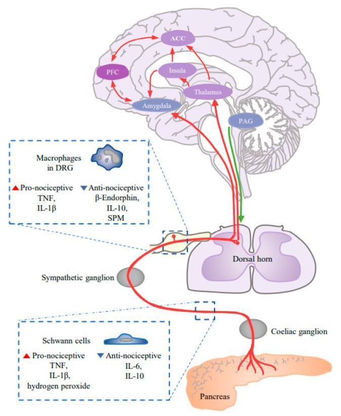
A pathophysiology diagram illustrating the pain conduction pathway in pancreatic ductal adenocarcinoma (PDAC), highlighting the transition from peripheral neurogenic inflammation to central sensitization. The pathway originates at the pancreas, where Schwann cells release pro-nociceptive (TNF, IL-1̠, hydrogen peroxide) and anti-nociceptive (IL-6, IL-10) factors. The signal traverses the coeliac ganglion and sympathetic ganglion to reach the dorsal horn of the spinal cord. Within the dorsal root ganglion (DRG), macrophages modulate the signal via pro-nociceptive (TNF, IL-1̠) and anti-nociceptive (B-Endorphin, IL-10, SPM) mediators. Ascending tracks project to the periaqueductal gray (PAG) in the brainstem, which then connects to a network of cortical and subcortical regions including the thalamus, amygdala, insula, anterior cingulate cortex (ACC), and prefrontal cortex (PFC). Red arrows denote ascending pain transmission, while green arrows indicate descending modulation. This diagram serves as an educational tool for understanding chronic pain mechanisms and synaptic remodeling in the central nervous system during pancreatic cancer progression.
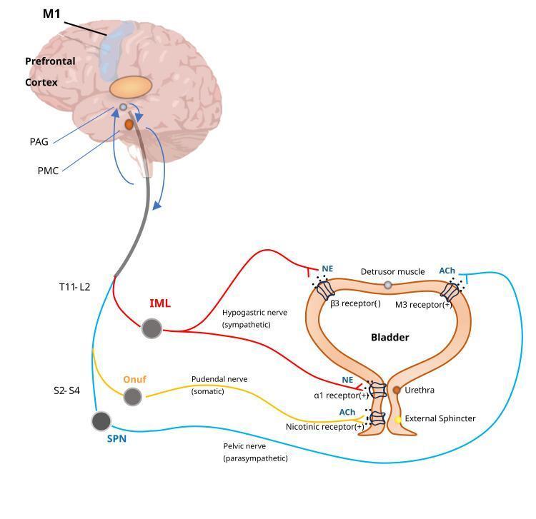
This medical illustration depicts the neural control of the lower urinary tract, specifically the micturition reflex and voluntary bladder control. It shows the hierarchical integration between the brain (primary motor cortex M1, prefrontal cortex, periaqueductal gray PAG, and pontine micturition center PMC) and the spinal cord. The diagram illustrates three distinct pathways: 1) The sympathetic pathway (red), originating from the T11-L2 intermediolateral cell column (IML) via the hypogastric nerve, releasing norepinephrine (NE) to inhibit the detrusor muscle (beta-3 receptors) and contract the bladder neck (alpha-1 receptors). 2) The parasympathetic pathway (blue), originating from the S2-S4 sacral parasympathetic nucleus (SPN) via the pelvic nerve, releasing acetylcholine (ACh) to contract the detrusor muscle (M3 receptors). 3) The somatic motor pathway (yellow), originating from Onuf's nucleus (S2-S4) via the pudendal nerve, releasing ACh to contract the external urethral sphincter (nicotinic receptors). This diagram serves as a pathophysiology and anatomical guide to neurogenic bladder and the physiological mechanisms of urination.
NEUROGENIC SHOCK
DEFINITION
"Neurogenic shock results from interrupted sympathetic and parasympathetic input from the spinal cord to the heart and peripheral vasculature, typically resulting from acute traumatic injury."
- Rosen's Emergency Medicine
"Neurogenic shock is defined as failure of the nervous system to provide effective peripheral vascular resistance, resulting in inadequate end-organ perfusion."
- Mulholland and Greenfield's Surgery, 7th Edition
IMPORTANT DISTINCTION: Neurogenic Shock vs Spinal Shock
| Feature | Neurogenic Shock | Spinal Shock |
|---|---|---|
| Nature | Hemodynamic/circulatory | Neurological |
| Definition | Loss of vasomotor tone -> hypotension, bradycardia | Temporary depression of all spinal cord reflexes below the injury level |
| Features | Hypotension, bradycardia, vasodilation | Flaccidity, absent reflexes, loss of voluntary movement |
| Duration | 24-48 hrs (vasopressors), up to 14 days for dysrhythmias | Days to weeks; up to 6 months |
| Key relevance | Can be fatal if untreated | May mimic complete cord injury |
FORENSIC CLASSIFICATION CONTEXT
| Forensic Type | Mechanism | Timing |
|---|---|---|
| Primary (Neurogenic) | Reflex neurovascular disturbance - vagal inhibition, sympathetico-adrenal stimulation | Immediate after injury/stimulus |
| Secondary (Haematogenic) | Circulatory failure from blood/plasma loss, progressive hypoperfusion | Develops gradually after injury |
CAUSES / ETIOLOGY
Traumatic (most common forensically)
- Cervical or high thoracic vertebral body fractures with cord disruption
- Spinal cord injury without bony fracture (e.g., epidural hematoma compressing cord)
- Penetrating neck/spine injuries
Non-traumatic
- Spinal/epidural anesthesia (especially when block extends too high) - sympathetic outflow blocked
- Deep general anesthesia - vasomotor center depression
- Brain injury/concussion - direct brainstem vasomotor center damage; prolonged cerebral ischemia (>5-10 min) causes vasomotor center paralysis (Guyton & Hall)
- Spinal cord neoplasm (Schwartz's Surgery)
Forensic "Primary Shock" Triggers (Parikh's; Dikshit's)
- Blow to a trigger/receptor area (throat, epigastrium, back of head, genitalia)
- Sudden intense pain, fear, grief, or emotional shock
- Sight of blood
- Holding/pressure on the neck (throat)
- Blow on the epigastrium
PATHOPHYSIOLOGY
Normal Anatomy
Mechanism in Spinal Cord Injury
- Injury to the cervical or high thoracic cord interrupts descending sympathetic vasomotor pathways
- Loss of basal vasoconstrictor tone to peripheral veins and arterioles
- Result: profound vasodilation of all microvascular beds below the level of injury
- Vasodilation of venous capacitance vessels -> massive peripheral blood pooling
- Decreased venous return -> decreased cardiac preload -> decreased stroke volume -> decreased cardiac output
- If injury at or above T4 (some sources say T1-T4), cardiac sympathetic fibers are also disrupted
- The heart is now under unopposed vagal (parasympathetic) tone
- Result: bradycardia (the hallmark distinguishing feature) and reduced cardiac contractility
- NO reflex tachycardia (which would normally compensate for hypotension) - this is why neurogenic shock is uniquely dangerous
- Combined low CO + low SVR -> profound systemic hypotension
- Peripheral oxygen delivery may still be preserved early (vasodilation keeps capillaries open)
- Failure to redirect blood from periphery to core -> excessive heat loss -> hypothermia
- Hypotension worsens ischemia to the already-injured spinal cord
- Further reduction in cord blood flow -> secondary cord injury mechanisms: vascular compromise, loss of autoregulation, vasospasm, thrombosis, free radical release, excitatory neurotransmitter accumulation
- This is why blood pressure management is critical for neurologic outcome
Mechanism in "Primary/Vagal" Neurogenic Shock (Forensic Context)
- Sudden stimulus of trigger areas activates the afferent pathway of a reflex arc
- Reflex vagal inhibition of cardiac and respiratory centres
- Sudden, intense sympathetico-adrenal stimulation -> neurogenic vasodilation -> blood pools in splanchnic and peripheral vascular bed
- Sudden drop in venous return -> cardiac arrest
- May also paradoxically cause a sudden rise in blood pressure momentarily, which can rupture atherosclerotic cerebral vessels, Berry aneurysms, or dissecting aortic aneurysms (Dikshit)
HEMODYNAMIC PROFILE
| Parameter | Neurogenic Shock | Hypovolemic Shock | Septic Shock (for comparison) |
|---|---|---|---|
| BP | Low | Low | Low |
| Heart Rate | Bradycardia (or absent tachycardia) | Tachycardia | Tachycardia |
| Cardiac Output | Low-normal | Low | High (early) |
| SVR | Very Low | High | Low |
| CVP | Low | Low | Low |
| Skin | Warm, dry, flushed | Cool, clammy, pale | Warm (early) |
| Temperature | Hypothermia (core cooling) | Normal/low | Fever |
CLINICAL FEATURES
- Tremors and anxiety
- Pallor
- Cold, clammy extremities
- Sighing respiration and repeated yawning
- Dilation of pupils
- Rapid pulse and slightly raised blood pressure (initial sympathetico-adrenal stimulation phase)
- Hypotension with relative bradycardia
- Warm, flushed, dry extremities (loss of vasoconstriction; no sweating)
- Motor and sensory deficits (paralysis/paresthesias) at and below the level of injury
- Loss of voluntary movement; flaccidity
- Hypothermia (loss of thermoregulation)
- Urinary retention, ileus (loss of autonomic reflexes)
- Priapism (loss of sympathetic tone to genital vasculature - a clinical sign of high SCI)
SEVERITY CORRELATION
- Complete motor injuries are >5 times more likely to require vasopressors for neurogenic shock compared to incomplete lesions (Schwartz's Principles of Surgery)
- Injuries at T4 or above produce the most severe cardiovascular dysfunction (both vasodilation AND bradycardia)
- Injuries below T6 typically produce only vasodilation without bradycardia (cardiac sympathetics preserved)
DIAGNOSIS
"Hypotension in the trauma patient can never be presumed to be caused by neurogenic shock until other possible sources of hypotension are excluded."
- Tintinalli's Emergency Medicine
Diagnostic criteria:
- History of acute spinal cord injury or triggering stimulus
- Hemodynamic: hypotension + bradycardia (or absent tachycardia) + warm extremities
- Neurological: motor/sensory deficits consistent with cord level
- Radiographic: vertebral fracture/dislocation
- Exclusion of: hemorrhagic shock, tension pneumothorax, cardiogenic shock, cardiac tamponade
TREATMENT
-
Airway and ventilation first - high SCI may impair respiratory muscles (phrenic nerve C3-C5; intercostals T1-T12)
-
IV fluid resuscitation - to fill the pathologically expanded vascular space; improves preload and CO; most patients respond to this alone
-
Vasopressors - only after hypovolemia is excluded:
- Dopamine (first-line; both alpha and beta effects)
- Phenylephrine (pure alpha-agonist) - for patients unresponsive to dopamine
- Norepinephrine - alternative
- Duration typically 24-48 hours; dysrhythmias may persist up to 14 days
-
Monitoring: Careful cardiac preload monitoring is essential - loss of pulmonary vasomotor tone predisposes to pulmonary edema if over-resuscitated
-
Target MAP >85-90 mmHg in acute SCI to minimize secondary cord ischemia
-
Atropine - for severe symptomatic bradycardia
-
Correct hypothermia - active external warming
FORENSIC RELEVANCE
1. Primary/Vagal Neurogenic Shock as Cause of Sudden Death
- Mechanism: Reflex vagal inhibition causing sudden cardiac/respiratory arrest from minimal or no externally visible injury
- Trigger areas (receptor spots):
- Neck/throat (carotid sinus, vagus nerve)
- Epigastrium (solar plexus)
- Genitalia (especially testes)
- Back of the head/occiput
- Larynx
- Forensic scenarios: Death from neck hold/chokehold, blow to epigastrium in a fight, pressure on the carotid sinus during a struggle
2. Autopsy Findings (Diagnosis by Exclusion)
- History of sudden death from blow/injury to receptor area
- Typically occurs in an emotionally labile, severely ill, elderly, or young person
- Absence of fatal wounds - no injury sufficient to cause death by direct tissue damage
- Absence of poisoning (toxicology negative)
- Absence of natural lethal disease
- The blow may have left no external mark on the body
- This is fundamentally a negative autopsy diagnosis - death is attributed to neurogenic shock when all other causes are excluded
"The cause of death is arrived at from negative findings: the history should be typical, viz. a blow on receptor area which may not have left a mark; instantaneous death; absence of fatal wounds; absence of poisoning; and no natural disease."
- Parikh's Textbook of Medical Jurisprudence
3. Medicolegal Significance
- Homicide cases: A blow to the throat during assault can cause instantaneous death from vagal inhibition with no visible injury - relevant in strangulation, choke-hold, and assault cases
- Accident cases: Death from a trivial blow (e.g., fall, elbow to the epigastrium in a sports event) that triggers vagal inhibition
- Pre-existing cardiac disease: Persons with chronic cardiac lesions are especially vulnerable - rapid death more likely; relevant in establishing whether the assault caused death
- Spinal cord injury deaths (RTA, assault): Neurogenic shock from cervical fractures is a recognized immediate cause of death in road traffic accidents and assaults
- Carotid sinus hypersensitivity: In elderly individuals with carotid body pathology, any pressure on the neck area (even a tight collar) can trigger fatal vagal inhibition - important in medicolegal investigation of unexplained sudden death
4. Challenge to the Forensic Pathologist
- The lack of visible injury makes cause-of-death certification difficult
- Must rely on circumstantial and historical evidence
- Must demonstrate a plausible reflex arc: stimulus -> receptor -> afferent nerve -> spinal cord/brainstem -> vagal efferent -> cardiac arrest
- Must exclude all other causes rigorously