
Biosynthesis of cholesterol
Reading File
Now let me search the medical library for cholesterol biosynthesis content, running parallel searches:
Finding Sources
Finding Sources
Excellent - found rich content in multiple textbooks. Let me read the main sections from Lippincott Biochemistry and Harper's simultaneously:
Reading File
Reading File
Excellent content retrieved. Let me now read the regulation section and remaining steps from both textbooks:
Reading File
Reading File
I have rich content now. Let me also fetch the key pathway diagrams to include in the answer:
Loading Image
Loading Image
Loading Image
Let me also read the remaining steps (mevalonate to squalene to cholesterol) from Lippincott and fetch the squalene/lanosterol pathway diagram:
Reading File
I now have all the content needed for a comprehensive answer. Let me compile the full response.
Biosynthesis of Cholesterol
Cholesterol is a 27-carbon sterol synthesized from acetyl-CoA through a multi-stage pathway. Virtually all nucleated cells can make cholesterol, but the liver, intestine, adrenal cortex, and reproductive tissues (ovaries, testes, placenta) are the largest contributors. About 700 mg/day is synthesized endogenously; the rest comes from diet. The pathway occurs in the cytosol and smooth endoplasmic reticulum (SER), and requires NADPH as the reducing agent and ATP as energy. All 27 carbons come from acetyl-CoA.
Overview: Five Major Stages
| Stage | Key Input | Key Output |
|---|---|---|
| 1 | Acetyl-CoA | Mevalonate |
| 2 | Mevalonate | Isopentenyl pyrophosphate (IPP) |
| 3 | IPP (x6) | Squalene |
| 4 | Squalene | Lanosterol |
| 5 | Lanosterol | Cholesterol |
Stage 1: Acetyl-CoA → Mevalonate
This is the most regulated stage, containing the rate-limiting step.
Step 1a - HMG-CoA synthesis (cytosol):
- Two molecules of acetyl-CoA condense via cytosolic thiolase to form acetoacetyl-CoA (4C)
- A third acetyl-CoA is added by HMG-CoA synthase (cytosolic isoform) to produce 3-hydroxy-3-methylglutaryl-CoA (HMG-CoA) (6C)
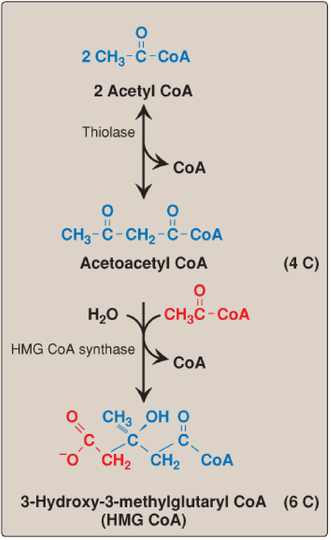
Note: There are two HMG-CoA synthase isoenzymes - the cytosolic one feeds cholesterol synthesis; the mitochondrial one feeds ketogenesis. The two pathways are distinct compartmentally.
Step 1b - Mevalonate synthesis (SER):
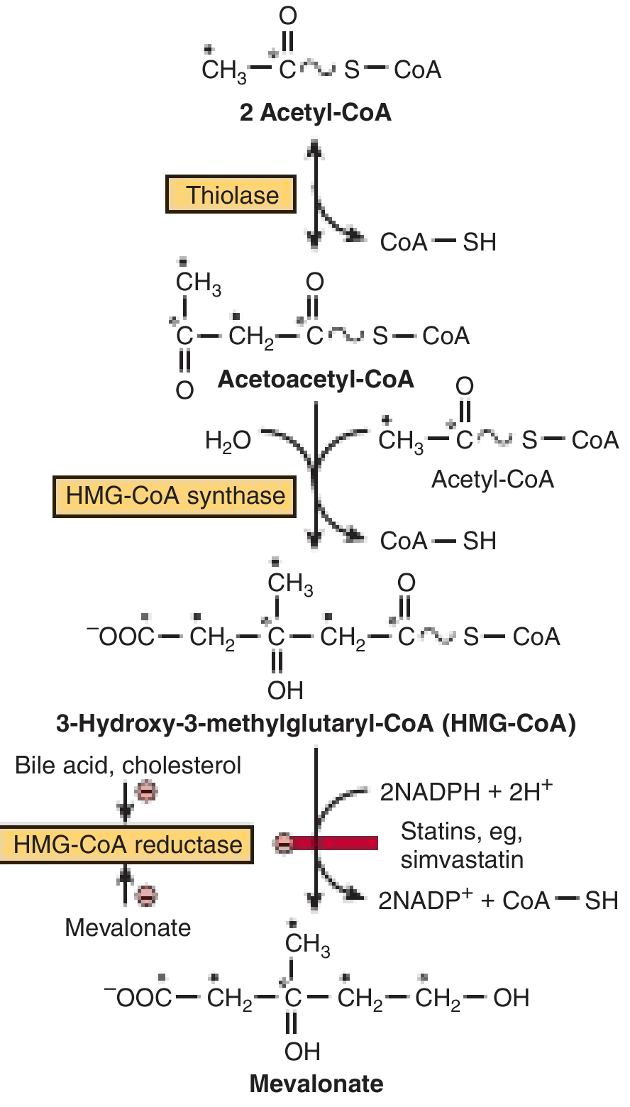
- HMG-CoA is reduced to mevalonate (6C) by HMG-CoA reductase (HMGCR)
- Reaction: HMG-CoA + 2 NADPH → Mevalonate + CoA
- This is irreversible, uses 2 NADPH, and is the rate-limiting, regulated step of the entire pathway
Stage 2: Mevalonate → Isopentenyl Pyrophosphate (IPP)
- Mevalonate undergoes two sequential phosphorylations by ATP kinases → 5-pyrophosphomevalonate
- Decarboxylation (requires ATP) produces isopentenyl pyrophosphate (IPP), the fundamental 5-carbon isoprene unit
- IPP is isomerized to dimethylallyl pyrophosphate (DMAPP / DPP) by IPP isomerase
IPP is the precursor of the entire isoprenoid family - cholesterol is a sterol isoprenoid; non-sterol isoprenoids produced here include dolichol, ubiquinone (CoQ10), and prenyl groups on proteins like Ras.
Stage 3: IPP → Squalene (30C)
Sequential head-to-tail condensations:
- IPP + DMAPP → geranyl pyrophosphate (GPP, 10C)
- GPP + IPP → farnesyl pyrophosphate (FPP, 15C)
- Two FPP molecules combine tail-to-tail (NADPH required) → squalene (30C)
Squalene synthesis requires 18 ATP total (3 ATP per mevalonate unit × 6 isoprene units).
FPP is also used in protein prenylation (farnesylation of Ras oncoproteins) - a key reason statins may have anti-tumor effects.
Stage 4: Squalene → Lanosterol (first sterol ring)
- Squalene epoxidase (SER) uses O₂ and NADPH to form squalene-2,3-epoxide
- The epoxide undergoes cyclization catalyzed by oxidosqualene cyclase (lanosterol cyclase), forming the tetracyclic steroid ring structure: lanosterol (30C)
This step generates the characteristic steroid ring (A-B-C-D ring system) in one concerted reaction.
Stage 5: Lanosterol → Cholesterol
A series of ~19 enzymatic steps in the SER convert lanosterol (30C) to cholesterol (27C):
- Three methyl groups are removed (losing 3C as CO₂)
- The side chain double bond is reduced
- The ring double bond migrates from C-8 to C-5
- Final step: 7-dehydrocholesterol reductase (DHCR7) reduces the Δ7 double bond to yield cholesterol
Smith-Lemli-Opitz syndrome is caused by a partial deficiency of DHCR7, leading to accumulation of 7-dehydrocholesterol, low cholesterol levels, and multiple developmental abnormalities.
Regulation of Cholesterol Synthesis
The principal regulatory point is HMG-CoA reductase. Four mechanisms operate:
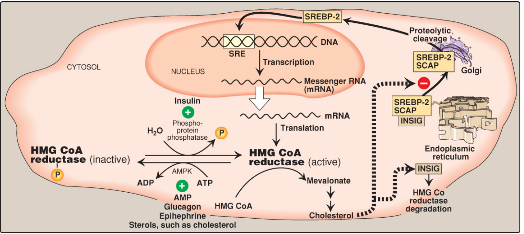
1. Transcriptional Regulation (SREBP-2 Pathway)
- When intracellular cholesterol is low, SCAP (SREBP cleavage-activating protein) escorts SREBP-2 from the ER to the Golgi
- Proteolytic cleavage releases the active transcription factor, which moves to the nucleus and binds SRE (sterol regulatory element)
- This upregulates transcription of HMG-CoA reductase and the LDL receptor
- When cholesterol is high, INSIG proteins retain the SCAP-SREBP complex in the ER - SREBP-2 is not released, transcription stops
2. Accelerated Enzyme Degradation
- When ER sterol levels are high, the reductase binds INSIG proteins, is ubiquitinated, and undergoes proteasomal degradation
3. Phosphorylation/Dephosphorylation
- AMP-activated protein kinase (AMPK) phosphorylates and inactivates HMG-CoA reductase
- Phosphoprotein phosphatase dephosphorylates and activates it
- When AMP (low energy) is high, AMPK is activated → reductase is inactivated → cholesterol synthesis decreases (similar to fatty acid synthesis control)
4. Hormonal Regulation
- Insulin → promotes dephosphorylation → activates reductase (fed state, anabolism)
- Glucagon / Epinephrine → promotes phosphorylation → inactivates reductase (fasted/stress state)
Clinical Relevance: Statins
Statins (atorvastatin, simvastatin, rosuvastatin, etc.) are structural analogs of HMG-CoA that act as reversible competitive inhibitors of HMG-CoA reductase - the rate-limiting enzyme. By lowering intracellular cholesterol, they trigger SREBP-2 activation, which also upregulates LDL receptors, increasing LDL clearance from plasma.
Key side effects include myopathy/rhabdomyolysis, partly because the mevalonate pathway also supplies heme A and ubiquinone (CoQ10), both needed for mitochondrial oxidative phosphorylation. A genetic polymorphism in the transporter OATP1B1 (SLCO1B1 521 T>C) is a biomarker for simvastatin-induced myopathy.
Summary Pathway
Acetyl-CoA (x3)
↓ [Thiolase + HMG-CoA synthase]
HMG-CoA (6C)
↓ [HMG-CoA reductase] ← RATE-LIMITING STEP (statin target)
Mevalonate (6C)
↓ [Kinases + decarboxylase, 3 ATP]
IPP / DMAPP (5C isoprene units)
↓ [Prenyltransferases]
Farnesyl-PP (15C) × 2
↓ [Squalene synthase, NADPH]
Squalene (30C)
↓ [Squalene epoxidase + Lanosterol cyclase, O₂ + NADPH]
Lanosterol (30C)
↓ [~19 SER reactions, NADPH]
Cholesterol (27C)
Sources:
- Biochemistry, 8th ed. Lippincott Illustrated Reviews, Chapter 18 (Cholesterol Synthesis, pp. 617-627)
- Harper's Illustrated Biochemistry, 32nd Ed., Chapter 26 (Cholesterol Biosynthesis)
Eye explanation with diagram
Reading File
This is a broad visual anatomy topic - searching the library in parallel for eye anatomy diagrams and content:
Finding Sources
Finding Sources
Found great sources. Reading content from Ganong's Physiology and Neuroscience simultaneously:
Reading File
Reading File
Now fetching all key diagrams in parallel:
Loading Image
Loading Image
Loading Image
Now let me also get the cross-sectional anatomy and retina/photoreceptor details:
Reading File
Finding Sources
Loading Image
Finding Sources
I have excellent content and four high-quality diagrams. Let me read the retina section from Ganong's to complete the photoreceptor content:
Reading File
I have all the material needed for a comprehensive, diagram-rich answer. Here it is:
The Human Eye - Anatomy and Function
Overview
The eye is the sensory organ of vision. It converts light energy (wavelengths 397-723 nm) into electrical signals (action potentials) carried by the optic nerve to the visual cortex. It consists of three concentric tissue layers (tunics), three fluid-filled chambers, and a refractive system that focuses sharp images onto the retina.
External View
Cross-Sectional Anatomy (Internal Structures)
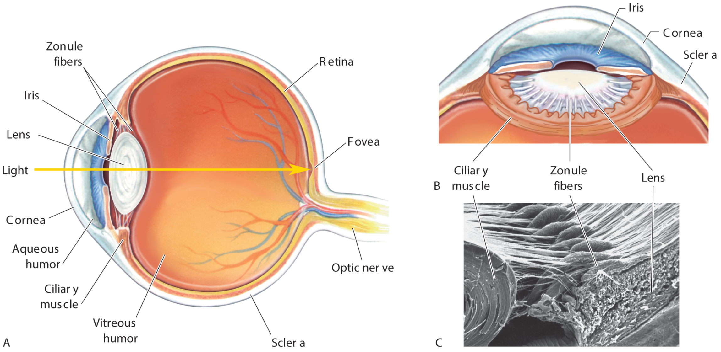
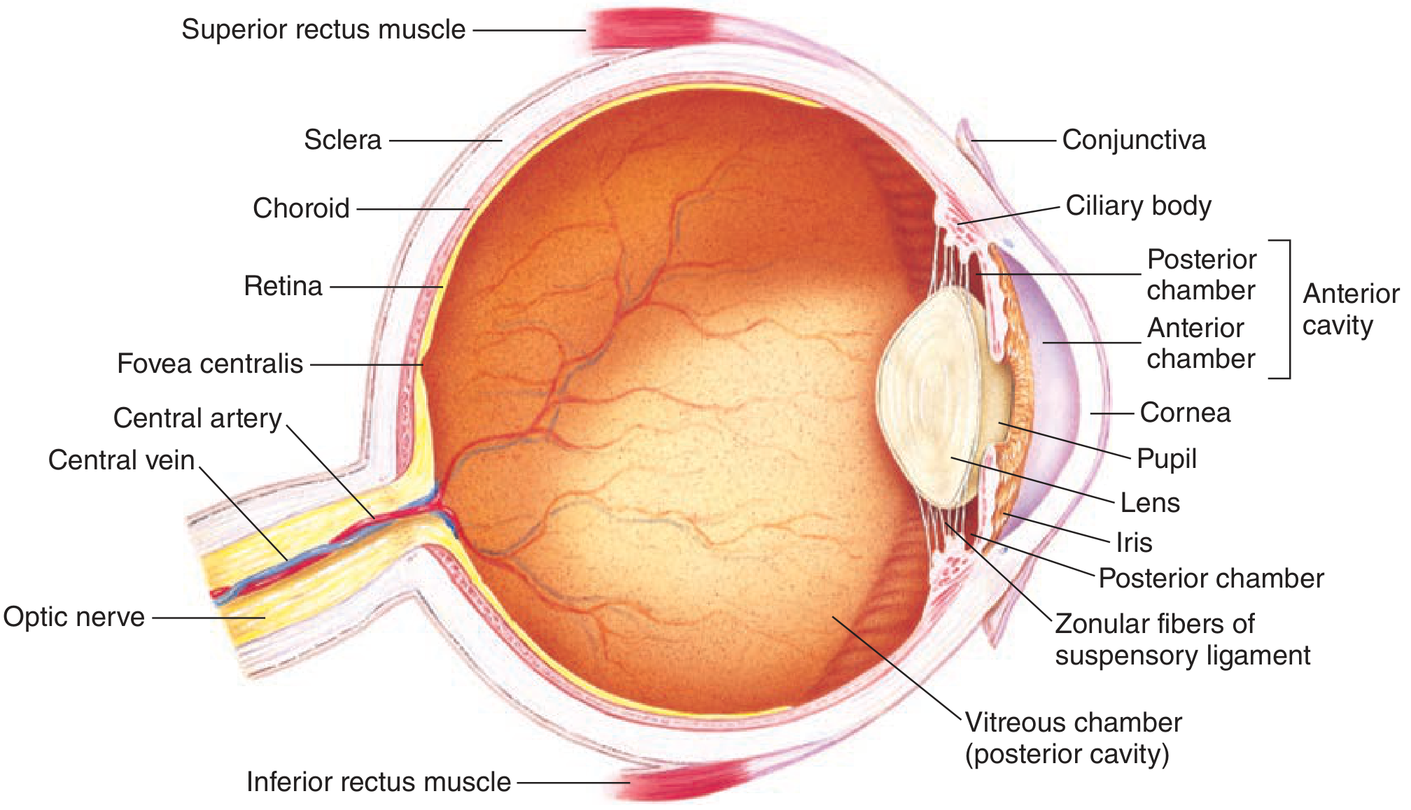
The Three Coats (Tunics) of the Eyeball
1. Fibrous Coat (Outer Layer)
| Structure | Description |
|---|---|
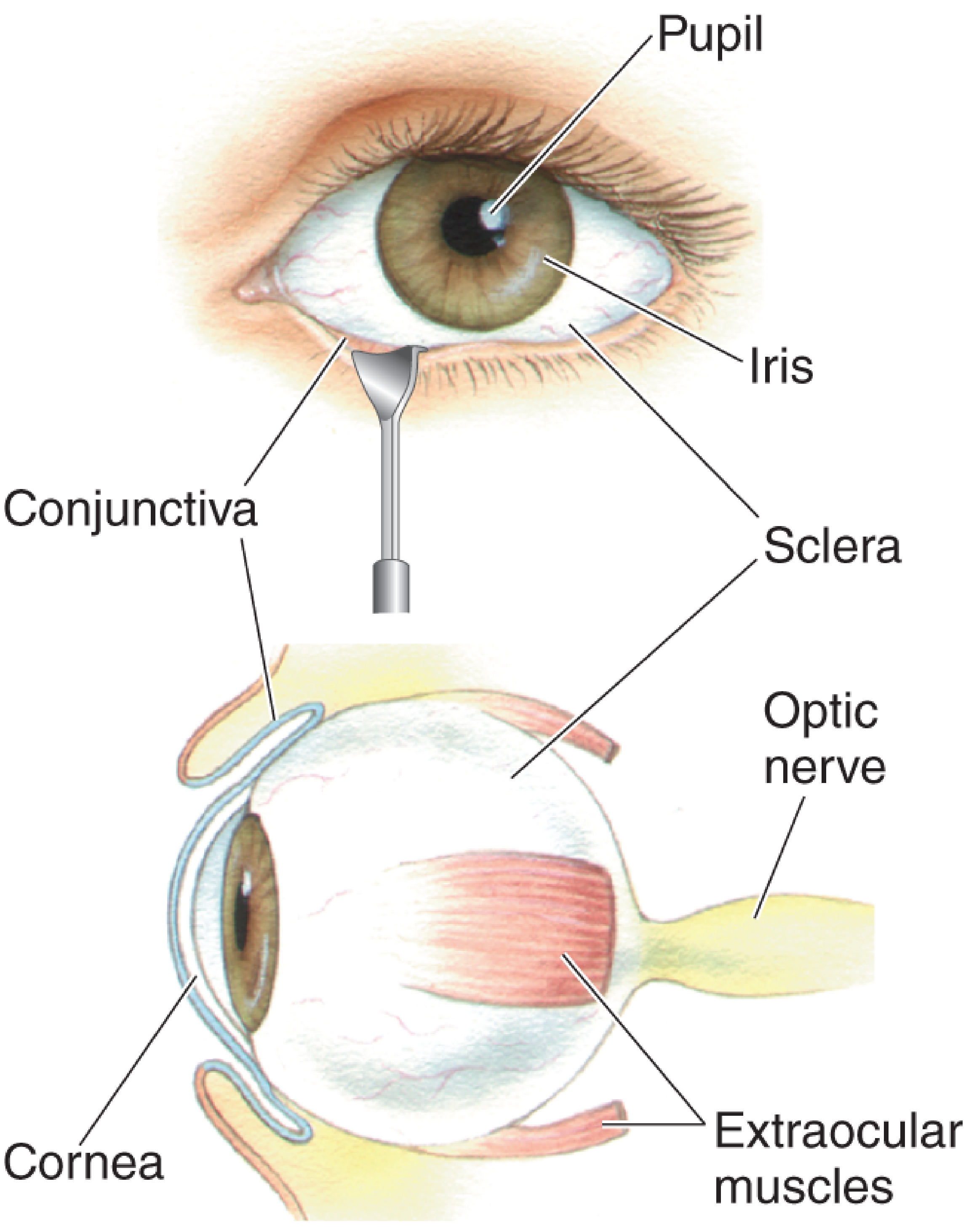
| Sclera | The tough, opaque, white outer coat. Forms the posterior 5/6 of the eyeball. Provides structural protection. No light passes through it. |
| Cornea | Transparent anterior modification of the sclera. Covers the front 1/6. The primary refractive surface of the eye (~70% of total refractive power). Avascular - nourished by aqueous humor. |
2. Vascular Coat - Uvea (Middle Layer)
| Structure | Description |
|---|---|
| Choroid | Highly vascular, pigmented layer lining the posterior 2/3 of the sclera. Supplies oxygen and nutrients to the outer retina. |
| Ciliary Body | Contains circular and longitudinal muscle fibers. Controls lens shape (accommodation) via zonule fibers. Also produces aqueous humor by active transport and diffusion from plasma. |
| Iris | Pigmented, opaque disc in front of the lens - gives the eye its color. Contains two muscles: sphincter pupillae (parasympathetic - miosis/constriction) and dilator pupillae (sympathetic - mydriasis/dilation). Pupil diameter changes can produce a 16-fold change in light reaching the retina. |
3. Neural Coat (Inner Layer)
| Structure | Description |
|---|---|
| Retina | Neural tissue lining the posterior 2/3 of the choroid. Contains photoreceptors (rods and cones), bipolar cells, and ganglion cells whose axons form the optic nerve. |
Chambers and Fluids
Anterior Cavity
- Anterior chamber - between the cornea and iris; filled with aqueous humor
- Posterior chamber - narrow space between the iris, zonule fibers, and lens; also contains aqueous humor
Aqueous humor is a clear, protein-free fluid produced by the ciliary body. It flows through the pupil from the posterior to anterior chamber, then drains via the canal of Schlemm (trabecular meshwork) at the iridocorneal angle. This drainage maintains intraocular pressure (IOP, normal 10-20 mmHg).
Blockage of aqueous drainage → raised IOP → glaucoma (open-angle: reduced trabecular permeability; closed-angle: iris balloons forward to block the angle)
Posterior Cavity (Vitreous Chamber)
- Between the lens and retina
- Filled with vitreous humor - a clear, jellylike gel that keeps the eye spherical and maintains retinal apposition
The Lens and Accommodation
The crystalline lens is a transparent, biconvex structure held in place by zonule fibers (suspensory ligament) attached to the ciliary muscle.
- Far vision (relaxed): Ciliary muscle relaxed → zonule fibers taut → lens is flat → less refractive power
- Near vision (accommodation): Ciliary muscle contracts → zonule fibers relax → lens bulges (becomes more convex) → greater refractive power
The refractive power of a lens is measured in diopters (= 1 / focal length in meters).
The Retina
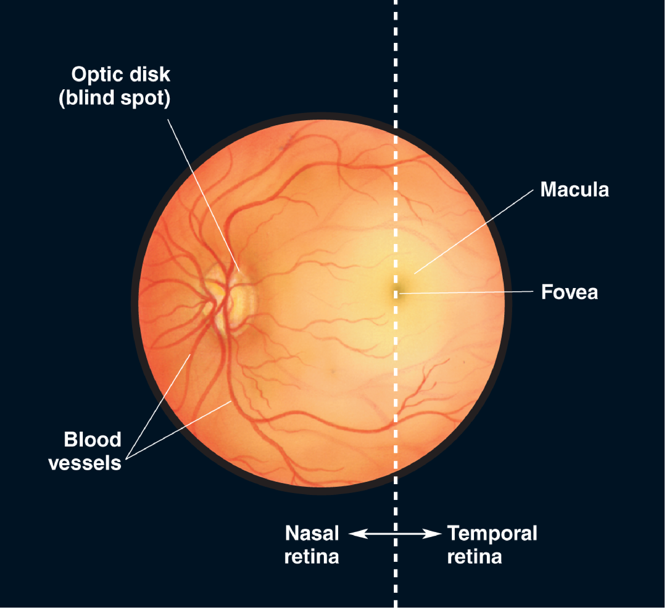
| Region | Function |
|---|---|
| Fovea centralis | Central pit in the macula. Densely packed with cones only. Site of maximum visual acuity and color vision. The point where focused light lands during direct gaze. |
| Macula lutea | Yellow pigmented area surrounding the fovea. Responsible for central, detailed vision. |
| Optic disc (optic nerve head) | Point where ganglion cell axons exit to form the optic nerve, and where central retinal artery/vein enter. Has NO photoreceptors → blind spot |
| Peripheral retina | Rich in rods. Responsible for peripheral vision and night vision. |
Photoreceptors
| Feature | Rods | Cones |
|---|---|---|
| Number | ~120 million | ~6 million |
| Location | Peripheral retina (absent from fovea) | Concentrated at fovea |
| Light sensitivity | Very high (function in dim/night light) | Low (require bright light) |
| Color discrimination | None (monochromatic) | Yes - 3 types (S, M, L = blue, green, red) |
| Pigment | Rhodopsin | Photopsins (iodopsins) |
The Light Path - From Entry to Perception
Light
→ Cornea (major refraction)
→ Aqueous humor (anterior chamber)
→ Pupil (regulates light quantity)
→ Lens (fine-tune focus / accommodation)
→ Vitreous humor
→ Retina (photoreceptors: rods and cones)
→ Bipolar cells
→ Ganglion cells → Optic nerve (CN II)
→ Optic chiasm
→ Lateral geniculate nucleus (thalamus)
→ Primary visual cortex (occipital lobe, V1)
Protection of the Eye
- Bony orbit - walls of the skull protect the eyeball from mechanical trauma
- Eyelids and blinking - physical protection; spread tears over the cornea
- Conjunctiva - clear mucous membrane lining the inner eyelids and covering the sclera; continuous with the corneal margin
- Lacrimal gland - in the upper outer orbit; produces tears that flow across the eye surface and drain into the nasolacrimal duct → nasal cavity. Tears keep the cornea moist, clear, and protected from infection
Clinical Correlations
| Condition | Structural Basis |
|---|---|
| Glaucoma | Impaired aqueous drainage → raised IOP → retinal ganglion cell death |
| Cataract | Clouding of the crystalline lens |
| Myopia (near-sightedness) | Eyeball too long or cornea too curved → image focused in front of retina |
| Hyperopia (far-sightedness) | Eyeball too short → image focused behind retina |
| Macular degeneration | Damage to fovea/macula → loss of central vision |
| Retinal detachment | Separation of neural retina from choroid → loss of photoreceptor nourishment |
Sources:
- Ganong's Review of Medical Physiology, 26th Ed., Chapter 10 (Vision)
- Neuroscience: Exploring the Brain, 5th Ed., Chapter 9 (The Eye)
This is a shared conversation. Sign in to Orris to start your own chat.