
DIC - syndrome. Etiopathogenesis and morphology
Reading File
Finding Sources
Finding Sources
Searching PubMed
"Disseminated Intravascular Coagulation"[MeSH Terms]
Reading File
Reading File
Reading File
Searching Images
disseminated intravascular coagulation fibrin thrombi glomerular capillaries
Reading File
query_images_db
Disseminated Intravascular Coagulation (DIC)
Definition
DIC is a syndrome of systemic activation of coagulation. It produces microthrombi throughout the microcirculation. Platelets and clotting factors are consumed, and fibrinolysis is activated secondarily. The result is one of two things, or both:
- tissue hypoxia and microinfarcts from microthrombi, or
- a bleeding disorder (consumptive coagulopathy).
DIC is always secondary to another disease. It probably causes bleeding more often than all the congenital coagulation disorders combined (Robbins & Kumar Basic Pathology, p. 426).
Etiology
DIC is most often associated with four groups of conditions:
| Group | Examples |
|---|---|
| Obstetric | Abruptio placentae, retained dead fetus, septic abortion, amniotic fluid embolism, eclampsia |
| Infections | Gram-negative and gram-positive sepsis, meningococcemia, Rocky Mountain spotted fever, malaria, histoplasmosis, aspergillosis |
| Neoplasms | Acute promyelocytic leukemia, and carcinomas of pancreas, prostate, lung and stomach |
| Massive tissue injury | Trauma (especially brain), burns, extensive surgery |
| Miscellaneous | Acute intravascular hemolysis, snakebite, giant hemangioma, shock, heat stroke, vasculitis, aortic aneurysm, liver disease |
The initiating events are often multiple and interrelated. In obstetric DIC, for example, placental or amniotic tissue factor enters the circulation. Coexisting shock, hypoxia and acidosis then injure the endothelium (Robbins & Kumar Basic Pathology, p. 427).
Pathogenesis
Normally, clotting is limited by three mechanisms:
- rapid hepatic clearance of activated factors,
- endogenous anticoagulants such as protein C, and
- concomitant fibrinolysis.
DIC begins with one of two triggers.
1. Release of tissue factor or other procoagulants into the circulation (extrinsic pathway)
- The sources are placenta, amniotic fluid, and tumor cells (especially acute promyelocytic leukemia and adenocarcinoma). Tumor cells can also release proteolytic enzymes.
- In sepsis, endotoxin or exotoxin induces tissue factor on monocytes.
- Activated monocytes release IL-1 and TNF. These raise tissue factor expression on endothelium and lower thrombomodulin, which activates protein C.
- The net effect is more thrombin generation and loss of the inhibitory pathways.
2. Widespread endothelial injury (intrinsic pathway and tissue factor)
- Injury exposes subendothelial collagen and vWF and releases tissue factor. Even subtle injury upregulates tissue factor and downregulates anticoagulants.
- Causes include antigen-antibody complexes (SLE), temperature extremes (heat stroke, burns), and infections (meningococci, rickettsiae).
- The systemic inflammatory response syndrome also injures endothelium.
- Brain trauma releases negatively charged membrane components that activate the intrinsic pathway.
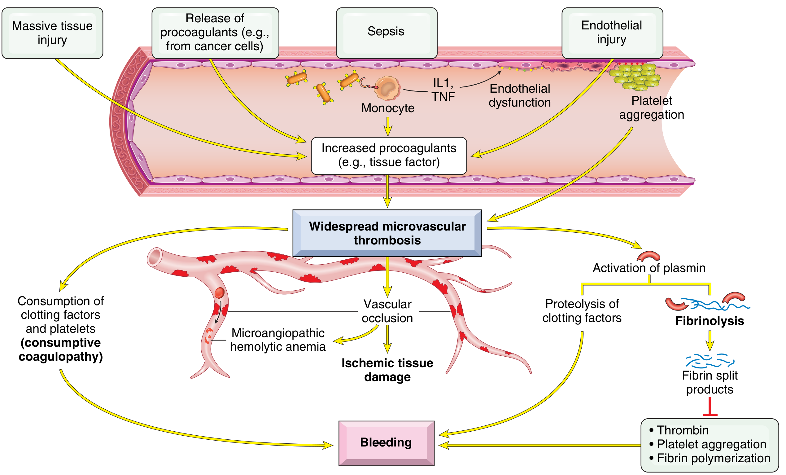
Two consequences
- Thrombosis.
- Widespread fibrin deposition in the microcirculation causes ischemia in vulnerable organs.
- Red cells fragment as they pass through fibrin-narrowed vessels. This is microangiopathic hemolytic anemia.
- Bleeding.
- Platelets and clotting factors are depleted.
- Plasminogen activators are released, so plasmin forms. Plasmin cleaves fibrin and also factors V and VIII.
- Fibrin degradation products (FDPs) inhibit platelet aggregation, have antithrombin activity, and impair fibrin polymerization.
Morphology
Microthrombi occur mainly in the arterioles and capillaries of the kidneys, adrenals, brain and heart, but no organ is spared.
- Kidney
- Glomerular capillaries contain small fibrin thrombi. These are associated with endothelial swelling or focal glomerulitis.
- Small cortical infarcts occur.
- In severe cases the whole cortex becomes ischemic, giving bilateral renal cortical necrosis.
- Adrenals: hemorrhagic necrosis can produce Waterhouse-Friderichsen syndrome.
- Brain: microinfarcts, often surrounded by microscopic or gross hemorrhage.
- Heart and anterior pituitary: similar microinfarcts. DIC can contribute to Sheehan postpartum pituitary necrosis.
- Bleeding manifestations
- Hemorrhages larger than expected near infarcts.
- Diffuse petechiae and ecchymoses of the skin.
- Hemorrhages on serosal linings of body cavities, epicardium, endocardium and lungs.
- Hemorrhage of the urinary tract mucosa.
- Blood film: schistocytes (helmet cells, fragmented red cells) and thrombocytopenia. This is my addition from general knowledge and is not from the retrieved text.
Clinical and laboratory correlates
- Acute DIC
- Fulminant, and dominated by bleeding. Examples are obstetric complications and sepsis.
- It may present with shock, acute renal failure, dyspnea, cyanosis, convulsions or coma.
- It often announces itself with petechiae and ecchymoses.
- Chronic DIC
- Mild laboratory abnormalities, with thrombosis predominating. An example is adenocarcinoma.
- Laboratory findings
- Thrombocytopenia.
- Prolonged PT and PTT.
- Low fibrinogen.
- Raised FDPs and D-dimer.
- Management
- Treat the underlying cause first.
- Support with platelets, fresh frozen plasma and cryoprecipitate as needed.
- Heparin may be used in selected thrombosis-dominant cases.
Recent evidence
A 2025 ISTH systematic review and meta-analysis on DIC mortality, diagnosis and etiology (PMID 40383152) and the 2025 Japanese DIC guidelines, part 4 (PMID 39890756) add recent data on diagnosis and management. I did not review them in full. Nothing in the abstracts' titles contradicts the pathogenesis above.
Source: Robbins & Kumar Basic Pathology, pp. 426-428.
This is a shared conversation. Sign in to Orris to start your own chat.