
Clinical tests for checking compression of sciatic nerve
Reading File
Clinical tests for sciatic nerve compression include straight leg raise (SLR/Lasegue's), Bragard's, Bowstring, slump test, and crossed SLR. Let me pull the authoritative textbook content and images for a thorough answer.
Searching sources...
Finding Sources
Finding Sources
Searching Images
straight leg raise test sciatic nerve compression clinical examination
Good sources found. Let me now read the detailed content from the best sections - Bailey & Love's (Special tests) and Rheumatology (Root tension signs).
Reading File
Reading File
Reading File
Excellent content retrieved. Let me now fetch the key images and search for additional tests (Slump test, Bowstring/Bragard's) in parallel.
Loading Image
Loading Image
Finding Sources
Good. Let me now read the slump test and bowstring sign content, and also the Rosen's EM passage on SLR.
Reading File
Reading File
I now have comprehensive content from multiple authoritative sources. Let me compile the full answer.
Clinical Tests for Sciatic Nerve Compression
The sciatic nerve is formed from the L4-S1 nerve roots. All the tests below aim to reproduce or provoke radicular pain (sciatica) by stretching the nerve and its dural attachments. These are collectively called nerve root tension signs or neural tension tests.
1. Straight Leg Raise (SLR) / Lasègue's Test
The most important and widely used test.
Technique:
- Patient lies supine, legs fully extended.
- Examiner places one hand under the ankle and one on the knee (to keep the knee straight).
- The leg is passively raised (hip flexed) with the knee extended, slowly and steadily.
- Each leg tested separately.
Positive result: Reproduction of radicular pain (shooting down the leg, typically below the knee) at 30-70° of elevation.
- Pain at < 30° is suspicious for non-organic causes (nerve root doesn't move in the foramen until > 30°).
- Pain at > 70° may simply reflect mechanical pain from muscle/ligament strain or joint disease.
- Sensitivity: 72-97%; Specificity: 11-66% - a negative SLR broadly rules out nerve root irritation. - Rosen's Emergency Medicine
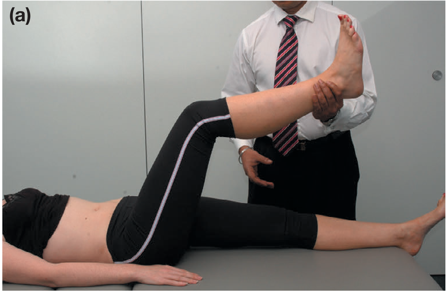
Fig. 35.8a - Lasègue's straight leg raise test (Bailey & Love's Surgery, 28th Ed.)
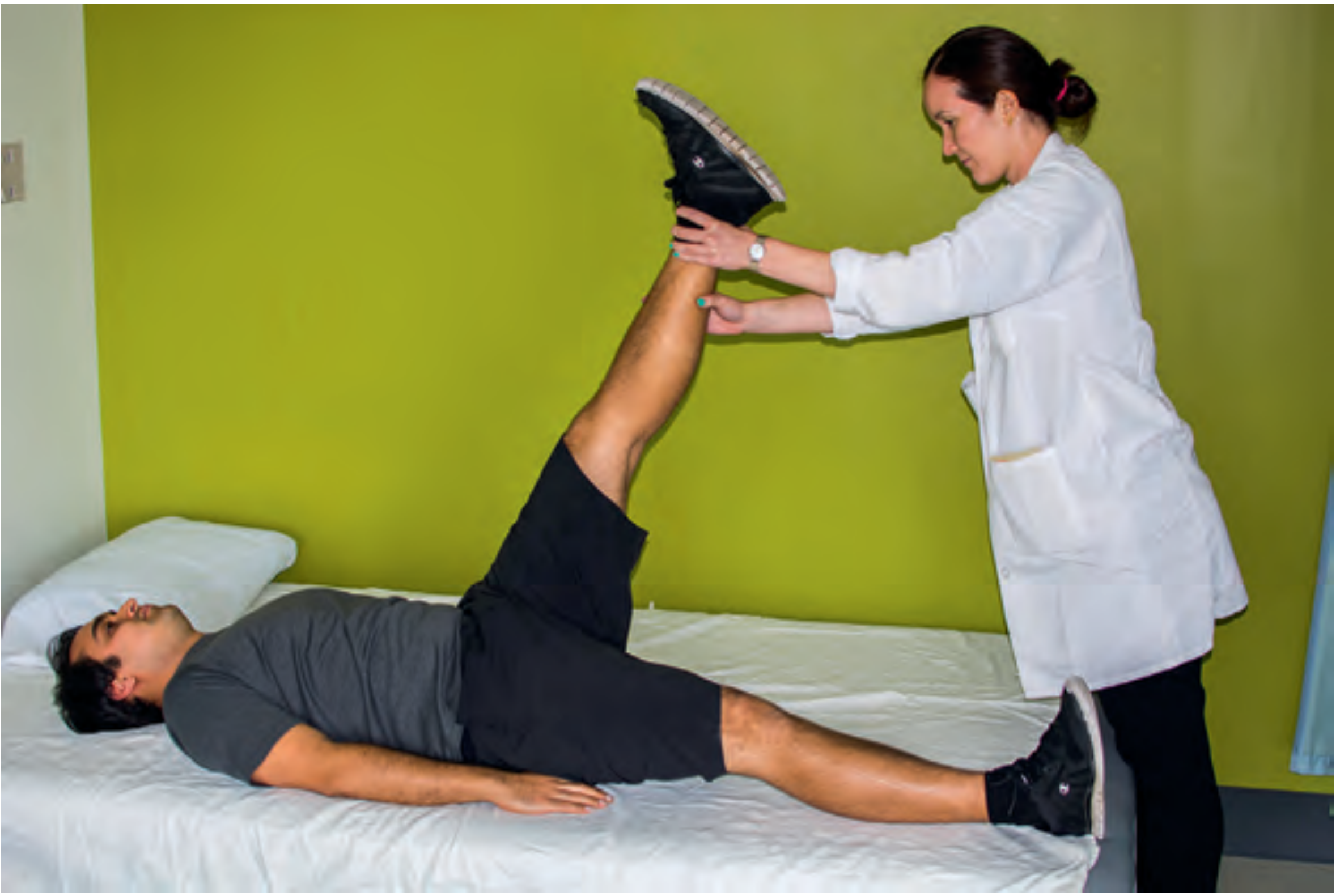
Straight-leg raise: pain between 20-70° indicates a positive result (Rheumatology, Elsevier 2022)
2. Bragard's Sign (Bragard's Test / Ankle Dorsiflexion Reinforcement)
A confirmatory adjunct to the SLR.
Technique:
- After a positive SLR, lower the leg slightly until the pain just eases.
- Then firmly dorsiflex the ankle - this further stretches the sciatic nerve through its course.
Positive result: Reproduction or intensification of radicular leg pain.
This increases the specificity of the SLR by confirming the pain is genuinely of neural origin (not just hamstring tightness). Bailey & Love's (Fig 35.8) and Rosen's EM both describe this step as an integral part of the full SLR sequence.
3. Crossed Straight Leg Raise (Crossed SLR / Well-Leg Raise / Contralateral SLR)
Technique:
- Raise the asymptomatic (contralateral) leg with the knee extended.
- Positive result: Pain is reproduced in the opposite (symptomatic) leg.
Clinical significance:
- Sensitivity: ~29%; Specificity: 85-100% - insensitive but highly specific.
- A positive crossed SLR is highly indicative of a herniated disc causing nerve root compression (positive in ~97% of disc herniations per Bailey & Love's).
4. Bowstring Sign (Cram Test / Popliteal Compression Test)
Technique:
- Perform an SLR to the point of pain.
- Then flex the knee slightly (to about 20°) - this usually relieves the pain by releasing hamstring tension.
- Then press firmly on the popliteal fossa (compressing the tibial nerve / sciatic nerve at the back of the knee).
Positive result: Reproduction of radicular pain down the leg when the popliteal nerve is compressed.
This confirms that the pain was genuinely due to sciatic nerve tension rather than hamstring tightness. Listed in Rheumatology (Elsevier) as a test for nerve root compression at L5/S1.
5. Slump Test (Seated Neural Tension Test)
Used when a patient is unable or unwilling to lie supine for SLR.
Technique (sequential steps):
- Patient sits at the edge of the table, hands behind back.
- Patient slumps forward (thoracic and lumbar flexion).
- Examiner applies overpressure to the cervical spine (chin to chest - neck flexion).
- Patient then extends the knee on the affected side.
- Patient then dorsiflexes the ankle.
Positive result: Reproduction of radicular leg pain. Releasing the neck flexion typically relieves the pain (confirms neural origin).
The slump test places the entire neural axis under tension. Per Rosen's EM, it is the preferred alternative when the supine SLR cannot be performed.
6. Distracted SLR (Sitting SLR)
Technique:
- Performed while the patient is seated and distracted (e.g., while appearing to examine the knee).
- The knee is passively extended.
Positive result: Radicular pain reproduced.
Used to detect symptom magnification or non-organic back pain (Waddell's signs) - a discrepancy between the supine SLR and distracted seated SLR suggests a non-organic component. - Rosen's Emergency Medicine
Summary Table
| Test | Position | Maneuver | Positive Finding | Nerve Roots |
|---|---|---|---|---|
| SLR / Lasègue's | Supine | Passive leg elevation, knee straight | Radicular pain at 30-70° | L5, S1 |
| Bragard's Sign | Supine (after SLR) | Lower leg to pain-free, then dorsiflex ankle | Pain reproduced | L5, S1 |
| Crossed SLR | Supine | Raise contralateral (normal) leg | Sciatica in opposite leg | L5, S1 |
| Bowstring Sign | Supine (after SLR) | Press popliteal fossa with knee slightly flexed | Radicular pain reproduced | L5, S1 |
| Slump Test | Sitting | Slump + neck flex + knee extension + dorsiflexion | Radicular pain reproduced | L5, S1 |
| Distracted SLR | Sitting | Extend knee while patient distracted | Radicular pain reproduced | L5, S1 |
Important Notes
- True positive SLR = pain radiating below the knee, following a dermatomal pattern (not just back or buttock pain).
- The SLR stretches L4, L5, and S1 roots. It is most sensitive for L5 and S1 root compression.
- Femoral stretch test (reverse SLR - patient prone, knee flexed, thigh lifted) tests the upper lumbar roots (L2, L3, L4) - not the sciatic nerve.
- A positive crossed SLR is the most specific single sign for disc herniation compressing a nerve root.
Sources: Bailey & Love's Short Practice of Surgery 28th Ed., Rheumatology 2-Volume Set (Elsevier 2022), Rosen's Emergency Medicine, Goldman-Cecil Medicine, Adams & Victor's Principles of Neurology 12th Ed.
This is a shared conversation. Sign in to Orris to start your own chat.