
Properties of smooth muscles
smooth muscle contraction mechanism calmodulin myosin light chain kinase diagram
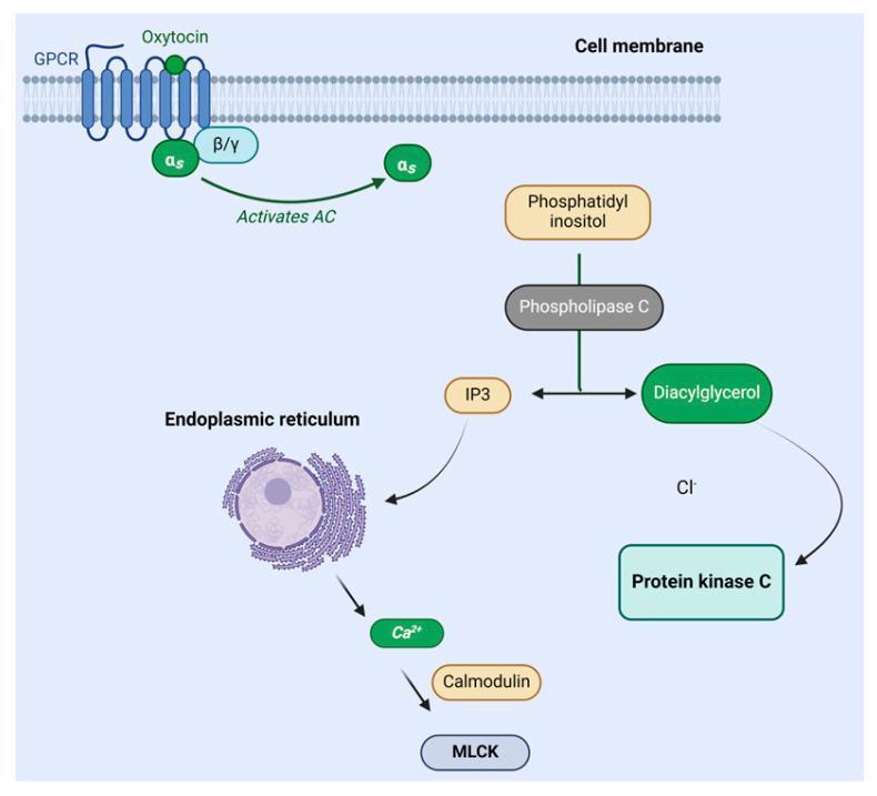
A pathophysiology diagram illustrating the oxytocin signaling pathway within a cell. The mechanism begins at the cell membrane where oxytocin binds to a G protein-coupled receptor (GPCR). This binding triggers the G-protein subunits (αs and β/γ), leading to the activation of adenylyl cyclase (AC). Downstream, phospholipase C (PLC) catalyzes the conversion of phosphatidylinositol into two secondary messengers: diacylglycerol (DAG) and inositol triphosphate (IP3). DAG is shown activating protein kinase C (PKC). Simultaneously, IP3 interacts with the endoplasmic reticulum, prompting the release of calcium ions (Ca2+). These calcium ions bind to calmodulin, which subsequently activates myosin light chain kinase (MLCK). This biochemical cascade is fundamental to understanding myometrial contraction and milk ejection. The diagram uses standard medical icons and labels to represent intracellular components and molecular interactions, suitable for undergraduate to advanced medical education.
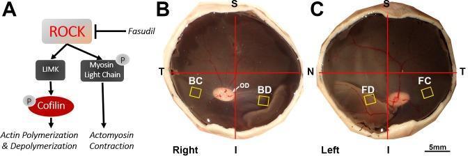
Educational figure illustrating the molecular mechanism and experimental model of retinal detachment and Rho-kinase (ROCK) inhibition. Panel A is a pathophysiology diagram showing that Fasudil inhibits ROCK, which normally activates LIMK and Myosin Light Chain. This inhibition prevents cofilin phosphorylation and actomyosin contraction, theoretically stopping photoreceptor axon retraction. Panels B and C are clinical specimen photographs of porcine eyecups (right and left eyes) used for in vivo studies. The images show the posterior segment with anatomical markers: S (Superior), I (Inferior), T (Temporal), N (Nasal), and OD (Optic Disc). Yellow squares indicate sample collection areas for morphological analysis: 'BC' and 'FC' represent attached retinal control areas, while 'BD' (control BSS) and 'FD' (Fasudil-treated) indicate areas of iatrogenic retinal detachment created in the inferior-nasal quadrant. The visual contrast between attached and detached retinal folds is evident. This material is designed for advanced ophthalmic research on neuroprotection and synaptic remodeling following retinal injury.
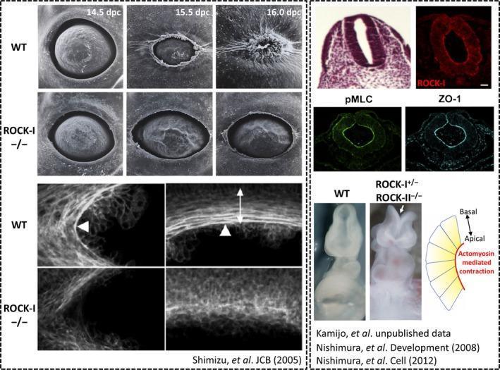
This composite educational image illustrates the role of Rho-associated coiled-coil containing kinase (ROCK) in embryonic development, specifically in eyelid and neural tube closure. The left panel features scanning electron micrographs (SEM) comparing eyelid morphogenesis in wild-type (WT) and ROCK-I−/− mouse embryos at 14.5, 15.5, and 16.0 days post-coitum (dpc). While WT eyelids successfully migrate and close, ROCK-I−/− embryos exhibit an 'eyes open at birth' phenotype. Below the SEMs, whole-mount immunofluorescence staining shows organized F-actin bundles (indicated by arrowheads) encircling the eye in WT embryos, which are significantly disrupted in ROCK-I mutants. The right panel demonstrates ROCK's involvement in neural tube closure. It includes H&E staining of a mouse embryo neural tube and immunofluorescence micrographs showing the localization of ROCK-I, phosphorylated myosin light chain (pMLC), and the apical marker ZO-1 along the apical surface of the neuroepithelium. A stereomicroscope comparison of 9.5 dpc embryos highlights impaired neural tube closure in ROCK-I+/−; ROCK-II−/− mutants. A schematic diagram summarizes the mechanism: ROCK-mediated actomyosin contraction at the apical surface facilitates tissue bending for morphogenetic closure.
Properties of Smooth Muscle
1. Structure and Morphology
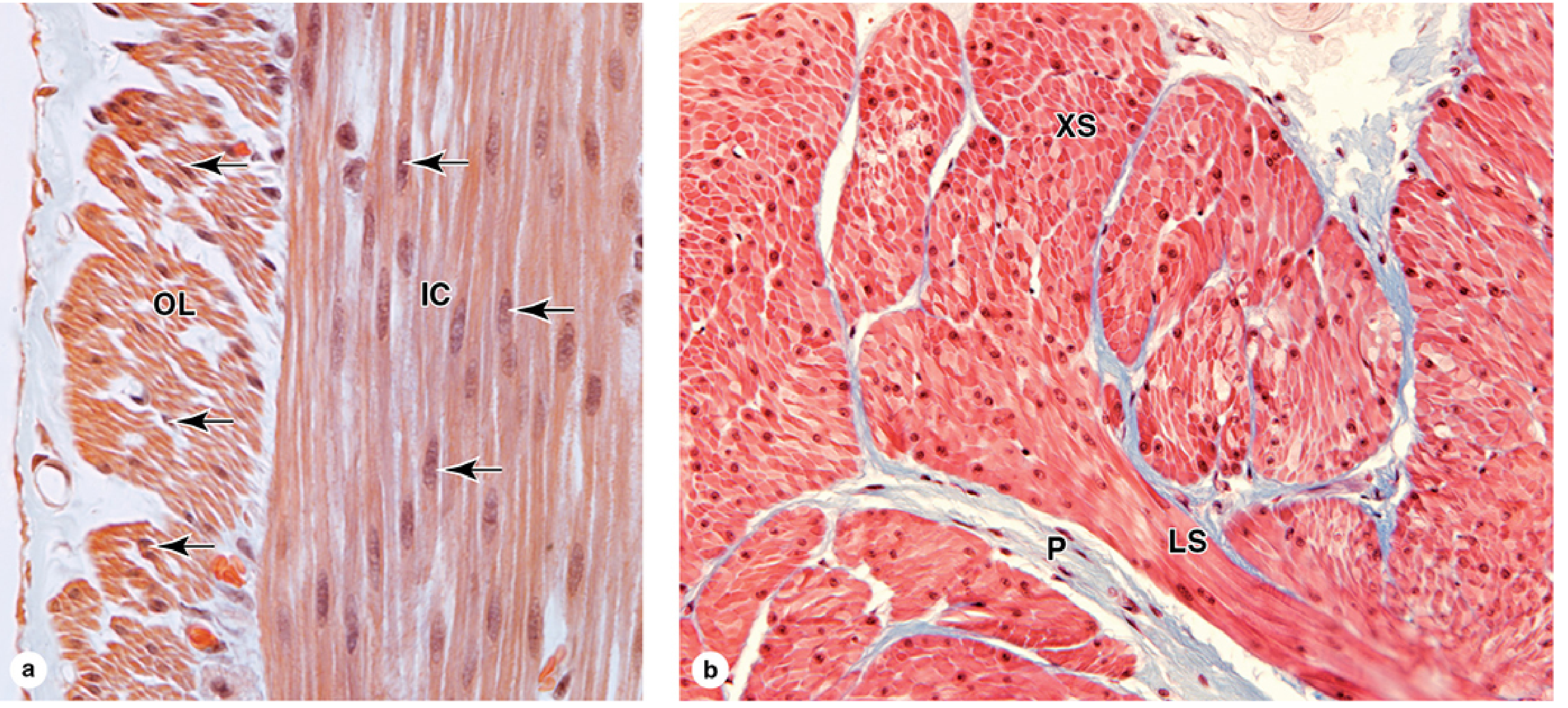
- Shape: Long, tapering cells with a single, centrally located, elongated nucleus at the cell's widest point. The nucleus becomes distorted (corkscrew appearance) during contraction.
- Size: Ranges from 20 µm (small blood vessels) to 500 µm (pregnant uterus). Diameter at the widest point is 5-10 µm.
- No striations: Unlike skeletal and cardiac muscle, smooth muscle lacks sarcomeres. Thick (myosin) and thin (actin) filaments are present but not organized into regular banding patterns.
- Packing arrangement: Narrow ends of each cell lie adjacent to the broad parts of neighboring cells - this achieves close packing.
- Organelles: Mitochondria, polyribosomes, RER, and Golgi apparatus are concentrated near the poles of the nucleus.
- Caveolae: Instead of T-tubules, smooth muscle has shallow invaginations of the plasma membrane called caveolae - these are lipid raft-enriched microdomains involved in signal transduction and local Ca²⁺ handling.
- Dense bodies: Actin filaments anchor to dense bodies (analogous to Z-discs) scattered through the cytoplasm and attached to the cell membrane.
- Sarcoplasmic reticulum (SR): Less developed than in skeletal muscle. A peripheral SR encircles the plasma membrane near caveolae; a central SR runs along the long axis and delivers Ca²⁺ to myofibrils.
2. Location and Function
- Walls of hollow organs: GI tract, bladder, uterus, ureter
- Vasculature (arteries, arterioles, veins)
- Airways (bronchioles)
- Eye (iris, ciliary muscles)
- Reproductive system, skin (arrector pili)
- Motility - propelling contents along luminal organs (e.g., peristalsis in the GI tract, urine along the ureter)
- Maintaining tension - regulating vascular tone, airway diameter
3. Types of Smooth Muscle
| Type | Location | Features |
|---|---|---|
| Unitary (single-unit) | GI tract, bladder, uterus, ureter | Gap junctions link cells; coordinated contraction; spontaneous pacemaker activity / slow waves |
| Multiunit | Iris, ciliary muscle, vas deferens | Each fiber acts independently; densely innervated by autonomic nerves; little or no cell-cell coupling |
| Vascular | Blood vessel walls | Combination of unitary and multiunit properties |
4. Excitation-Contraction (E-C) Coupling
Steps:
-
Depolarization opens voltage-gated Ca²⁺ channels (Cav1.2) in the sarcolemma → Ca²⁺ enters the cell. (Even subthreshold depolarization - without a full action potential - can trigger this in smooth muscle.)
-
Additional Ca²⁺ sources:
- Ca²⁺ entry triggers Ca²⁺-induced Ca²⁺ release (CICR) from SR via RYR3
- Hormones/neurotransmitters activate phospholipase C (PLC) → IP₃ → IP₃R-mediated Ca²⁺ release from SR
- Ligand-gated Ca²⁺ channels (opened by agonists) contribute further Ca²⁺
-
Ca²⁺ binds calmodulin (4 ions cooperatively) → Ca²⁺-calmodulin complex activates myosin light-chain kinase (MLCK)
-
MLCK phosphorylates the myosin light chain → changes conformation of the myosin head → enables cross-bridge cycling with actin
-
Relaxation: Ca²⁺ falls → calmodulin dissociates from MLCK → myosin light-chain phosphatase (MLCP) dephosphorylates myosin → cross-bridges detach → relaxation
Latch State:
5. Electrical Properties
| Property | Detail |
|---|---|
| Action potential type | Spikes, plateaus, OR graded membrane potential changes |
| Slow waves | Spontaneous oscillations in membrane potential (pacemaker activity) - characteristic of unitary smooth muscle |
| Threshold | Lower than skeletal muscle; subthreshold depolarization can still cause Ca²⁺ entry and contraction |
| Regulation | Also via Ca²⁺-activated K⁺ channels and Ca²⁺-activated Cl⁻ channels (affect repolarization) |
6. Mechanical Properties
- Slow, sustained contractions - compared to the quick contractions of skeletal and cardiac muscle
- Involuntary - under autonomic nervous system and hormonal control
- Large length-tension range: Can generate force over a wide range of lengths (important for hollow organs that change volume, like the bladder)
- Plasticity: Smooth muscle can adapt its resting length - this is the stress-relaxation or plastic property
- Low energy consumption: The latch mechanism allows prolonged tone with minimal ATP expenditure
7. Regulation (Autonomic and Hormonal)
- Autonomic innervation: Sympathetic and parasympathetic inputs (note: the neurotransmitter is released diffusely from varicosities, not at discrete end-plates)
- Hormones: Oxytocin (uterus), angiotensin II, epinephrine, etc.
- Local factors: Stretch, local pH, PO₂, PCO₂, metabolites
8. Plasticity and Phenotypic Adaptability
- Uterus (myometrium) undergoes transformation during pregnancy: hypertrophy, increased gap junction formation, isoform changes in contractile proteins, new ion channel and hormone receptor expression to allow coordinated labor contractions.
- Smooth muscle cells express a wider variety of contractile protein isoforms than striated muscle - this accounts for the tissue-specific diversity in contraction characteristics.
- Smooth muscle can also transdifferentiate into a synthetic phenotype (producing extracellular matrix) vs. a contractile phenotype - especially relevant in vascular disease (atherosclerosis).
Summary Comparison Table
| Property | Smooth Muscle | Skeletal Muscle | Cardiac Muscle |
|---|---|---|---|
| Striations | Absent | Present | Present |
| Control | Involuntary | Voluntary | Involuntary |
| Ca²⁺ sensor | Calmodulin | Troponin C | Troponin C |
| EC Coupling | CICR + IP₃R + MLCK pathway | Mechanical coupling (RYR1) | CICR via RYR2 |
| Speed | Slow | Fast | Intermediate |
| Nuclei per cell | 1 (central) | Multiple (peripheral) | 1-2 (central) |
| Gap junctions | Yes (unitary type) | No | Yes (intercalated discs) |
| Fatigue | Very resistant | Yes | Resistant |