
Trigeminal neuralgia
"trigeminal neuralgia"[MeSH Terms] AND treatment
trigeminal neuralgia nerve anatomy distribution face
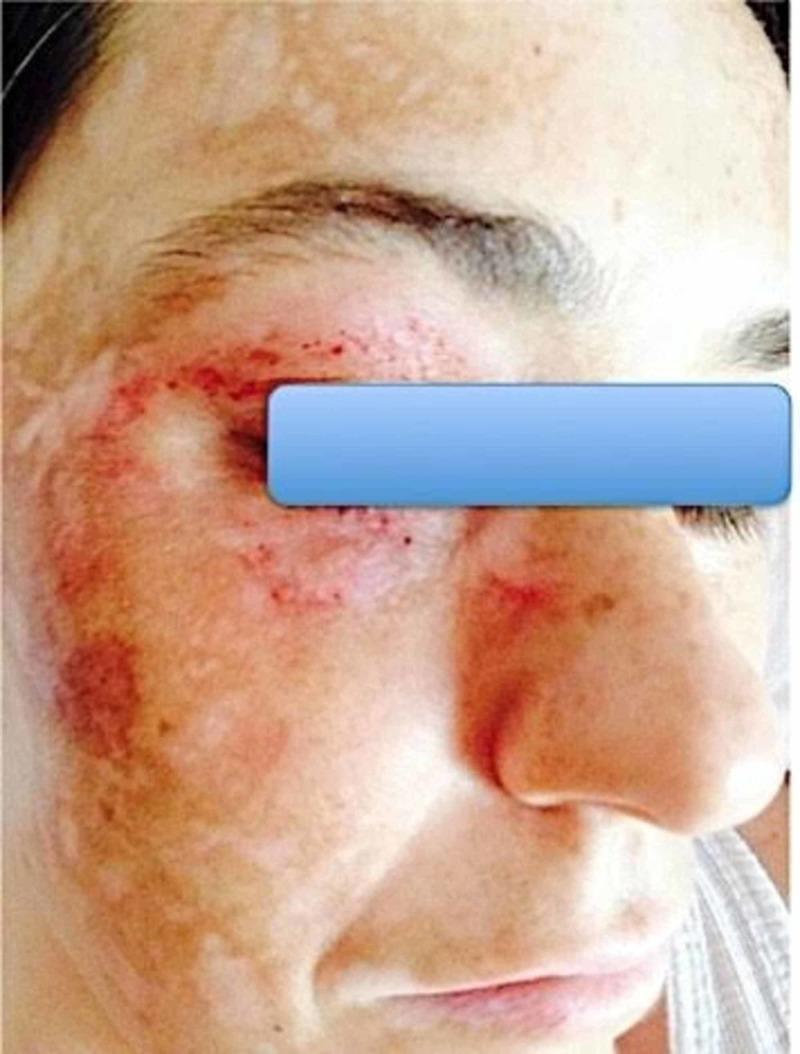
This clinical photograph shows a close-up frontal view of a patient's face, primarily focusing on the right side. The skin exhibits complex dermatological changes following stereotactic radiosurgery for trigeminal neuralgia. The lesions are localized in a distribution corresponding to the branches of the right trigeminal nerve (V1, V2, and V3). Morphological features include multiple erythematous, petechial-like punctate lesions and maculopapular eruptions around the orbital region and right cheek. Additionally, there are significant areas of dyschromia, characterized by large, irregular patches of hypopigmentation (leukoderma) interspersed with hyperpigmented macules, giving the skin a mottled appearance. The skin texture appears uneven with areas of thinning and possible scarring. This image demonstrates the dermatological manifestations of Trigeminal Trophic Syndrome (TTS), a rare complication where self-inflicted trauma or dysregulated sensory feedback leads to chronic skin lesions in the anesthetic distribution of the nerve. It serves as an educational example of the clinical relationship between neuropathic pain disorders and secondary cutaneous pathology.

Diagnostic diagnostic image showing a Stereotactic Radiosurgery (SRS) treatment plan for refractory right trigeminal neuralgia in axial, coronal, and sagittal planes. The imaging modality is MRI-based planning software. The primary anatomical focus is the Trigeminal Nerve Root (TNR) at its exit from the lateral pons (brainstem) toward Meckel's cave. Key contours include the brainstem outlined in green and the TNR target contoured in red. Superimposed on the anatomy are dose distribution curves (isodose lines): the 40 Gy isodose line (50% of the prescription dose) is shown in light green, encompassing the TNR, while the 16 Gy isodose line is shown in blue, demonstrating the steep dose gradient designed to spare the adjacent brainstem (Organs at Risk). A plus sign indicates the treatment isocenter. This visual illustrates the clinical application of high-precision radiation oncology to target small cranial nerves while maintaining a maximum brainstem dose below 16 Gy, optimizing the therapeutic ratio for pain relief while avoiding neurological deficits.
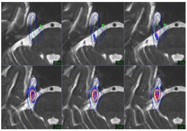
This diagnostic image sequence consists of six axial slices (284–291) showing fused heavy T2-weighted MRI and thin-slice CT for radiosurgery planning in a case of trigeminal neuralgia. The images visualize the trigeminal nerve complex, including the pre-ganglion portion, Gasserian ganglion, and retro-ganglion portion at the petrous apex. Superimposed on the anatomy are color-coded isodose lines representing radiation dose distribution. The prescribed target area is the retro-ganglion portion, indicated by high-intensity red and orange central isodose lines that expand and intensify from slice 289 to 291. Surrounding blue and light blue lines delineate lower dose gradients. A critical anatomical feature, the abducens nerve (highlighted in green), is visible and positioned outside the prescribed isodose range to ensure functional preservation. The fused imaging provides the high-contrast soft tissue detail of T2-MRI for nerve identification and the high-density bone detail of CT for spatial orientation, facilitating precise stereotactic targeting for neurosurgical treatment.
Trigeminal Neuralgia
Definition
- Goldman-Cecil Medicine, p. 1072
- Bradley and Daroff's Neurology in Clinical Practice, p. 2498
Classification (ICHD-3)
| Type | Feature |
|---|---|
| Classical TN | Vascular compression of the trigeminal nerve root confirmed on MRI or surgery, with nerve atrophy or displacement |
| Secondary TN | Due to an identifiable disease - multiple sclerosis, space-occupying lesion, etc. May present with both paroxysmal and continuous pain |
| Idiopathic TN | No causative lesion; no morphological change from vascular contact |
- Bradley and Daroff's Neurology, p. 2498
Epidemiology
- Incidence: ~4 per 100,000 population
- Onset after age 40 in 90% of patients; most common between ages 50-70
- Slightly more common in women (F:M ratio ~1.5:1)
- Incidence increases with age
- Rare familial cases exist; in younger patients, think of multiple sclerosis
Pathogenesis
- Focal hyperexcitability of axons
- Ectopic and repetitive neuronal discharges
- The characteristic paroxysmal pain
- Bradley and Daroff's Neurology, p. 2498
Clinical Features
Pain Characteristics
- Quality: Sharp, lancinating (shooting), electric shock-like, stabbing
- Duration: Each attack lasts only seconds (up to 2 minutes)
- Pattern: Repetitive attacks at short intervals; attacks may blur together
- Location: Unilateral; V2 (cheek) and V3 (chin, lower teeth) most common; V2+V3 combination is the most frequent; V1 (ophthalmic) involvement alone is extremely rare
- Interictal period: A refractory period after each volley; most patients are pain-free between attacks, though some have a residual dull continuous pain
Triggers
- Touching the face or nasolabial fold
- Chewing, teeth brushing, talking
- Cool breeze or air movement across the face
- Eating (mastication)
Physical Findings (Classical TN)
- No sensory deficit - this is key
- Motor division of the nerve is intact
- Presence of sensory loss or masticatory weakness suggests secondary TN or trigeminal neuropathy, and warrants further investigation
Associated Features (Severe Cases)
- Weight loss and dehydration (avoidance of eating/drinking due to trigger)
- Depression
Diagnosis
- Paroxysmal attacks of pain lasting 1 second to 2 minutes
- Affects one or more divisions of the trigeminal nerve
- Pain is intensely sharp, stabbing, or precipitated by a trigger
- Each attack is stereotypical
- No neurological deficits (in classical/idiopathic TN)
Investigations
- MRI is recommended in all patients to:
- Identify structural lesions (pontine lacunar infarct, demyelinating plaque, meningioma, schwannoma, skull base malignancy)
- High-resolution MRI/MRA may demonstrate vascular compression
- EMG and nerve conduction studies (e.g., blink reflex) are normal in idiopathic TN
Differential Diagnosis
- Trigeminal autonomic cephalalgias (have autonomic accompaniments)
- Atypical facial pain
- Idiopathic stabbing headache
- Tolosa-Hunt syndrome (inflammatory, anterior cavernous sinus)
- Dental pain
- Postherpetic neuralgia (history of herpes zoster, continuous burning pain)
Treatment
Medical Management
| Drug | Dose | Notes |
|---|---|---|
| Carbamazepine | 400-1200 mg/day | Drug of choice; start low (50-100 mg), titrate slowly |
| Oxcarbazepine | 300-1800 mg/day | May be better tolerated; watch for hyponatremia |
- Highly favorable response in the majority of patients with sodium channel blockers
- Monitor: CBC (agranulocytosis), LFTs, serum sodium for first few months and annually thereafter
- Gabapentin (900-1800 mg) - favorable side-effect profile, can be tried as alternative to carbamazepine
- Pregabalin
- Phenytoin (200-300 mg)
- Baclofen (50-60 mg)
- Lamotrigine (100-400 mg)
- Valproic acid (500-1500 mg)
- Clonazepam (2-6 mg)
- Topiramate (50-200 mg)
- Levetiracetam (2-4 g)
- Botulinum toxin (emerging evidence)
- IV fosphenytoin 15-20 mg PE/kg for severe acute attacks
- Topical ophthalmic anesthetic (proparacaine) to ipsilateral conjunctival sac - can provide hours to days of relief
Surgical Management
| Procedure | Mechanism | Notes |
|---|---|---|
| Microvascular decompression (MVD) | Separates compressing vessel from trigeminal root | Best long-term pain relief; preserves sensory function; requires craniotomy |
| Percutaneous radiofrequency thermocoagulation | Heat lesion to gasserian ganglion | Good for elderly or medically unfit patients |
| Percutaneous glycerol rhizotomy | Chemical destruction of gasserian ganglion | Less invasive |
| Gamma Knife radiosurgery (stereotactic) | Focused radiation to trigeminal root entry zone | Minimally invasive; delayed onset of effect |
| Peripheral nerve block (alcohol injection) | Blocks peripheral branch (mental, infraorbital, supraorbital nerve) | Simple, low morbidity; relief lasts 6-18 months; useful bridge |
Choosing the Surgical Option
- Depends on: patient age, medical comorbidities, risk tolerance, desire for sensory preservation
- MVD has the highest long-term success and preserves sensation but carries craniotomy risk
- Percutaneous procedures are preferred for elderly/frail patients
- Radiosurgery is preferred for patients who prefer a non-invasive approach or have contraindications to open surgery
Course and Prognosis
- TN tends to be a relapsing-remitting condition; spontaneous remissions occur
- Medical therapy controls pain in most patients but long-term dependence on medications is common
- Surgical outcomes are generally favorable, with MVD offering the best durable pain relief
Key Points Summary
| Feature | Detail |
|---|---|
| Most common division | V2 + V3 |
| Trigger | Touch, chewing, talking, wind |
| Pathology | Neurovascular compression → focal demyelination at root entry zone |
| First-line drug | Carbamazepine |
| Best surgical option (long-term) | Microvascular decompression (MVD) |
| Red flag (suggests secondary TN) | Sensory loss, motor weakness, young patient |
| Key investigation | MRI brain (with MRA) |