
Louis Pasteur and contribution
Louis Pasteur contributions germ theory pasteurization rabies vaccine official history
Pasteur germ theory
Louis Pasteur (1822-1895) and his contributions
-
Germ theory of disease
- He showed that microorganisms are present in air and can cause fermentation, spoilage, and disease.
- His experiments disproved the idea of spontaneous generation, the belief that living organisms arise from non-living matter.
- This work helped establish that many infectious diseases are caused by specific microbes. Park's Textbook of Preventive and Social Medicine, section “Germ theory of disease,” lines 413-430.
-
Pasteurization
- He developed controlled heating of liquids to destroy harmful or spoilage-causing microorganisms without greatly altering the product.
- It was first applied to wine and beer, and later became widely used for milk and other foods.
- Pasteurization improved food safety and reduced foodborne infection.
-
Fermentation
- Pasteur demonstrated that fermentation is caused by living microorganisms, such as yeast, rather than being only a chemical process.
- This was important for the wine, beer, and dairy industries.
-
Vaccination
- Pasteur developed or advanced vaccines against:
- Chicken cholera
- Anthrax
- Rabies
- In 1885, he treated Joseph Meister, a boy bitten by a rabid dog, with a rabies vaccine. This became a landmark in post-exposure prevention. Sherris & Ryan's Medical Microbiology, “Rabies,” lines 1470-1474.
- Pasteur developed or advanced vaccines against:
-
Public health and aseptic practice
- His discoveries supported sterilization, cleanliness, infection prevention, and later antiseptic surgical methods.
- They changed medical understanding by emphasizing that infections can be prevented by controlling microbes.
-
Silkworm disease
- Pasteur identified microbial causes of silkworm epidemics and developed methods to prevent spread, helping save France's silk industry.
Robert coach contribution
Robert Koch contributions microbiology Koch postulates tuberculosis cholera official history
Robert Koch tuberculosis
Robert Koch (1843-1910) and his contributions
-
Proved specific microbes cause specific diseases
- Koch provided strong experimental evidence that infectious diseases are caused by particular microorganisms, helping establish the germ theory of disease. Janeway's Immunobiology, “Basic Concepts in Immunology,” lines 28-31.
-
Koch’s postulates
- He formulated principles to link a microorganism to a disease:
- The organism should be associated with the disease.
- It should be isolated and grown in pure culture.
- The cultured organism should cause the same disease in a susceptible host.
- The same organism should be re-isolated from that host.
- These principles became the classic framework for identifying disease-causing microbes, although they have limitations for viruses and organisms that cannot be cultured easily.
- He formulated principles to link a microorganism to a disease:
-
Anthrax
- Koch showed that Bacillus anthracis causes anthrax and studied its spores.
- This was a major early demonstration that a specific bacterium can cause a specific disease.
-
Tuberculosis
- In 1882, Koch identified the tubercle bacillus, now called Mycobacterium tuberculosis, as the cause of tuberculosis. Tietz Textbook of Laboratory Medicine, “Mycobacterium tuberculosis,” line 750.
- This discovery made diagnosis, prevention, and later treatment of TB more scientific.
- He received the Nobel Prize in Physiology or Medicine in 1905, principally for this work.
-
Cholera
- He identified the bacterium responsible for cholera, Vibrio cholerae, supporting control measures related to contaminated water and sanitation.
-
Laboratory methods
- Koch advanced pure-culture methods and bacterial staining and microscopy.
- His laboratory work helped establish the use of solid culture media, which allowed bacteria to be separated into individual colonies and studied accurately.
Koch postulates
Koch's postulates limitations microbiology CDC
Koch’s Postulates
-
The microorganism must be present in every case of the disease and absent from healthy individuals.
-
The microorganism must be isolated from the diseased host and grown in pure culture.
-
The cultured microorganism must cause the same disease when introduced into a healthy, susceptible host.
-
The same microorganism must be re-isolated from the experimentally infected host and shown to be identical to the original organism.
Importance
Limitations
- Some organisms cannot be grown in artificial culture, such as Treponema pallidum.
- Viruses require living cells for replication.
- Healthy carriers may carry pathogens without disease.
- Some diseases result from multiple organisms or host factors.
- It may be unethical to infect healthy humans deliberately.
- The same organism can cause different diseases depending on host immunity.
Zn stain
Ziehl Neelsen stain acid fast bacilli procedure CDC
Ziehl Neelsen acid fast staining
Ziehl-Neelsen (ZN) stain
Principle
Reagents and procedure
| Step | Reagent/action | Purpose |
|---|---|---|
| 1 | Prepare and heat-fix smear | Fixes specimen onto the slide |
| 2 | Flood with strong carbol fuchsin and gently heat | Primary stain enters the waxy cell wall |
| 3 | Wash with water | Removes excess stain |
| 4 | Decolorize with acid-alcohol | Removes stain from non-acid-fast organisms |
| 5 | Wash with water | Stops decolorization |
| 6 | Counterstain with methylene blue | Stains background and non-acid-fast cells |
| 7 | Examine under oil immersion, 100× objective | Detects AFB |
Result
- Acid-fast bacilli: bright red or pink slender rods
- Background and non-acid-fast organisms: blue
Uses
- Screening sputum and other specimens for AFB in suspected pulmonary tuberculosis.
- Detection of mycobacteria in suitable clinical specimens.
- Modified acid-fast methods can also help detect organisms such as Nocardia and certain coccidian parasites.
Advantages
- Simple, inexpensive, and rapid.
- Useful in laboratories with limited resources.
Limitations
- A negative smear does not exclude tuberculosis because its sensitivity is limited, particularly when the bacterial load is low.
- It detects AFB but does not by itself identify the species or determine drug susceptibility.
- Culture and molecular tests such as NAAT/PCR are needed for confirmation and further testing.
Hydatid disease saq microbiology
WHO echinococcosis hydatid disease diagnosis treatment
cystic echinococcosis diagnosis treatment
Hydatid disease (Cystic echinococcosis) - SAQ
Host and life cycle
- Definitive host: Dog and other canids, which harbor adult worms in the intestine.
- Intermediate hosts: Sheep, cattle, goats, etc.
- Humans: Accidental intermediate host.
- Infection: Ingestion of eggs from dog feces through contaminated food, water, or hands.
- Eggs hatch in the intestine, larvae enter portal circulation and form hydatid cysts, mainly in liver and lungs.
Sites affected
- Liver: Most common, about 70%
- Lungs: Second most common
- Less commonly: brain, bone, kidney, spleen, heart, and other organs.
Structure of hydatid cyst
- Pericyst: Outer fibrous host reaction.
- Ectocyst: Laminated, acellular membrane.
- Endocyst: Inner germinal layer which produces brood capsules and daughter cysts.
- Hydatid sand: Free scolices, hooklets, and brood capsules in cyst fluid.
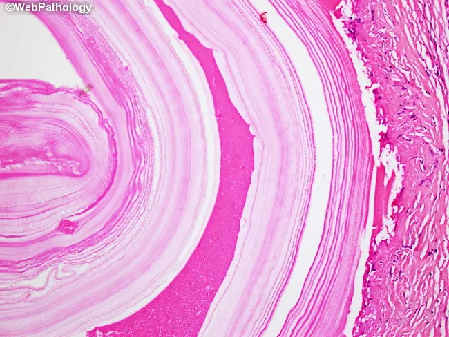
Clinical features
- Often asymptomatic for years.
- Hepatic cyst: Right upper abdominal pain, hepatomegaly, obstructive jaundice if bile ducts are compressed or involved.
- Pulmonary cyst: Cough, chest pain, dyspnea, hemoptysis.
- Rupture of cyst: May cause urticaria, eosinophilia, secondary cyst formation, or severe anaphylaxis.
Laboratory diagnosis
- Imaging is central: Ultrasound for liver cysts; CT or MRI to define site, stage, and complications.
- Serology: IgG ELISA, indirect hemagglutination, or immunoblot may support diagnosis, but negative results do not exclude disease.
- Microscopy of cyst contents, if obtained safely during surgery or a planned percutaneous procedure: hydatid sand, scolices, or hooklets.
- Peripheral blood eosinophilia may occur, but is not diagnostic.
Treatment
- Observation for selected inactive uncomplicated cysts.
- Albendazole as antiparasitic therapy.
- PAIR: Puncture, aspiration, injection of a scolicidal agent, and re-aspiration for selected cysts.
- Surgery for large, complicated, ruptured, superficially located, infected cysts, or cysts communicating with the biliary tract.
Prevention
- Deworm dogs regularly.
- Prevent dogs from eating infected raw offal.
- Proper disposal of infected animal viscera.
- Hand hygiene and safe food and water practices.
- Meat inspection and control of stray dogs.
Malaria laq microbiology
- Causative species and epidemiology
- Life cycle and pathogenesis
- Clinical features and complications
- Laboratory diagnosis, including peripheral smear findings
- Treatment principles
- Prevention and vector control
Which malaria LAQ section should I write in detail?
Louis Pasteur (1822-1895) and his contributions
-
Germ theory of disease
- He showed that microorganisms are present in air and can cause fermentation, spoilage, and disease.
- His experiments disproved the idea of spontaneous generation, the belief that living organisms arise from non-living matter.
- This work helped establish that many infectious diseases are caused by specific microbes. Park's Textbook of Preventive and Social Medicine, section “Germ theory of disease,” lines 413-430.
-
Pasteurization
- He developed controlled heating of liquids to destroy harmful or spoilage-causing microorganisms without greatly altering the product.
- It was first applied to wine and beer, and later became widely used for milk and other foods.
- Pasteurization improved food safety and reduced foodborne infection.
-
Fermentation
- Pasteur demonstrated that fermentation is caused by living microorganisms, such as yeast, rather than being only a chemical process.
- This was important for the wine, beer, and dairy industries.
-
Vaccination
- Pasteur developed or advanced vaccines against:
- Chicken cholera
- Anthrax
- Rabies
- In 1885, he treated Joseph Meister, a boy bitten by a rabid dog, with a rabies vaccine. This became a landmark in post-exposure prevention. Sherris & Ryan's Medical Microbiology, “Rabies,” lines 1470-1474.
- Pasteur developed or advanced vaccines against:
-
Public health and aseptic practice
- His discoveries supported sterilization, cleanliness, infection prevention, and later antiseptic surgical methods.
- They changed medical understanding by emphasizing that infections can be prevented by controlling microbes.
-
Silkworm disease
- Pasteur identified microbial causes of silkworm epidemics and developed methods to prevent spread, helping save France's silk industry.
-
Proved specific microbes cause specific diseases
- Koch provided strong experimental evidence that infectious diseases are caused by particular microorganisms, helping establish the germ theory of disease. Janeway's Immunobiology, “Basic Concepts in Immunology,” lines 28-31.
-
Koch’s postulates
- He formulated principles to link a microorganism to a disease:
- The organism should be associated with the disease.
- It should be isolated and grown in pure culture.
- The cultured organism should cause the same disease in a susceptible host.
- The same organism should be re-isolated from that host.
- These principles became the classic framework for identifying disease-causing microbes, although they have limitations for viruses and organisms that cannot be cultured easily.
- He formulated principles to link a microorganism to a disease:
-
Anthrax
- Koch showed that Bacillus anthracis causes anthrax and studied its spores.
- This was a major early demonstration that a specific bacterium can cause a specific disease.
-
Tuberculosis
- In 1882, Koch identified the tubercle bacillus, now called Mycobacterium tuberculosis, as the cause of tuberculosis. Tietz Textbook of Laboratory Medicine, “Mycobacterium tuberculosis,” line 750.
- This discovery made diagnosis, prevention, and later treatment of TB more scientific.
- He received the Nobel Prize in Physiology or Medicine in 1905, principally for this work.
-
Cholera
- He identified the bacterium responsible for cholera, Vibrio cholerae, supporting control measures related to contaminated water and sanitation.
-
Laboratory methods
- Koch advanced pure-culture methods and bacterial staining and microscopy.
- His laboratory work helped establish the use of solid culture media, which allowed bacteria to be separated into individual colonies and studied accurately.
-
The microorganism must be present in every case of the disease and absent from healthy individuals.
-
The microorganism must be isolated from the diseased host and grown in pure culture.
-
The cultured microorganism must cause the same disease when introduced into a healthy, susceptible host.
-
The same microorganism must be re-isolated from the experimentally infected host and shown to be identical to the original organism.
Importance
Limitations
- Some organisms cannot be grown in artificial culture, such as Treponema pallidum.
- Viruses require living cells for replication.
- Healthy carriers may carry pathogens without disease.
- Some diseases result from multiple organisms or host factors.
- It may be unethical to infect healthy humans deliberately.
- The same organism can cause different diseases depending on host immunity.
Principle
Reagents and procedure
| Step | Reagent/action | Purpose |
|---|---|---|
| 1 | Prepare and heat-fix smear | Fixes specimen onto the slide |
| 2 | Flood with strong carbol fuchsin and gently heat | Primary stain enters the waxy cell wall |
| 3 | Wash with water | Removes excess stain |
| 4 | Decolorize with acid-alcohol | Removes stain from non-acid-fast organisms |
| 5 | Wash with water | Stops decolorization |
| 6 | Counterstain with methylene blue | Stains background and non-acid-fast cells |
| 7 | Examine under oil immersion, 100× objective | Detects AFB |
Result
- Acid-fast bacilli: bright red or pink slender rods
- Background and non-acid-fast organisms: blue
Uses
- Screening sputum and other specimens for AFB in suspected pulmonary tuberculosis.
- Detection of mycobacteria in suitable clinical specimens.
- Modified acid-fast methods can also help detect organisms such as Nocardia and certain coccidian parasites.
Advantages
- Simple, inexpensive, and rapid.
- Useful in laboratories with limited resources.
Limitations
- A negative smear does not exclude tuberculosis because its sensitivity is limited, particularly when the bacterial load is low.
- It detects AFB but does not by itself identify the species or determine drug susceptibility.
- Culture and molecular tests such as NAAT/PCR are needed for confirmation and further testing.
Host and life cycle
- Definitive host: Dog and other canids, which harbor adult worms in the intestine.
- Intermediate hosts: Sheep, cattle, goats, etc.
- Humans: Accidental intermediate host.
- Infection: Ingestion of eggs from dog feces through contaminated food, water, or hands.
- Eggs hatch in the intestine, larvae enter portal circulation and form hydatid cysts, mainly in liver and lungs.
Sites affected
- Liver: Most common, about 70%
- Lungs: Second most common
- Less commonly: brain, bone, kidney, spleen, heart, and other organs.
Structure of hydatid cyst
- Pericyst: Outer fibrous host reaction.
- Ectocyst: Laminated, acellular membrane.
- Endocyst: Inner germinal layer which produces brood capsules and daughter cysts.
- Hydatid sand: Free scolices, hooklets, and brood capsules in cyst fluid.
Clinical features
- Often asymptomatic for years.
- Hepatic cyst: Right upper abdominal pain, hepatomegaly, obstructive jaundice if bile ducts are compressed or involved.
- Pulmonary cyst: Cough, chest pain, dyspnea, hemoptysis.
- Rupture of cyst: May cause urticaria, eosinophilia, secondary cyst formation, or severe anaphylaxis.
Laboratory diagnosis
- Imaging is central: Ultrasound for liver cysts; CT or MRI to define site, stage, and complications.
- Serology: IgG ELISA, indirect hemagglutination, or immunoblot may support diagnosis, but negative results do not exclude disease.
- Microscopy of cyst contents, if obtained safely during surgery or a planned percutaneous procedure: hydatid sand, scolices, or hooklets.
- Peripheral blood eosinophilia may occur, but is not diagnostic.
Treatment
- Observation for selected inactive uncomplicated cysts.
- Albendazole as antiparasitic therapy.
- PAIR: Puncture, aspiration, injection of a scolicidal agent, and re-aspiration for selected cysts.
- Surgery for large, complicated, ruptured, superficially located, infected cysts, or cysts communicating with the biliary tract.
Prevention
- Deworm dogs regularly.
- Prevent dogs from eating infected raw offal.
- Proper disposal of infected animal viscera.
- Hand hygiene and safe food and water practices.
- Meat inspection and control of stray dogs.
- Causative species and epidemiology
- Life cycle and pathogenesis
- Clinical features and complications
- Laboratory diagnosis, including peripheral smear findings
- Treatment principles
- Prevention and vector control
- Full malaria LAQ
- Life cycle
- Laboratory diagnosis
- Treatment and prevention
Cooms test
direct indirect antiglobulin Coombs test interpretation official clinical laboratory
direct antiglobulin test
Coombs test / Antiglobulin test
Principle
Types
| Feature | Direct Coombs test (DAT) | Indirect Coombs test (IAT) |
|---|---|---|
| Detects | IgG and/or complement already bound to the patient's RBCs | Free anti-RBC antibodies in the patient's serum |
| Specimen | Patient's washed RBCs | Patient's serum |
| Positive result | Agglutination after anti-human globulin is added | Agglutination after serum is incubated with test RBCs and anti-human globulin is added |
| Main use | Diagnosing immune hemolysis | Antibody screening and compatibility testing before transfusion |
1. Direct Coombs test
- Collect patient RBCs and wash them to remove unbound serum proteins.
- Add anti-human globulin reagent.
- Agglutination = positive DAT.
- Autoimmune hemolytic anemia
- Hemolytic disease of the fetus and newborn
- Hemolytic transfusion reaction
- Drug-induced immune hemolytic anemia
2. Indirect Coombs test
- Mix the patient's serum with reagent RBCs of known antigen profile.
- Incubate to allow any antibodies in serum to attach to RBCs.
- Wash cells and add anti-human globulin.
- Agglutination = positive IAT.
- Screening for irregular antibodies before blood transfusion
- Cross-matching donor and recipient blood
- Antenatal screening of Rh-negative pregnant women for anti-D antibodies
Interpretation
- DAT positive: RBC-bound antibody/complement is present. It supports immune hemolysis but should be interpreted with hemoglobin, reticulocyte count, bilirubin, LDH, haptoglobin, and clinical findings.
- IAT positive: Circulating antibodies against RBC antigens are present. This may indicate risk of transfusion reaction or hemolytic disease of the fetus/newborn.
Direct test = antibody on patient's RBCs.
Indirect test = antibody in patient's serum.
Graft vs host reaction
graft versus host disease prevention treatment
graft versus host disease NIH symptoms prevention treatment
Graft-versus-host disease (GVHD)
Conditions required for GVHD
- The graft contains viable, immunocompetent donor T cells.
- The recipient possesses antigens, especially HLA antigens, that differ from those of the donor.
- The recipient is unable to reject the donor lymphocytes because of immunosuppression or immunodeficiency.
Pathogenesis
- Recipient tissue damage from the conditioning regimen releases inflammatory cytokines.
- Donor T cells recognize recipient HLA/alloantigens as foreign.
- Donor T cells become activated, proliferate, and release cytokines.
- Cytotoxic T cells and inflammatory mediators damage recipient tissues.
Types
| Feature | Acute GVHD | Chronic GVHD |
|---|---|---|
| Typical onset | Usually within first 100 days after transplant, though it may occur later | Usually after 100 days |
| Main mechanism | Donor T-cell-mediated inflammation and cytotoxicity | More complex immune dysregulation, with T cells and B cells involved |
| Principal organs | Skin, liver, gastrointestinal tract | Skin, eyes, mouth, liver, lungs, gastrointestinal tract, genital tract, fascia |
Acute GVHD: clinical features
- Skin: Maculopapular erythematous rash, often beginning on palms, soles, ears, face, or trunk. Severe disease may cause blistering or epidermal necrolysis.
- Gastrointestinal tract: Anorexia, nausea, abdominal pain, profuse watery diarrhea, ileus, or bleeding.
- Liver: Cholestatic jaundice, raised bilirubin and alkaline phosphatase.
Chronic GVHD: clinical features
- Lichenoid or scleroderma-like skin lesions
- Dry eyes and dry mouth
- Oral ulcers or mucosal changes
- Joint stiffness and fascial fibrosis
- Cholestatic liver disease
- Bronchiolitis obliterans causing cough, breathlessness, and obstructive lung disease
- Genital involvement
Diagnosis
- Timing after allogeneic transplant
- Characteristic skin, liver, or gastrointestinal symptoms
- Exclusion of infection, drug reactions, and other causes
- Biopsy of affected skin or gastrointestinal mucosa when needed
Prevention
- Careful donor-recipient HLA matching
- Immunosuppressive prophylaxis, commonly a calcineurin inhibitor such as tacrolimus or cyclosporine combined with other agents according to transplant protocol
- T-cell depletion or selected graft manipulation in some settings
- Irradiation of cellular blood components for patients at risk of transfusion-associated GVHD
Treatment principles
- Mild localized disease: topical treatment may be used.
- Moderate to severe acute GVHD: systemic corticosteroids are generally first-line treatment.
- Steroid-refractory disease requires specialist-directed second-line immunosuppressive therapy.
- Chronic GVHD treatment is individualized according to organs involved and severity, together with supportive care for eyes, skin, mouth, nutrition, and infection prevention.
Post prophylaxis of hepatitis virus
CDC hepatitis B postexposure prophylaxis healthcare personnel HBIG vaccine hepatitis C follow up testing hepatitis A post exposure prophylaxis
hepatitis B postexposure prophylaxis
Post-exposure prophylaxis (PEP) of viral hepatitis
1. Hepatitis B PEP
- HBsAg status of source
- Whether the exposed person is vaccinated
- Documented response to vaccination, that is anti-HBs ≥10 mIU/mL
| Exposed person | Source HBsAg-positive or unknown | Source HBsAg-negative |
|---|---|---|
| Documented vaccine responder - anti-HBs ≥10 mIU/mL | No PEP needed | No PEP needed |
| Unvaccinated or incompletely vaccinated | HBIG 1 dose + start/complete Hepatitis B vaccine series at different injection sites | Start/complete Hepatitis B vaccine series |
| Vaccinated, response unknown | Test anti-HBs. If anti-HBs <10 mIU/mL: HBIG 1 dose + revaccination | If anti-HBs <10 mIU/mL: give booster/revaccination, no HBIG |
| Documented non-responder after two full vaccine series | HBIG 2 doses, 1 month apart | No PEP needed |
Hepatitis B vaccine: gives long-term active immunity.
- Give HBIG and first dose of Hepatitis B vaccine within 12 hours of birth, at separate sites.
- Complete the vaccine schedule and perform follow-up testing as recommended.
2. Hepatitis A PEP
- Hepatitis A vaccine: preferred for healthy persons aged ≥12 months.
- Immune globulin (IG): consider or add for people at greater risk of severe disease, especially:
- Immunocompromised persons
- Persons with chronic liver disease
- Older adults, depending on risk assessment
- Infants aged <12 months, who cannot receive vaccine
3. Hepatitis C PEP
- No vaccine
- No immune globulin
- No antiviral PEP is recommended
- Test source for HCV infection, if possible.
- Obtain baseline testing of exposed person.
- Do HCV RNA test at 3-6 weeks after exposure.
- If negative, perform anti-HCV test at 4-6 months, with HCV RNA testing if antibody positive.
4. Hepatitis D and E
- Hepatitis D: occurs only with HBV infection. Thus, Hepatitis B vaccination prevents HDV infection. There is no separate HDV PEP.
- Hepatitis E: no routine PEP is available.
One-line exam answer
Chemical disinfectant used in hospital
CDC chemical disinfectants healthcare facilities high intermediate low level disinfectants
hospital disinfectants
Chemical disinfectants used in hospitals
Common hospital disinfectants
| Group | Examples | Common uses | Key points |
|---|---|---|---|
| Halogens | Sodium hypochlorite (bleach), chlorine compounds, iodophors | Blood spills, floors, surfaces, water | Chlorine is inexpensive and broad spectrum. Hypochlorite at 0.5% (5,000 ppm) is used for blood/body-fluid spills and sporicidal cleaning. |
| Alcohols | 70% ethyl alcohol, 70% isopropyl alcohol | Skin preparation, thermometers, vial tops, small equipment | Rapid action, but no sporicidal activity. Not suitable for large dirty surfaces because it evaporates quickly. |
| Aldehydes | 2% glutaraldehyde, ortho-phthalaldehyde (OPA) | High-level disinfection of heat-sensitive equipment, e.g. endoscopes | Active against bacteria, mycobacteria, fungi, viruses, and with adequate exposure, spores. Toxic fumes mean careful handling is required. |
| Oxidizing agents | Hydrogen peroxide, peracetic acid, accelerated hydrogen peroxide | Environmental surfaces; high-level disinfection of some instruments | Broad antimicrobial action. Peracetic acid or hydrogen-peroxide systems are used for heat-sensitive medical devices. |
| Phenolics | Phenol derivatives | Floors, walls, furniture, noncritical surfaces | Not sporicidal. Avoid use on neonatal equipment and food-contact surfaces. |
| Quaternary ammonium compounds | Benzalkonium chloride, cetrimide | Floors, furniture, bedside tables, other low-risk environmental surfaces | Low-level disinfectants. Poor activity against spores, mycobacteria, and some non-enveloped viruses. |
| Biguanides | Chlorhexidine | Mainly skin and hand antisepsis, not environmental disinfection | Persistent action on skin. Usually classified as an antiseptic rather than a surface disinfectant. |
Classification by level of disinfection
-
High-level disinfectants
- Kill all organisms except high numbers of bacterial spores.
- Examples: glutaraldehyde, OPA, hydrogen peroxide, peracetic acid.
- Used for semicritical items that contact mucous membranes, such as endoscopes.
-
Intermediate-level disinfectants
- Kill vegetative bacteria, mycobacteria, fungi, and most viruses, but not reliably spores.
- Examples: alcohols, iodophors, phenolics, chlorine compounds.
- Used for contaminated surfaces and some noncritical equipment.
-
Low-level disinfectants
- Kill vegetative bacteria, fungi, enveloped viruses, and some non-enveloped viruses.
- Examples: quaternary ammonium compounds and some phenolics.
- Used for routine cleaning of floors, furniture, and noncritical surfaces.
Important exam points
- 2% glutaraldehyde: high-level disinfectant for endoscopes and other heat-sensitive instruments.
- 70% alcohol: commonly used for skin preparation and small equipment.
- Sodium hypochlorite: used for blood spills, environmental surfaces, and Clostridioides difficile contamination.
- Chlorhexidine: antiseptic for hand hygiene and skin preparation.
- Always clean visible organic matter first, because blood and protein reduce the action of many disinfectants.