Explain Lymphoid organ and it's function
Reading File
Finding Sources
Searching the Web
lymphoid organs primary secondary structure function
Finding Sources
Finding Sources
Reading File
Reading File
Reading File
Finding Sources
Finding Sources
Reading File
Reading File
Reading File
Loading Image
Loading Image
Here is a detailed, textbook-based explanation of lymphoid organs and their functions.
Lymphoid Organs and Their Functions
Lymphoid organs are the structural foundation of the immune system. They are broadly divided into two categories: primary (generative) lymphoid organs and secondary (peripheral) lymphoid organs.
"The organs of the immune system may be divided into the primary, or generative, lymphoid organs (bone marrow and thymus), where lymphocytes mature, and the secondary, or peripheral, lymphoid organs (lymph nodes, spleen, and the mucosal and cutaneous immune systems), where lymphocytes are activated by antigens."
- Robbins & Kumar Basic Pathology
1. Primary (Generative) Lymphoid Organs
These are where lymphocytes develop from immature precursors and become immunologically competent.
A. Bone Marrow
- The source of all blood cells, including lymphocytes (haematopoiesis)
- Site of B lymphocyte maturation - B cells complete their development here and become mature naive B cells
- Releases mature B and T cell precursors into the circulation
- T cell precursors leave the bone marrow early and migrate to the thymus to finish maturing
- Contains ~50 x 10⁹ lymphocytes in a healthy adult
B. Thymus
- Located in the upper anterior mediastinum (chest)
- The exclusive site of T lymphocyte maturation
- T cell precursors from bone marrow migrate here and undergo:
- Positive selection - cells that can recognize self-MHC molecules survive
- Negative selection - self-reactive cells are eliminated (clonal deletion) to prevent autoimmunity
- The thymus environment transforms precursors into functional, self-tolerant T cells
- Mature T cells are then released into the circulation to populate secondary lymphoid organs
- The thymus contains 100% T lymphocytes and 0% B lymphocytes (unlike all other lymphoid organs)
2. Secondary (Peripheral) Lymphoid Organs
These organs concentrate antigens, antigen-presenting cells (APCs), and lymphocytes to optimize immune responses. They are where adaptive immunity is actually initiated.
"The secondary lymphoid organs are organized to concentrate antigens, APCs, and lymphocytes in a way that optimizes interactions among these cells and the development of adaptive immune responses."
- Robbins & Kumar Basic Pathology
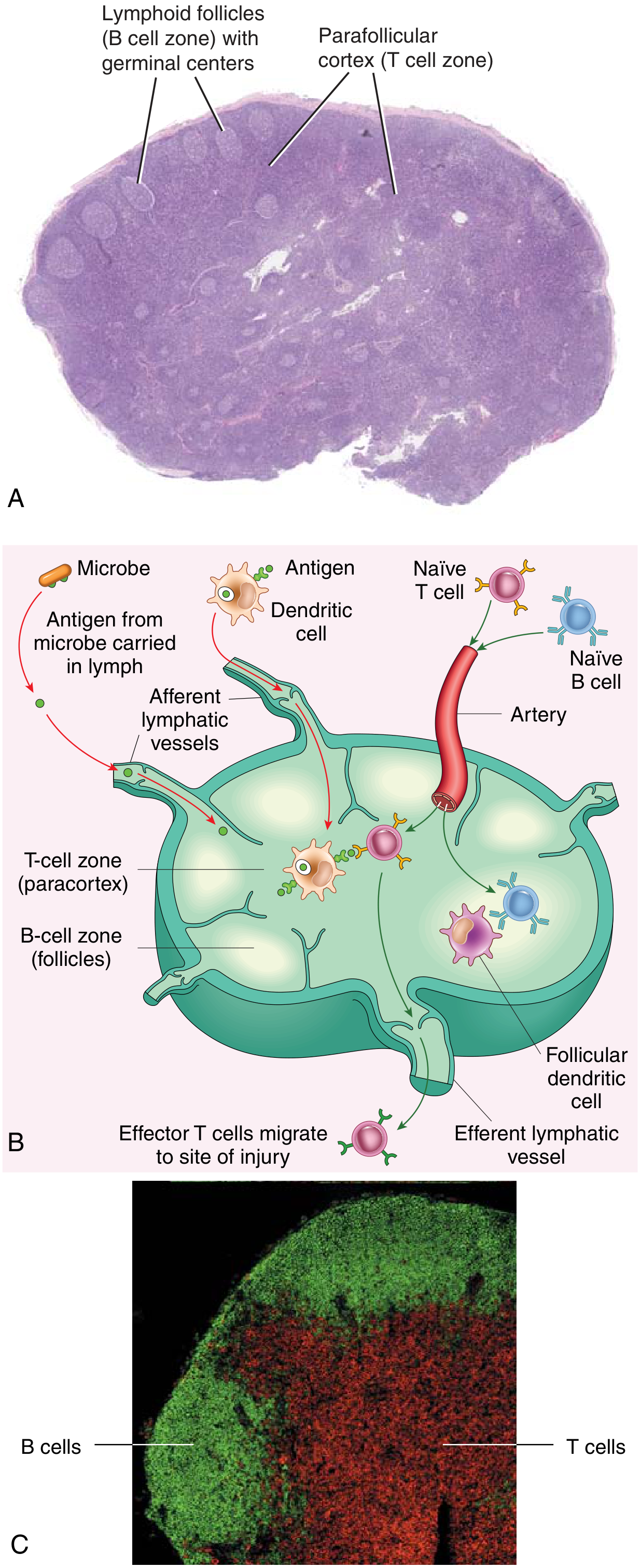
A. Lymph Nodes
Lymph nodes are the most numerous secondary lymphoid organs. They are encapsulated structures located throughout the body along lymphatic channels.
Structure:
- Cortex - contains B cell follicles (primary follicles = naive B cells; secondary follicles = germinal centers where B cells are actively responding to antigen)
- Paracortex (T cell zone) - rich in T lymphocytes and dendritic cells
- Medulla - contains plasma cells and macrophages
Functions:
- Antigen surveillance - as lymph drains from tissues, APCs (especially dendritic cells) carry antigens to the node via afferent lymphatics
- Immune activation - naive T and B cells circulating through the node encounter antigen-bearing dendritic cells and become activated
- Effector cell production - activated lymphocytes proliferate and differentiate; effector T cells leave via the efferent lymphatic vessel and return to bloodstream to fight infection
- Antibody production - activated B cells form germinal centers in follicles and differentiate into antibody-secreting plasma cells
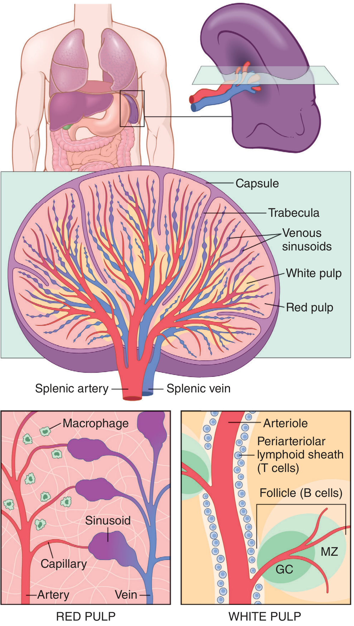
B. Spleen
The spleen is the largest secondary lymphoid organ, located in the left upper abdomen. It is specialized to respond to bloodborne antigens (while lymph nodes handle tissue antigens).
Structure:
- Red pulp - a network of vascular sinusoids filled with blood; site of red blood cell destruction and blood filtration
- White pulp - lymphoid tissue organized around central arterioles:
- Periarteriolar lymphoid sheath (PALS) - T cell zone surrounding the central arteriole
- Follicles - B cell zones; secondary follicles contain germinal centers
- Marginal zone - transition zone between red and white pulp; important for trapping blood-borne antigens
Functions:
- Immune surveillance of blood - blood entering the spleen flows through sinusoids where DCs and macrophages trap bloodborne antigens and microbes
- Initiation of immune responses - antigen-bearing DCs present antigens to T and B cells in the white pulp
- Blood filtration - removes old, damaged, or opsonized red blood cells (red pulp macrophages phagocytose them)
- Blood reservoir - the red pulp stores concentrated red blood cells that can be mobilized by the sympathetic nervous system (up to 50 mL released during stress)
- Defense against encapsulated bacteria - asplenic patients are highly susceptible to infections with organisms like Streptococcus pneumoniae
C. Mucosa-Associated Lymphoid Tissue (MALT)
MALT refers to lymphoid tissue embedded beneath the epithelial surfaces of the gastrointestinal, respiratory, and genitourinary tracts.
- Gut-associated lymphoid tissue (GALT) includes tonsils, adenoids, appendix, and Peyer's patches
- Peyer's patches (in the small intestine) are the most organized MALT structures; they collect antigens from the gut via specialized M cells (microfold cells) in the epithelium
- The mucosal immune system is estimated to contain as many lymphocytes as all the rest of the body combined
- Lymphocytes generated in mucosal lymphoid tissue preferentially home back to mucosal surfaces ("homing")
3. Distribution of Lymphocytes Across Organs (Healthy Adult)
| Tissue | Lymphocytes (×10⁹) |
|---|---|
| Lymph nodes | 190 |
| Spleen | 70 |
| Intestines (MALT) | 50 |
| Bone marrow | 50 |
| Lungs | 30 |
| Skin | 20 |
| Blood | 10 |
| Liver | 10 |
(Source: Robbins & Kumar Basic Pathology)
4. Key Differences: Primary vs. Secondary Lymphoid Organs
| Feature | Primary (Generative) | Secondary (Peripheral) |
|---|---|---|
| Examples | Bone marrow, Thymus | Lymph nodes, Spleen, MALT |
| Main role | Lymphocyte development and maturation | Lymphocyte activation by antigen |
| Antigen dependence | Antigen-independent | Antigen-dependent |
| Time of development | Early embryonic life | Late fetal / postnatal |
| Effect of removal | Loss of lymphocytes; no immune responses | Minimal or modest effect on lymphocyte production |
Summary
Lymphoid organs form a network that produces, matures, and deploys lymphocytes. Primary organs (bone marrow and thymus) generate the naive lymphocyte repertoire. Secondary organs (lymph nodes, spleen, MALT) trap antigens and bring them into contact with naive lymphocytes, triggering the adaptive immune responses that protect against infection and disease. The organized segregation of T and B cell zones within each secondary lymphoid organ ensures efficient cell-cell interactions needed to mount effective antibody and cell-mediated immune responses.
- Robbins & Kumar Basic Pathology, p. 161
- Janeway's Immunobiology 10e, p. 40-46
- Cellular and Molecular Immunology, p. 128-129
- Guyton and Hall Textbook of Medical Physiology
Lymphoid organ and it's functions define Histology
Finding Sources
Searching the Web
lymphoid organ histology structure microscopy
Finding Sources
Finding Sources
Finding Sources
Finding Sources
Your credits are exhausted. You can purchase a top-up or subscribe to a plan to continue using Orris. If you're already subscribed, your daily credits will reset tomorrow.
This is a shared conversation. Sign in to Orris to start your own chat.