
Gangrenous cholecystitis
gangrenous cholecystitis
gangrenous cholecystitis pathology gross specimen histology
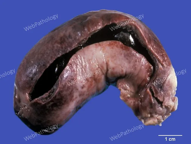
Gross pathology photograph of a gallbladder specimen showing gangrenous cholecystitis. The organ is enlarged and markedly distended, opened to reveal intraluminal gallstones. The serosal surface is dusky red-brown from congestion and hemorrhage, with transmural inflammation and necrosis evident in the gallbladder wall. The walls are friable and may show edema while the mucosa is disrupted by necrotic debris. External serosa appears thickened and mottled; the lumen contains pigmented bile and debris. Overall, features are consistent with acute calculous cholecystitis complicated by gangrene, with possible early perforation risk. This macroscopic appearance correlates with ischemic injury to the gallbladder wall and extensive inflammatory infiltration. The image illustrates a classic teaching example for gross surgical pathology and hepatobiliary disease. Clinically, gangrenous cholecystitis represents a severe, life-threatening progression from simple cholecystitis, often in patients with comorbidities such as diabetes, cardiovascular disease, and advanced age. The photo supports differential considerations including emphysematous cholecystitis or perforated cholecystitis and emphasizes urgent surgical management and histopathologic confirmation of transmural necrosis. Note the scale is conveyed by a contrasting background and a centimeter ruler present in the image, supporting gross measurements and education. This representation facilitates correlation with surgical findings and guides learning in medical school, pathology residency, and radiology-pathology conferences.
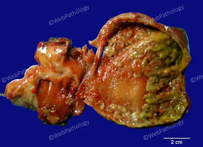
Gross pathology of a gallbladder with acute calculous cholecystitis. The specimen shows a distended, inflamed gallbladder wall with mucosal edema and erythema. The mucosa is congested and irregular, with focal necrosis and yellow-green exudate admixed with fibrinous material. Patchy greenish-yellow pseudomembranes overlying necrotic mucosa are evident, consistent with pseudomembranous cholecystitis. The serosa may be variably irritated, and adherent inflammatory adhesions can be seen along the gallbladder bed. The exterior surface demonstrates hyperemia; the wall may be thickened due to edema. A portion of the lumen is exposed, showing necrotic debris and exudate filling the lumen interface. The 2 cm scale bar provides context for dimensions; overall changes correlate with acute inflammatory insult, often precipitated by gallstone obstruction of the cystic duct. The appearance aligns with fulminant cholecystitis and may precede gangrenous transformation if infection progresses. These macroscopic findings are supported by typical histology of mucosal necrosis, neutrophilic infiltrate, edema, and fibrinous exudate on the surface. This image highlights hallmark features of pseudomembranous cholecystitis with inflammatory exudate forming pseudomembranes, mucosal ulceration, and mural necrosis within the gallbladder wall. Correlation with clinical history of gallstones, fever, RUQ pain, leukocytosis, and imaging findings reinforces diagnosis and guides emergent management for surgical intervention planning.
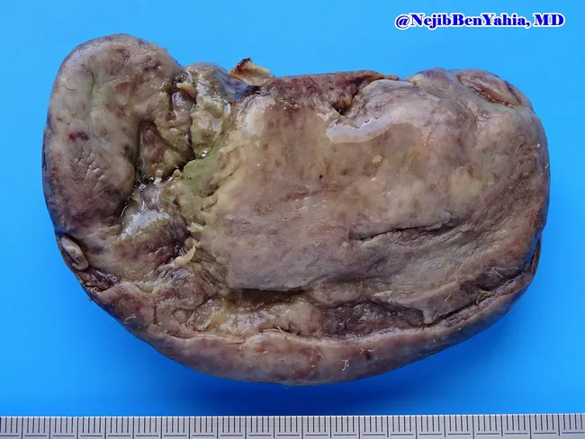
Gross pathology photograph illustrating gangrenous cholecystitis. The gallbladder is enlarged and distended, with a thickened wall and friable consistency. The serosal surface appears dull and dusky red‑brown, reflecting profound congestion and ischemia. Depressed necrotic foci are present on the mucosal and serosal surfaces, consistent with transmural necrosis typical of gallbladder gangrene. The organ shows irregular, hemorrhagic, devitalized areas interspersed with relatively preserved tissue. Exterior faces mild edema with focal surface ulcers at the fundus and body. The lumen contents are not visible in this specimen, as processing often removes bile. This macroscopic image was captured from an excised gallbladder to illustrate severe inflammatory biliary disease in surgical pathology. The blue background and centimeter scale provide reference for size and orientation. Imaging technique includes gross pathology photography, color‑calibrated macro imaging, and high‑resolution digital capture under standardized lighting. Diagnostic significance lies in confirming advanced gallbladder necrosis with gangrene, which elevates risk of perforation, peritonitis, and sepsis; it supports a clinical diagnosis of acute gangrenous cholecystitis. Educationally, this image aids recognition of gross features, correlates with clinical presentation, and enhances training in surgical pathology interpretation. This image is suitable for medical students, residents, and fellows studying gallbladder pathology, inflammatory hepatobiliary disease, and surgical specimen assessment.
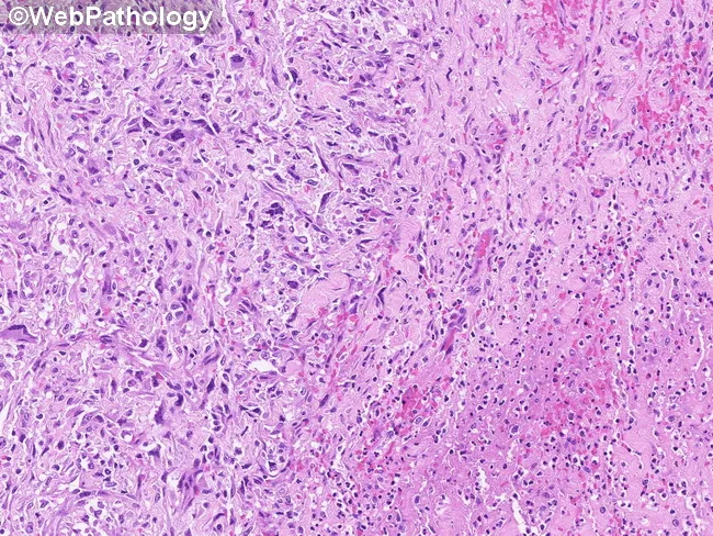
This image depicts a high-magnification light microscopy view of gallbladder wall in a case of gangrenous cholecystitis. Prepared from routine formalin-fixed paraffin-embedded tissue and stained with Hematoxylin and Eosin, the specimen is examined at high power to highlight cellular detail. In the left half, a pseudosarcomatous myofibroblastic proliferation is evident: plump spindle-shaped cells with elongated, hyperchromatic nuclei arranged in short fascicles within a loose myxoid-to-fibrous stroma, admixed inflammatory cells, and areas of necrosis consistent with gangrenous change. The right half shows more densely packed inflammatory infiltrates and vascular congestion with surrounding edema. The overall pattern can simulate a high-grade sarcoma when taken out of context, owing to marked nuclear atypia in a myofibroblastic component. Clinically, this histology corresponds to severe gallbladder inflammation with tissue destruction rather than a primary mesenchymal neoplasm. Diagnostic significance lies in recognizing a reactive, post-inflammatory pseudosarcomatous pattern to avoid overcalling sarcoma; ancillary studies, including immunohistochemistry for smooth muscle actin and desmin, and correlation with gross pathology, aid confirmation. This image is relevant for surgical pathology education, differential diagnosis of spindle cell proliferations, and radiologic-pathologic correlation in biliary disease. It informs differential diagnoses: inflammatory myofibroblastic reaction vs sarcoma; guides management.
Gangrenous Cholecystitis
Pathophysiology
- The fundus is the most common site of necrosis, as it is the farthest from the blood supply.
- In 90% of cases, an obstructing gallstone is present in the neck or cystic duct.
- Full-thickness necrosis is usually present, but this does not always result in frank perforation.
Gross and Histological Pathology
Risk Factors
- Diabetic patients (impaired vascular supply and immune response)
- Elderly patients
- Cardiovascular disease / microvascular disease
- Male sex (also associated with emphysematous cholecystitis)
- Delayed presentation or treatment of acute cholecystitis
Clinical Features
Clinical findings may paradoxically diminish as the disease progresses to gangrene - as necrosis destroys stretch receptors in the gallbladder wall, pain may actually lessen, masking the severity of disease. (Grainger & Allison's Diagnostic Radiology)
- Severe RUQ or epigastric pain (may plateau or decrease)
- High fever (>38.5°C), rigors
- Leukocytosis (often >15,000/mm³, sometimes with left shift)
- Signs of systemic toxicity or sepsis
- Absent or diminished sonographic Murphy's sign (due to necrosis of sensory fibers - noted in Yamada's Textbook of Gastroenterology)
Imaging
Ultrasound (US)
- Pronounced irregularity or asymmetrical thickening of the gallbladder wall
- Internal membranous echoes - representing sloughed necrotic mucosa (intraluminal membrane/flap sign)
- Pericholecystic fluid
- Absent Murphy's sign on probe pressure
CT / MRI (recommended when gangrenous cholecystitis is suspected)
| CT/MRI Finding | Notes |
|---|---|
| Interrupted rim sign - discontinuous/irregular mucosal enhancement | Most sensitive (70.6%) and highly specific (100%) |
| Gas in the wall or lumen | Also suggests emphysematous component |
| Intraluminal membranes (intraluminal flap) | Sloughed mucosal debris |
| Irregular thickening of the gallbladder wall | |
| Increased density of pericholecystic fat | |
| Pericholecystic abscess | Suggests perforation |
Tokyo Guidelines Severity Grading
Complications
- Gallbladder perforation - occurs in 5-10% of acute cholecystitis; risk is highest with gangrenous change. May manifest as:
- Free spillage into the peritoneal cavity (biliary peritonitis)
- Localised pericholecystic abscess
- Fistula formation (cholecystoenteric fistula - to duodenum or transverse colon most commonly)
- Gallstone ileus (if fistula develops and large stone passes into bowel)
- Sepsis / multiorgan failure
- Superimposed emphysematous cholecystitis
Management
General measures
- IV fluid resuscitation and electrolyte correction
- IV antibiotics: Bile/gallbladder wall cultures are positive for bacteria in >40% of patients
- Mild-moderate: cefoxitin (cephalosporin)
- Severe / suspected gangrenous: piperacillin-tazobactam OR third-generation cephalosporin + metronidazole (anaerobic coverage is mandatory)
- Continue antibiotics postoperatively in gangrenous cholecystitis (unlike mild disease where they are stopped after source control)
Surgical management
- Urgent/emergent cholecystectomy is the treatment of choice
- Early cholecystectomy is preferred over delayed: combined data from 7 RCTs shows early surgery results in 0% mortality vs 2% delayed, and total hospital stay of 9.6 vs 17.8 days; 26% of delayed patients required early surgery anyway due to worsening (Sleisenger and Fordtran's)
- Laparoscopic approach is attempted; high conversion to open is expected given severe inflammation
- Subtotal/partial cholecystectomy may be required when anatomy is distorted
- For unstable/high-risk patients: percutaneous cholecystostomy tube as bridge to definitive surgery
Comparison with Emphysematous Cholecystitis
| Feature | Gangrenous | Emphysematous |
|---|---|---|
| Mechanism | Ischemic wall necrosis | Gas-forming bacterial infection |
| Gas on imaging | Possible (late) | Hallmark finding |
| Stones | Present in 90% | Present in <50% |
| Sex | No gender reversal | More common in men |
| Diabetes | Risk factor | ~50% diabetic |
| Mortality | High | Relatively high |
| CT modality | CT/MRI preferred | CT is modality of choice |
Key Teaching Points
- Paradoxical improvement in pain despite clinical deterioration is a warning sign - necrosis blunts the pain response.
- The interrupted rim sign on contrast CT/MRI is the most specific imaging finding (100% specificity).
- Absent sonographic Murphy's sign in a sick patient should raise suspicion, not reassurance.
- Anaerobic antibiotic coverage is mandatory in suspected gangrenous cholecystitis.
- Postoperative antibiotics should be continued (unlike uncomplicated acute cholecystitis).
- Tokyo Guidelines Grade II - requires early surgical intervention.