
Which organelle is responsible for formation of carcinogen
The smooth endoplasmic reticulum (SER) is the organelle primarily responsible for the formation (activation) of carcinogens. It contains cytochrome P450 enzymes that convert procarcinogens into their active carcinogenic forms through oxidation reactions.
Searching sources...
Reading File
Finding Sources
Searching the Web
which organelle is responsible for carcinogen formation activation cytochrome P450
Finding Sources
Finding Sources
Reading File
Reading File
The answer is well-supported by authoritative pathology textbooks. Here is the complete explanation:
Organelle Responsible for Carcinogen Formation
The Smooth Endoplasmic Reticulum (SER) is the organelle responsible for the formation (metabolic activation) of carcinogens.
How It Works
Most chemical carcinogens are not directly active - they exist as procarcinogens (indirect-acting agents) that must first undergo metabolic conversion to become the ultimate carcinogen. This conversion happens in the SER via its resident enzyme system:
- The SER houses cytochrome P450-dependent mono-oxygenases (mixed-function oxidases)
- These enzymes oxidize procarcinogens into highly reactive electrophilic metabolites
- The reactive electrophiles then form covalent adducts with DNA, causing mutations that can lead to cancer
Classic Example
Benzo[a]pyrene (found in tobacco smoke and charred food) is metabolized by the CYP1A1 enzyme (a cytochrome P450) in the SER into a reactive epoxide that binds DNA and initiates carcinogenesis.
Why This Matters Clinically
- Polymorphisms in cytochrome P450 genes alter the rate of procarcinogen activation
- Individuals with a highly inducible CYP1A1 form have a sevenfold increased risk of lung cancer when they smoke
- This is why not all smokers develop lung cancer - individual enzyme variation matters
Summary Table
| Category | Example | Requires SER Activation? |
|---|---|---|
| Direct-acting carcinogens | Alkylating agents (chemotherapy drugs) | No - already reactive |
| Indirect-acting (procarcinogens) | Benzo[a]pyrene, aflatoxin, azo dyes | Yes - SER/CYP450 required |
So in short: the Smooth Endoplasmic Reticulum - through its cytochrome P450 enzyme system - converts procarcinogens into their ultimate active carcinogenic forms.
(Robbins Pathologic Basis of Disease; Sabiston Textbook of Surgery, p. 1217)
Which tumor market increased in pancreatic tumor Results in which jaundice
CA 19-9 is the primary tumor marker elevated in pancreatic cancer. When a pancreatic head tumor obstructs the common bile duct, it causes obstructive (painless) jaundice.
Searching sources...
Finding Sources
Searching the Web
pancreatic tumor marker CA 19-9 jaundice obstructive pancreatic head cancer
Reading File
Finding Sources
Reading File
Here is the complete, textbook-backed answer:
Tumor Marker in Pancreatic Cancer with Jaundice
Primary Tumor Marker: CA 19-9 (Carbohydrate Antigen 19-9)
CA 19-9 is the most important and widely used tumor marker for pancreatic cancer. It is a carbohydrate antigen (a sialylated Lewis blood group antigen) that is elevated in the majority of pancreatic ductal adenocarcinomas (PDAC).
- Normal: < 37 U/mL
- Sensitivity: ~81% | Specificity: ~90% for pancreatic cancer
- Used primarily for monitoring treatment response and detecting recurrence
- Also elevated (but less so) in biliary, gastric, and colorectal cancers
Why Jaundice Occurs - The Mechanism
Pancreatic cancer of the head of the pancreas directly compresses or invades the common bile duct as it passes through the pancreatic head, causing obstructive (cholestatic) jaundice.
"Obstructive jaundice is associated with most cases involving the pancreatic head, as these tend to block the common bile duct."
- Robbins Pathologic Basis of Disease
This is painless jaundice - a hallmark presentation, because unlike gallstones (which cause colicky pain), the obstruction here is slow, progressive, and painless.
Courvoisier Sign (Key Exam Point)
When pancreatic head cancer blocks the bile duct, the gallbladder distends but remains non-tender and palpable - this is called the Courvoisier sign:
Painless jaundice + Palpable, non-tender gallbladder = Courvoisier Sign = Think pancreatic head carcinoma
Important Caveat about CA 19-9
CA 19-9 can be falsely elevated in obstructive jaundice itself (even from benign causes). So in a jaundiced patient, a high CA 19-9 alone does not confirm cancer - imaging (CT "pancreas protocol") is essential for diagnosis.
Additionally, 10-30% of patients cannot synthesize CA 19-9 (Lewis antigen-negative individuals), making it unreliable as a standalone screening test.
Quick Summary
| Feature | Detail |
|---|---|
| Tumor marker | CA 19-9 |
| Type of cancer | Pancreatic ductal adenocarcinoma (PDAC) |
| Location causing jaundice | Head of pancreas |
| Mechanism of jaundice | Compression of common bile duct |
| Type of jaundice | Painless obstructive jaundice |
| Associated sign | Courvoisier sign |
| Other marker also elevated | CEA (less specific) |
(Robbins Pathologic Basis of Disease; Fischer's Mastery of Surgery, 8th ed.)
In ascitis what happens to albumin level
In ascites, serum albumin levels are decreased (hypoalbuminemia). The key diagnostic concept is the SAAG (Serum-Ascites Albumin Gradient) - the difference between serum albumin and ascitic fluid albumin.
Searching sources...
Finding Sources
Searching the Web
ascites albumin level SAAG serum ascites albumin gradient hypoalbuminemia
Reading File
Reading File
Excellent textbook sources obtained. Here is the complete answer:
Albumin in Ascites
The question has two important dimensions - what happens to serum albumin and what happens to ascitic fluid albumin, and how the relationship between them (the SAAG) is used clinically.
1. Serum Albumin - DECREASED
In the most common cause of ascites (cirrhosis), serum albumin is low (hypoalbuminemia) because:
- The damaged liver produces less albumin
- The low oncotic pressure allows fluid to leak into the peritoneal cavity
- This is a contributing cause of ascites formation
2. The SAAG - The Key Diagnostic Tool
SAAG = Serum Albumin - Ascitic Fluid Albumin (Both samples must be taken on the same day)
The SAAG reflects hepatic sinusoidal pressure and is based on Starling forces - it correlates strongly with the hepatic venous pressure gradient (r = 0.72).
| SAAG Value | Interpretation | Causes |
|---|---|---|
| ≥ 1.1 g/dL (High gradient) | Portal hypertension present | Cirrhosis, Cardiac failure, Budd-Chiari syndrome, Massive liver metastases |
| < 1.1 g/dL (Low gradient) | Non-portal hypertensive | Peritoneal carcinomatosis, TB peritonitis, Nephrotic syndrome, Pancreatic ascites |
3. Why SAAG Works - The Physiology
When portal hypertension raises hydrostatic pressure in sinusoids, fluid is forced into the peritoneal cavity. The body compensates by keeping albumin concentrated in the blood (high serum albumin relative to ascitic fluid albumin) - creating a large gradient (≥1.1). The ascitic fluid in portal hypertension is therefore low in albumin (transudate-like).
In non-portal causes (e.g., malignancy, TB peritonitis), fluid leaks from an inflamed/tumor-laden peritoneum and carries protein with it - so ascitic albumin is high, narrowing the gradient to <1.1.
4. Second Step - Ascitic Protein Level (after SAAG)
When SAAG ≥1.1, the ascitic total protein further narrows the cause:
| Ascitic Protein | Interpretation |
|---|---|
| ≥ 2.5 g/dL | Normal hepatic sinusoids - Cardiac ascites, early Budd-Chiari |
| < 2.5 g/dL | Damaged/scarred sinusoids - Cirrhosis, late Budd-Chiari |
Summary
- Serum albumin in ascites (especially cirrhotic) = Decreased
- SAAG ≥ 1.1 = Portal hypertension (cirrhosis is #1 cause)
- SAAG < 1.1 = Non-portal cause (malignancy, TB, nephrotic syndrome)
- SAAG accuracy is reduced when serum albumin is very low, samples are not simultaneous, or ascites is chylous
(Harrison's Principles of Internal Medicine 22e; Yamada's Textbook of Gastroenterology, 7th ed.)
Lesch nyhan syndrome
Lesch-Nyhan syndrome is an X-linked recessive disorder caused by deficiency of HGPRT (hypoxanthine-guanine phosphoribosyltransferase), leading to excess uric acid production. It presents with gout, intellectual disability, and self-mutilation behavior.
Searching sources...
Finding Sources
Searching the Web
Lesch-Nyhan syndrome HGPRT deficiency pathophysiology clinical features treatment
Reading File
Reading File
Let me fetch the pathway diagram and the lip lesion image from the textbook:
Loading Image
Loading Image
Here is a complete, textbook-backed overview of Lesch-Nyhan Syndrome:
Lesch-Nyhan Syndrome
Basics
| Feature | Detail |
|---|---|
| Inheritance | X-linked recessive |
| Affected | Almost exclusively males |
| Enzyme deficient | HGPRT (Hypoxanthine-Guanine PhosphoRibosylTransferase) |
| Gene | HPRT gene on X chromosome |
| Defect type | Virtually complete (partial deficiency = milder variant) |
The Purine Salvage Pathway - What Goes Wrong
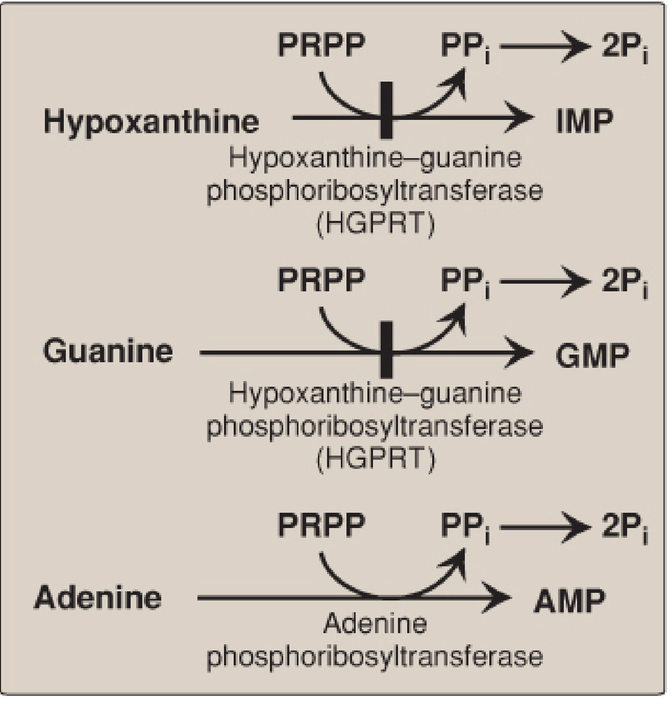
Normally, HGPRT salvages (recycles) purines:
- Hypoxanthine → IMP (via HGPRT)
- Guanine → GMP (via HGPRT)
When HGPRT is absent, these reactions are blocked. The consequences cascade:
- Hypoxanthine and guanine cannot be recycled - they are degraded to uric acid
- PRPP accumulates (it is normally consumed by HGPRT)
- IMP and GMP levels fall - releasing feedback inhibition on GPAT (the regulated/rate-limiting step of de novo purine synthesis)
- De novo purine synthesis is massively upregulated
- More purines are made - more are degraded - more uric acid is produced
Result: Double hit of decreased salvage + increased de novo synthesis = severe hyperuricemia
Clinical Features (Triad)
1. Hyperuricemia and Its Consequences
- Uric acid kidney stones (urolithiasis) - often first sign in infants (orange-red "sand" in diapers)
- Gouty arthritis - urate crystal deposition in joints
- Tophi - subcutaneous urate deposits
2. Neurological Dysfunction
- Hypotonia in infancy, followed by spasticity
- Dystonia and choreoathetosis
- Dysarthria (difficulty speaking)
- Developmental delay and intellectual disability
3. Behavioral/Psychiatric - The Pathognomonic Feature
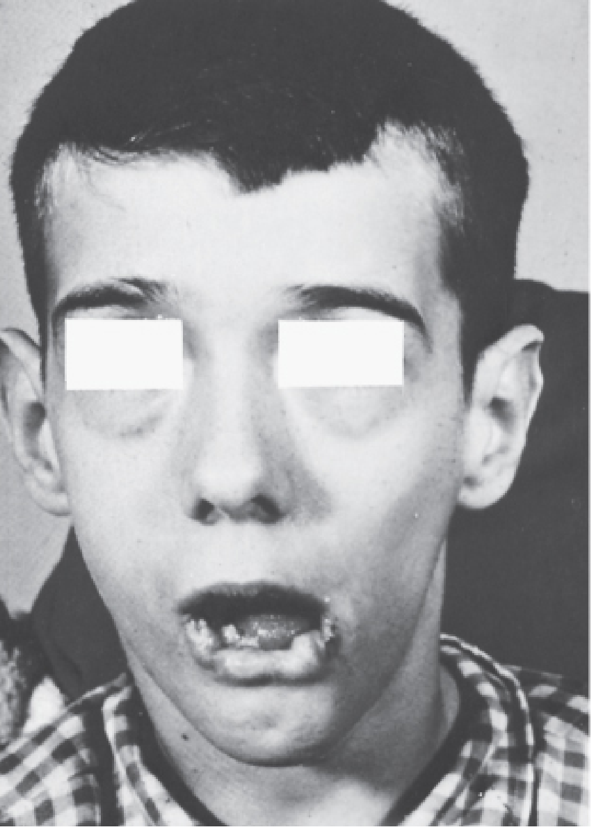
- Self-mutilation - compulsive biting of lips, fingers, and tongue
- Aggression toward others
- The self-injury is involuntary and distressing to the patient (they do not want to hurt themselves)
Diagnosis
| Test | Finding |
|---|---|
| Serum uric acid | Markedly elevated |
| Urine uric acid | Markedly elevated |
| HGPRT enzyme assay (RBCs) | Absent or near-absent activity |
| Genetic testing | Mutation in HPRT gene on X chromosome |
| Ames test / fibroblast culture | Can confirm enzymatic deficiency |
Treatment
| Target | Drug/Approach |
|---|---|
| Hyperuricemia | Allopurinol (xanthine oxidase inhibitor - reduces uric acid production) |
| Dystonia/spasticity | Baclofen, benzodiazepines |
| Self-injury prevention | Physical restraints, dental extraction (controversial), behavioral therapy |
| No cure | No treatment reverses the neurological features - allopurinol only addresses the metabolic component |
Key point: Allopurinol corrects the gout and kidney stones but does NOT improve the neurological or behavioral symptoms.
Key Exam Points
- Deficient enzyme = HGPRT (not APRT, not adenosine deaminase)
- Inheritance = X-linked recessive (males affected, females carriers)
- Self-mutilation (lip/finger biting) = pathognomonic
- Mechanism = failed salvage → excess PRPP → excess de novo synthesis → hyperuricemia
- Treatment of choice for metabolic component = Allopurinol
- "Orange sand" in diapers (urate crystals) = early clue in infants
(Lippincott Biochemistry 8th ed.; Basic Medical Biochemistry 6th ed.; Robbins Basic Pathology)
Screening and confirmation test for phenylketonuria
The Guthrie test (bacterial inhibition assay) is the classic newborn screening test for PKU, while plasma phenylalanine quantification (tandem mass spectrometry) serves as both the modern screening and confirmation method.
Searching sources...
Finding Sources
Searching the Web
phenylketonuria PKU screening Guthrie test confirmation tandem mass spectrometry newborn
Finding Sources
Reading File
Reading File
Finding Sources
Finding Sources
Finding Sources
Reading File
Finding Sources
I have comprehensive information from the textbooks. Here is the complete answer:
Phenylketonuria (PKU) - Screening and Confirmation Tests
Background
PKU is an autosomal recessive disorder caused by deficiency of phenylalanine hydroxylase (PAH) - the enzyme that converts phenylalanine to tyrosine. Incidence is approximately 1 in 10,000-20,000 births. Early detection is critical because a low-phenylalanine diet started in infancy prevents irreversible intellectual disability.
Screening Tests
1. Guthrie Test (Bacterial Inhibition Assay) - Classic/Traditional Screening
| Feature | Detail |
|---|---|
| Sample | Heel-prick blood on filter paper (day 6-10 of life) |
| Principle | Bacillus subtilis is inhibited by β-2-thienylalanine in culture; excess phenylalanine in the blood spot overcomes this inhibition, allowing bacterial growth - a positive result |
| Timing | Performed 6-10 days after birth (after the baby has been fed - phenylalanine must accumulate) |
| Use today | Classic method; still used in many countries including India as mass screening |
Important: Blood phenylalanine may not be detectable until 3-4 days postpartum, which is why the test is not done on day 1. False positives occur in premature infants due to delayed enzyme maturation.
2. Ferric Chloride (FeCl₃) Test - Older Urine Screening
| Feature | Detail |
|---|---|
| Sample | Urine |
| Principle | FeCl₃ reacts with urinary phenylpyruvate (an overflow metabolite of phenylalanine) to give a green/blue-green color |
| Timing | After 3-6 weeks of age (phenylpyruvate appears in urine later than blood phenylalanine) |
| Limitation | Older and less reliable - misses cases if done too early; can miss mild hyperphenylalaninemias |
3. Tandem Mass Spectrometry (MS/MS) - Modern Screening
| Feature | Detail |
|---|---|
| Sample | Dried blood spot (same heel-prick card) |
| Principle | Directly quantifies blood phenylalanine AND phenylalanine:tyrosine ratio |
| Advantage | Highly sensitive and specific; simultaneously screens for 30+ metabolic disorders |
| Status | Has largely supplanted the Guthrie test in the USA and developed countries |
Confirmation Tests
After a positive screen, confirmatory testing is required:
| Test | Detail |
|---|---|
| Plasma phenylalanine quantification (fluorometry/HPLC) | Gold standard confirmation - phenylalanine >1200 μmol/L (>20 mg/dL) = classic PKU |
| Phenylalanine:Tyrosine ratio | Ratio >3 is abnormal (tyrosine is low because PAH is non-functional) |
| BH4 loading test | To distinguish classic PKU from BH4-cofactor deficiency variants (2% of cases) |
| Urine pterin analysis | To identify biopterin synthesis/recycling defects |
| PAH gene mutation analysis (DNA) | Definitive molecular confirmation; also used for prenatal diagnosis |
| DHPR enzyme assay | To rule out dihydrobiopterin reductase deficiency |
Why Ruling Out BH4 Deficiency Matters
~2% of hyperphenylalaninemias are due to BH4 cofactor deficiency (not PAH deficiency). These patients:
- Are clinically identical to classic PKU on newborn screen
- Deteriorate neurologically despite dietary phenylalanine control (because BH4 is also needed for tyrosine and tryptophan hydroxylases → dopamine and serotonin deficiency)
- Require BH4 supplementation + neurotransmitter precursors (L-Dopa, 5-OH-tryptophan), not just diet
Summary Table
| Test | Purpose | Sample | When |
|---|---|---|---|
| Guthrie test | Screening | Heel-prick blood (filter paper) | Day 6-10 |
| FeCl₃ test | Older screening | Urine | After 3-6 weeks |
| Tandem MS/MS | Modern screening | Dried blood spot | Day 2-3 (or with Guthrie) |
| Plasma Phe quantification | Confirmation | Venous blood | After positive screen |
| BH4 loading test | Differential diagnosis | Blood + urine | After confirmation |
| Gene mutation analysis | Definitive / prenatal | Blood DNA | Anytime |
(Harper's Biochemistry 32nd ed.; Park's Preventive and Social Medicine; Tietz Textbook of Laboratory Medicine 7th ed.)
Precipitating factor for porphyrias
The classic precipitating factors for porphyrias (especially acute intermittent porphyria) include drugs (barbiturates, sulfonamides), fasting/starvation, alcohol, infections, hormones (progesterone), and stress.
Searching sources...
Finding Sources
Searching the Web
precipitating factors acute porphyria AIP drugs fasting hormones list
Reading File
Finding Sources
Finding Sources
Finding Sources
Reading File
Finding Sources
Excellent content obtained. Here is the complete, well-organized answer:
Precipitating Factors for Porphyrias
The Core Mechanism
All precipitating factors share a common final pathway: they induce hepatic δ-ALAS (delta-aminolevulinic acid synthase) - the rate-limiting enzyme of heme biosynthesis. In porphyria patients, there is already a partial enzyme block downstream. When ALAS is induced, it drives excess substrate through the already-blocked pathway, causing overproduction and overflow of toxic porphyrin precursors (ALA and PBG).
Precipitating factors → ↑ ALAS activity → ↑ ALA + PBG accumulation → Acute attack
Precipitating Factors - "FAST CHAMP" Mnemonic
| Category | Specific Agents/Situations |
|---|---|
| F - Fasting / Starvation | Low caloric intake, crash diets, prolonged vomiting, anorexia, intercurrent illness with poor intake |
| A - Alcohol | Direct ALAS inducer |
| S - Stress | Surgery, infections, intercurrent illness, physical/emotional stress |
| T - Toxins & Drugs | See detailed drug list below |
| C - Cycle (Menstrual) | Premenstrual phase (luteal phase); progesterone is the main hormonal trigger |
| H - Hormones | Progesterone, oral contraceptives (especially progestin-containing), exogenous sex hormones |
| A - Attacks more common in... | Women (5x more common and severe), 2nd-4th decade |
| M - Medications (cytochrome P450 inducers) | Inducers consume heme → feedback increases ALAS |
| P - Pregnancy | Hormonal surges + nutritional demands |
Drugs - Most Important Category
UNSAFE Drugs (Precipitate Attack - induce CYP450 / ALAS)
| Class | Examples |
|---|---|
| Barbiturates | Phenobarbital, thiopental - classic trigger |
| Anticonvulsants | Phenytoin, carbamazepine, valproate |
| Sulfonamide antibiotics | Sulfamethoxazole (co-trimoxazole) |
| Rifampicin | Strong CYP inducer |
| Griseofulvin | Antifungal |
| Ergot alkaloids | Ergotamine |
| Progestins / OCP | Especially progesterone-dominant pills |
| Alcohol | |
| Chloroquine | Antimalarial |
| Danazol | Androgen |
SAFE Drugs (Can be used in porphyria)
| Class | Examples |
|---|---|
| Analgesics | Aspirin, paracetamol (acetaminophen), opioids |
| Antibiotics | Penicillins, cephalosporins, erythromycin |
| Antihypertensives | Propranolol (beta-blockers safe) |
| Antiemetics | Ondansetron, promethazine |
| Anesthetics | Propofol, fentanyl (NOT thiopental/barbiturates) |
Why Women Are More Affected
- Attacks are 5x more common and severe in women
- Progesterone is a potent ALAS inducer
- Attacks classically cluster in the premenstrual (luteal) phase
- Cyclical porphyria - monthly attacks correlating with the menstrual cycle
- Pregnancy can worsen or trigger attacks
- Treatment: GnRH analogues to suppress ovarian cycles in recurrent cases
Clinical Triad of Acute Attack (for context)
- Abdominal pain (most common - recurrent, severe, colicky - often misdiagnosed as surgical abdomen)
- Neurological dysfunction (motor neuropathy, weakness, autonomic - tachycardia, hypertension)
- Psychiatric disturbances (anxiety, depression, psychosis)
Urine turns port-wine/dark red on standing (oxidation of PBG to porphobilin)
Types of Acute (Hepatic) Porphyria
| Type | Enzyme Deficient | Photosensitivity |
|---|---|---|
| AIP (Acute Intermittent Porphyria) | PBG deaminase (HMBS) | None |
| Variegate Porphyria | Protoporphyrinogen oxidase | Present |
| Hereditary Coproporphyria | Coproporphyrinogen oxidase | Present |
| Plumboporphyria | ALA dehydratase | None |
All four share the same precipitating factors and acute neurological presentation.
Management of Acute Attack
- Remove/stop the precipitating factor immediately
- High-carbohydrate loading (glucose 300-400 g/day IV) - suppresses ALAS via insulin
- Hemin (hematin) infusion - directly replenishes heme pool → feedback inhibits ALAS - most effective
- Supportive: pain control (opioids safe), propranolol for tachycardia, ondansetron for nausea
(Bradley and Daroff's Neurology in Clinical Practice)
This is a shared conversation. Sign in to Orris to start your own chat.