
GBS
"Guillain-Barre Syndrome"[MeSH Terms]
Guillain-Barre syndrome
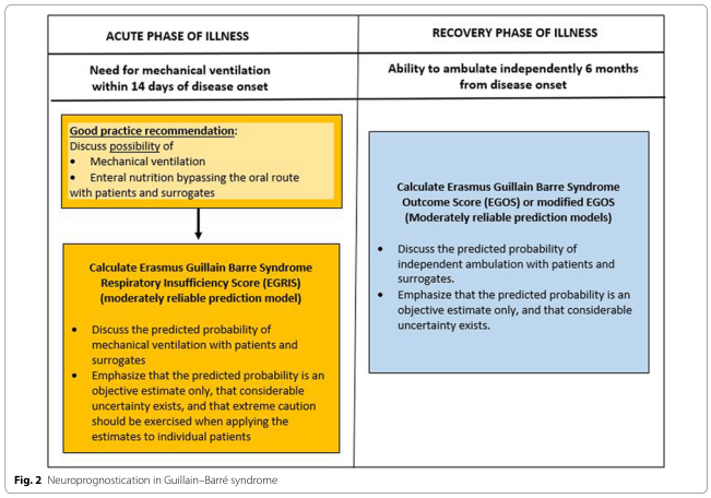
Summary : This figure presents a neuroprognostication framework for Guillain–Barré syndrome, dividing the process into the acute phase (need for mechanical ventilation within 14 days of onset) and the recovery phase (ability to ambulate independently at 6 months), with recommendations and prediction models for each phase.
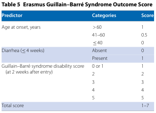
Table 5 Erasmus Guillain–Barré Syndrome Outcome Score
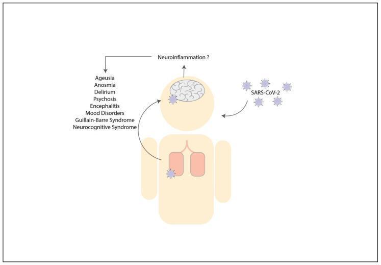
This pathophysiology diagram illustrates the potential link between respiratory SARS-CoV-2 infection and neurological complications in humans. A schematic figure is shown with stylized lungs and a brain highlighted. SARS-CoV-2 virions are depicted entering the body and localizing in both the pulmonary system and the central nervous system. An arrow leads from the brain to the text 'Neuroinflammation ?', suggesting a hypothesized inflammatory response. This central concept is further linked via arrows to a list of clinical neurological and neuropsychiatric manifestations, including ageusia, anosmia, delirium, psychosis, encephalitis, mood disorders, Guillain-Barré Syndrome, and neurocognitive syndrome. The diagram serves as an educational summary of the indirect or direct impact of COVID-19 on the nervous system, potentially mediated by systemic cytokine storms or neuroinflammation.
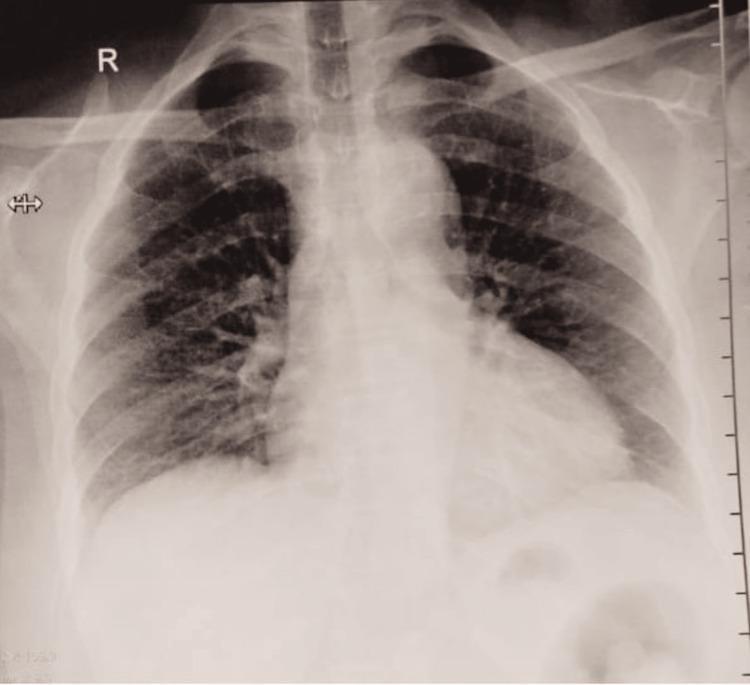
This diagnostic image is a posteroanterior (PA) view chest radiograph of an adult patient. The lung fields are clear bilaterally with no evidence of focal consolidation, masses, or pleural effusions. Pulmonary vascular markings are within normal limits and demonstrate a symmetric distribution. The cardiac silhouette is normal in size and shape, with a cardiothoracic ratio within expected parameters. The mediastinum is midline without widening or hilar adenopathy. The diaphragm appears smooth and well-defined with sharp costophrenic and cardiophrenic angles. Visible osseous structures, including the ribs, clavicles, and vertebrae, appear intact without acute fracture or aggressive lesions. This radiograph represents a normal chest study used in clinical practice to rule out cardiopulmonary pathology in patients presenting with systemic neurological symptoms, such as those associated with Miller Fisher Syndrome or Guillain-Barré Syndrome.
Guillain-Barré syndrome diagnosis and treatment guideline IVIG plasma exchange second IVIG dose
Guillain-Barré Syndrome (GBS)
Epidemiology
- Incidence is about 1.8 per 100,000 per year. It rises with age, from 0.8 in people under 18 to 3.2 in those 60 and over.
- Males are affected more than females (about 1.5:1).
- Mortality was about 33% before positive-pressure ventilation. It is now about 1-5% (Bradley and Daroff's).
Subtypes
- AIDP (acute inflammatory demyelinating polyradiculoneuropathy) is the most common form in Europe and North America.
- AMAN (acute motor axonal neuropathy) is a pure motor axonal form. It was first described in northern China, in summer epidemics in children and young adults.
- AMSAN (acute motor-sensory axonal neuropathy) involves motor and sensory fibres. It is severe and recovers poorly.
- Variants:
- Miller Fisher syndrome (ophthalmoplegia, ataxia, areflexia), about 6% of cases in the West.
- Pharyngeal-cervical-brachial variant.
- Facial diplegia with paresthesias.
- Acute pandysautonomia.
Clinical features
- Weakness with or without paresthesias, often worse in the hands and fingers. It typically starts in the legs and ascends over hours to days.
- It may involve the face, oropharynx, and respiratory muscles.
- Areflexia or hyporeflexia is the hallmark, though it may be absent early.
- Autonomic dysfunction and bifacial palsy are common and support the diagnosis.
- Progression lasts up to 4 weeks. Recovery starts 2-4 weeks after progression stops.
- A preceding infection is common. Campylobacter jejuni is the classic trigger, especially for AMAN. This is from general knowledge, not from the textbook passages I retrieved.
Diagnosis (Asbury-Cornblath criteria)
- Required: progressive weakness of arms and legs, plus areflexia or hyporeflexia.
- Supportive:
- Relative symmetry.
- Mild sensory signs.
- Absence of fever at onset.
- CSF protein raised with fewer than 10 cells/µL (albuminocytologic dissociation).
- Nerve conduction slowing or block.
- CSF protein can be normal in the first week. Nerve conduction studies can also be normal early.
Management (from general knowledge and the web sources below)
- Supportive care is the priority.
- Serial bedside respiratory monitoring (FVC, NIF).
- Telemetry for autonomic instability.
- DVT prophylaxis, pain control, and early rehabilitation.
- Intubate early if the patient is deteriorating or bulbar function is poor.
- Immunotherapy is started when the patient cannot walk unaided, or when weakness is rapidly progressing or severe. It is most effective within 2 weeks of onset and is considered up to 4 weeks. Either option is equivalent:
- IVIG 0.4 g/kg daily for 5 days (2 g/kg total).
- Plasma exchange of about 40-50 mL/kg over 5 sessions (the 5-7 session range comes from the web sources).
- Corticosteroids are not effective.
- Do not combine IVIG and plasma exchange. Do not routinely repeat IVIG in non-responders. In the SID-GBS trial, a second IVIG dose in patients with a poor predicted outcome gave no benefit and more serious adverse events, including thromboembolism.
- Treatment-related fluctuation (relapse after initial improvement) is a separate situation. It is usually managed with a further course. If there are repeated relapses, consider acute-onset CIDP.
Prognosis
- Most patients recover, but recovery can take months. Some are left with residual weakness or fatigue.
- Prognostic tools:
- EGOS (Erasmus GBS Outcome Score) estimates whether the patient will walk at 6 months.
- mEGOS and EGRIS (Erasmus GBS Respiratory Insufficiency Score) estimate the risk of needing ventilation.
- Poor prognostic factors from the literature include older age, preceding diarrhoea, severe weakness at nadir, and need for ventilation. A 2025 systematic review of prognostic factors covers this.
Recent evidence check
- A 2026 rehabilitation review (PMID 41824781).
- A 2025 review of GBS after bariatric surgery (PMID 40897982).
- The 2025 prognostic factors review (PMID 39377571).
Ivig
"Immunoglobulins, Intravenous"[MeSH Terms] AND "Guillain-Barre Syndrome"[MeSH Terms]
IVIG adverse effects thrombosis renal failure IgA deficiency aseptic meningitis infusion rate precautions
IVIG (intravenous immunoglobulin), with a focus on GBS
Mechanism
- Anti-idiotypic antibodies, and interference with complement and cytokines.
- Cytolysis of target cells, and apoptosis induced through Fc receptors.
- Modulation of costimulatory molecules (Firestein & Kelley's Textbook of Rheumatology, Intravenous Immunoglobulin section).
- B-cell apoptosis and altered B-cell signalling at high doses. It also blocks complement activation (Lippincott Illustrated Reviews: Pharmacology, p. 1286).
- Half-life of about 3-4 weeks (Lippincott).
Use in GBS
- Indication: the patient cannot walk unaided, or weakness is severe or rapidly progressing.
- Timing: start within 2 weeks of onset. It is considered up to 4 weeks.
- Dose: 0.4 g/kg/day for 5 days, a total of 2 g/kg. A UIC drug information summary of the guidelines gives this regimen.
- Compared with plasma exchange: efficacy is equal. IVIG is easier to give and has fewer logistical problems. It is usually preferred when access to plasma exchange or vascular access is limited.
- Do not combine IVIG with plasma exchange.
- Corticosteroids are not effective in GBS.
Poor responders and retreatment
- A second IVIG dose is not routinely recommended. The SID-GBS trial found no benefit in patients with a poor predicted outcome. They had more serious adverse events, including thromboembolism.
- A 2024 systematic review (PMID 38553651) looked at IVIG retreatment in GBS patients who responded poorly to the first cycle. I read only its title, not the abstract, so I can't say what it concluded.
- Treatment-related fluctuation is a relapse after initial improvement, usually within weeks. It is often given another course. If relapses keep happening, consider acute-onset CIDP.
Adverse effects
- Aseptic meningitis. It causes severe headache, photophobia, and neck stiffness within about 72 hours. CSF shows neutrophilic pleocytosis. It is more common in people with migraine and usually resolves in 24-48 hours.
- Acute kidney injury. This is mainly with sucrose-containing products (Firestein). Risk factors are pre-existing renal disease, diabetes, age over 64, volume depletion, sepsis, and nephrotoxic drugs.
- Thromboembolism (DVT, MI, stroke, PE). It is more likely in older patients with atherosclerotic risk factors. GBS patients are already immobile and prothrombotic, so thromboprophylaxis matters.
- Haemolysis. It occurs in roughly 1-2% of patients, especially with non-O blood groups and high doses (review of immunoglobulin adverse effects).
- Anaphylaxis. It is rare and mostly seen in IgA deficiency. Fitzpatrick's Dermatology recommends checking serum IgA before infusion in at-risk patients.
- Other: TRALI, arrhythmia, and transient hyponatraemia (pseudohyponatraemia).
Practical precautions
- Hydrate the patient before the infusion.
- Start slowly (the first 30 minutes slower), then step up if tolerated.
- Use the slowest feasible rate in high-risk patients.
- Monitor blood pressure and urine output.
- Check baseline creatinine.
- Choose a sucrose-free product in at-risk patients.
- Live vaccines are less effective after IVIG, so defer them.