
IV fluids
Iv fluids from millers
IV Fluids - Miller's Anesthesia (10e)
1. Classification of IV Fluids
Crystalloids
| Fluid | Osmolality | Key Features |
|---|---|---|
| Normal Saline (0.9% NaCl) | 308 mOsm/L | Slightly hyperosmolar vs. plasma (295 mOsm/L); large volumes cause hyperchloremic metabolic acidosis |
| Lactated Ringer's (LR) | 273 mOsm/L | Slightly hypo-osmolar; very reasonable compromise for replacement of blood/third-space losses; may theoretically lower serum osmolarity in large volumes |
| Plasma-Lyte / NormosolR | ~294 mOsm/L | Buffered crystalloid (pH 7.4); physicochemically closest to plasma; no calcium; favorable strong ion difference - may be advantageous in acidosis from hemorrhagic shock |
Colloids
- Albumin (4%): Reasonable choice for volume expansion; 4% solution is hypo-osmolar (274 mOsm/L). In the SAFE trial subset of severe TBI patients, albumin was associated with increased mortality, though this finding has been questioned due to randomization imbalances. Potentially beneficial in subarachnoid hemorrhage (SAH).
- Hydroxyethyl Starches (HES): Interfere with platelets and the factor VIII complex in addition to causing dilutional reduction of coagulation factors. Effects proportional to molecular weight and hydroxyethyl substitution ratio. Should be used cautiously; respect manufacturer dosage limits. Concerns about adverse renal effects in critical care settings have made many clinicians reluctant to use these.
- Dextrans: Generally avoided due to effects on platelet function.
Hypertonic Saline (HS)
- Draws fluid into the vascular space from the interstitium, enhancing volume restoration relative to an equivalent volume of isotonic solution.
- Studied extensively in hemorrhagic shock resuscitation - results in trauma patients are inconclusive overall.
- Greatest apparent benefit in polytrauma with concurrent TBI - improved neurologic status demonstrated in some studies.
- Commonly used as an osmotic agent to manage raised ICP. In one systematic review of 13 studies (593 patients), HS was associated with decreased all-cause mortality vs. mannitol.
- Neurocritical Care Society guidelines support hyperosmolar therapy for ICP reduction in TBI, SAH, ischemic stroke, and ICH - though neurologic outcomes are not clearly affected.
2. Principles of Fluid Selection
For Neurosurgery
- Maintain normovolemia - to support normal MAP
- Avoid reduction of serum osmolarity - free water lowers serum osmolarity and causes cerebral edema in both normal and abnormal brain
- Normal saline and balanced salt solutions (LR, Plasma-Lyte) are most commonly used intraoperatively.
- In large-volume administration (e.g., massive trauma, aneurysm rupture), the authors recommend alternating LR and normal saline liter-by-liter to balance the risks of both.
- For crystalloid vs. colloid: reducing colloid oncotic pressure (COP) produces only very small transcapillary gradients compared to osmolarity changes. For most elective craniotomies, colloid is not needed. In massive resuscitation, a combination of isotonic crystalloid and colloid may be appropriate.
For Trauma / Hemorrhagic Shock
- Isotonic crystalloids (NS, LR, Plasma-Lyte) are the initial resuscitative fluids.
- Damage Control Resuscitation (DCR): The modern paradigm - limit crystalloids, favor blood component therapy (RBCs + plasma + platelets in balanced ratios). DCR has been associated with improved outcomes for over two decades.
- Early administration of plasma reduces risk of dilutional coagulopathy.
- RBCs remain the mainstay of treatment for hemorrhagic shock (hematocrit ~50-60% per unit; restores O2-carrying capacity as effectively as colloids).
3. Perioperative Fluid Quantity
Traditional (Weight-Based) Approach
- Based on the 4-2-1 rule (Holliday-Segar) for calculating maintenance fluid needs.
- Crystalloid used to replace blood loss in a 3:1 ratio to account for redistribution to extravascular compartment.
- Much of the physiologic basis has been questioned.
Liberal vs. Restrictive Fluids
- Giving >3,500-5,000 mL of crystalloid intraoperatively (when not targeting physiologic endpoints) is associated with: weight gain, cardiopulmonary dysfunction, impaired wound healing, delayed GI recovery, and longer hospital stay.
- The RELIEF trial (3,000 patients, major abdominal surgery) compared restrictive (median 3.7 L) vs. liberal (median 6.1 L) fluid regimens - no difference in disability-free survival at 1 year, but restrictive group had higher rates of surgical site infection (16.5% vs. 13.6%) and renal replacement therapy (0.9% vs. 0.3%). This warns against excessive restriction.
- A post-operative weight gain of approximately +1.5 kg is a useful benchmark.
Goal-Directed Therapy (GDT)
- Measures key hemodynamic variables (cardiac output, O2 delivery) and uses fluids +/- inotropes/vasopressors to optimize tissue perfusion.
- Targets physiologic endpoints (cardiac index >4.5 L/min/m², DO2I >600 mL/min/m²) rather than giving preset volumes.
- Evidence supports GDT reducing postoperative morbidity in high-risk major surgery.
- Stroke volume variation (SVV) - variation in stroke volume with the respiratory cycle during positive-pressure ventilation - is a reliable predictor of decreased intravascular volume and is used to guide fluid therapy.
4. Key Clinical Pearls
- Hyperchloremic metabolic acidosis is a known consequence of large-volume NS; clinical significance is debated, but may adversely affect renal blood flow and GFR.
- Glucose-containing fluids are generally avoided intraoperatively in neuro patients - hyperglycemia aggravates cerebral ischemia. The threshold for treatment is ~250 mg/dL, targeting <200 mg/dL.
- Temperature: RBCs are stored at 4°C and can cause hypothermia if not warmed before infusion.
- Hyperbaric environment: In hyperbaric chambers, IV fluid administration requires special pumps capable of handling pressure differentials up to 3 ATA; glass bottles should be excluded due to risk of explosive rupture during decompression.
In detail from theory and viva point of view, give classification based in tonicity, indications for using different types id fluids and their contents
IV Fluids - Classification, Contents, and Indications
(Miller's Anesthesia, 10e - Theory & Viva)
PART 1: CLASSIFICATION BY TONICITY
| Class | Osmolality | Examples |
|---|---|---|
| Hypotonic | < 280 mOsm/L | 0.45% NaCl ("half normal saline"), 5% Dextrose (D5W), 0.18% NaCl + 4% Dextrose, Lactated Ringer's (273 mOsm/L) |
| Isotonic | 280-310 mOsm/L | 0.9% NaCl ("normal saline", 308 mOsm/L), Plasma-Lyte/NormosolR (294 mOsm/L), Hartmann's solution |
| Hypertonic | > 310 mOsm/L | 3% NaCl, 7.5% NaCl (hypertonic saline), 20% Mannitol, 20% Albumin, Dextran 70 |
Viva point: Lactated Ringer's (LR) is technically slightly hypotonic (273 mOsm/L) but is used as an isotonic replacement fluid. Normal saline at 308 mOsm/L is slightly hypertonic. Plasma-Lyte at 294 mOsm/L is the most physiologically matched to plasma.
PART 2: DETAILED CLASSIFICATION WITH CONTENTS
A. CRYSTALLOIDS
1. Normal Saline (0.9% NaCl) - ISOTONIC
| Component | Amount |
|---|---|
| Na⁺ | 154 mEq/L |
| Cl⁻ | 154 mEq/L |
| Osmolality | 308 mOsm/L |
| pH | 5.0 |
- Initial resuscitation in trauma/hemorrhagic shock
- Hypovolemia (any cause)
- Hypochloremic metabolic alkalosis (e.g., from vomiting/nasogastric losses)
- Hypercalcemia treatment (volume expansion + loop diuretics)
- Diluent for blood products (LR causes RBC clumping due to calcium content)
- Hyponatremia correction
- Replacing upper GI losses (high Cl⁻ content matches gastric secretions)
- Large volumes cause hyperchloremic metabolic acidosis (due to excess Cl⁻ lowering the strong ion difference/SID)
- Excess NaCl and water load is excreted more slowly than balanced crystalloids
- May adversely affect renal blood flow and GFR (hyperchloremia)
- Contains no potassium, no bicarbonate, no calcium
2. Lactated Ringer's (LR) / Hartmann's Solution - MILDLY HYPOTONIC
| Component | Amount |
|---|---|
| Na⁺ | 130 mEq/L |
| K⁺ | 4 mEq/L |
| Ca²⁺ | 3 mEq/L |
| Cl⁻ | 109 mEq/L |
| Lactate (as HCO₃⁻ precursor) | 28 mEq/L |
| Osmolality | 273 mOsm/L |
| pH | 6.5 |
- Most intraoperative fluid replacement (blood/third-space losses)
- Burns resuscitation (Parkland formula uses LR)
- Acute blood loss replacement
- Replacing lower GI losses (high K⁺ and HCO₃⁻ match small/large bowel secretions)
- General fluid resuscitation where large volumes needed
- Hypotonic (273 mOsm/L) - can lower serum osmolarity with very large volumes, potentially worsening cerebral edema in TBI/neurosurgery
- Contains calcium - cannot be used to dilute blood products (calcium chelates citrate anticoagulant, may cause clotting)
- Lactate metabolism may be impaired in severe liver failure
- In animal models, LR increases hepatic and intestinal apoptosis after hemorrhagic resuscitation (via reperfusion injury) - unlike whole blood or hypertonic saline
3. Plasma-Lyte / NormosolR - ISOTONIC (BALANCED)
| Component | Amount |
|---|---|
| Na⁺ | 140 mEq/L |
| K⁺ | 5 mEq/L |
| Mg²⁺ | 3 mEq/L |
| Cl⁻ | 98 mEq/L |
| Acetate | 27 mEq/L |
| Gluconate | 23 mEq/L |
| Osmolality | 294 mOsm/L |
| pH | 7.4 |
- Preferred balanced crystalloid in patients at risk of acidosis (hemorrhagic shock)
- When normal saline-related hyperchloremic acidosis is a concern
- General perioperative maintenance and replacement
- Suitable for patients needing large-volume crystalloid resuscitation
- pH of 7.4 - closest to plasma
- No calcium - safe to use with blood products
- Acetate and gluconate (not lactate) - metabolized peripherally, not just in the liver; safer in hepatic dysfunction
- Favorable strong ion difference (SID) - does not cause metabolic acidosis
4. 5% Dextrose (D5W) - HYPOTONIC (effectively free water)
| Component | Amount |
|---|---|
| Dextrose | 50 g/L (5 g/100 mL) |
| Osmolality (pre-metabolism) | 252 mOsm/L |
| Effective tonicity after glucose metabolism | = free water (0 mOsm/L effective) |
| Na⁺, K⁺, Cl⁻ | None |
- Pure maintenance fluid to replace insensible losses (free water losses)
- Hypernatremia correction
- Hypoglycemia (though 10-50% dextrose preferred for frank hypoglycemia)
- Postoperative maintenance component (in combination fluids)
- Vehicle for IV drug infusions
- After glucose is metabolized, this is effectively free water - distributes throughout total body water (2/3 intracellular, 1/3 extracellular)
- Never use for resuscitation - no electrolytes, no intravascular volume expansion
- Can worsen cerebral edema (lowers serum osmolarity)
- Intraoperative hyperglycemia aggravates cerebral ischemia - avoided routinely in neuroanesthesia
- Can cause dilutional hyponatremia postoperatively
5. 0.18% NaCl + 4% Dextrose ("Dextrose-Saline") - HYPOTONIC
| Component | Amount |
|---|---|
| Na⁺ | 30 mEq/L |
| Cl⁻ | 30 mEq/L |
| Dextrose | 40 g/L |
| Osmolality | ~284 mOsm/L (but hypotonic after dextrose metabolism) |
- Pediatric maintenance fluid (traditionally)
- Part of adult postoperative maintenance (alongside isotonic fluids)
- Provides free water + minimal electrolytes for daily maintenance needs
6. Hypertonic Saline (3%, 7.5% NaCl) - HYPERTONIC
| Concentration | Osmolality | Na⁺ content |
|---|---|---|
| 3% NaCl | ~1026 mOsm/L | 513 mEq/L |
| 7.5% NaCl | ~2566 mOsm/L | 1283 mEq/L |
- Severe symptomatic hyponatremia (3% NaCl, controlled infusion)
- Raised intracranial pressure (ICP) - draws free water from brain via osmotic gradient (superior to or equivalent to mannitol)
- Polytrauma with concurrent TBI - improved neurologic outcomes in some studies
- Hemorrhagic shock resuscitation under austere/military conditions (small volume, enhanced effect)
B. COLLOIDS
1. Albumin - ISOTONIC (4%) or HYPERTONIC (20-25%)
| Preparation | Oncotic Pressure | Osmolality | Key Features |
|---|---|---|---|
| 4% Albumin | Low | 274 mOsm/L (hypo-osmolar) | Iso-oncotic |
| 20-25% Albumin | High | Hypertonic | Hyperoncotic; pulls fluid intravascularly |
- Intravascular volume expansion when crystalloids alone are inadequate
- Spontaneous bacterial peritonitis (SBP) in cirrhosis (prevents hepatorenal syndrome)
- Large-volume paracentesis replacement
- Hypoalbuminemia with symptomatic edema
- Subarachnoid hemorrhage (SAH) - potential neuroprotective benefit
- Burns (delayed phase, after 24h)
- The SAFE trial: in patients with severe TBI (GCS 3-8), 4% albumin was associated with increased mortality - but the finding is questioned due to randomization imbalances and the hypo-osmolarity of the 4% preparation
- Albumin has no albumin-specific hazard from a physiologic standpoint - if edema formation is a concern, it applies equally to all colloids (FFP, starches)
- ALIAS trial (acute stroke): albumin did not improve neurologic outcome; associated with increased CHF rates
- Most expensive of the colloid options
2. Hydroxyethyl Starch (HES) - ISOTONIC or HYPERTONIC
- Derived from amylopectin (plant starch); available in different molecular weights (MW) and molar substitution ratios (MS)
- The higher the MW and MS, the greater the coagulopathy
- Perioperative intravascular volume expansion
- Should be avoided in sepsis/critical illness (proven AKI and mortality increase in VISEP, 6S, CHEST trials)
- Interferes with platelets and factor VIII complex (von Willebrand factor) - dilutional + direct effect
- Coagulopathy proportional to MW and degree of hydroxyethyl substitution
- Reports of neurosurgical bleeding when dosage limits exceeded or used on successive days (accumulation effect)
- Multiple RCTs (VISEP, 6S, CHEST) show increased AKI and need for renal replacement therapy in critical illness/sepsis
- Miller's: use with caution; respect manufacturer dosage limits; use additional restraint when there are other causes of coagulopathy
3. Dextrans - HYPERTONIC
- Polysaccharide glucose polymers (Dextran 40, Dextran 70)
4. Gelatins - ISOTONIC
- Derived from bovine collagen (Gelofusine, Haemaccel)
- Less data available compared to starches
- Shorter intravascular half-life than starches
- Less data on renal safety than starches
- Some risk of anaphylaxis (bovine origin)
- Miller's notes: limited comparative data with crystalloids; no consistent clinical benefit over crystalloids
PART 3: INDICATIONS BY CLINICAL SCENARIO (Viva-Ready)
| Clinical Situation | Recommended Fluid |
|---|---|
| Acute hemorrhagic shock (initial) | Isotonic crystalloid (NS, LR, Plasma-Lyte), then blood products (DCR) |
| Neurosurgery / TBI (ICP raised) | Hypertonic saline or Mannitol; avoid hypotonic fluids |
| Neurosurgery (maintenance) | Normal saline or alternate NS/LR; avoid free water |
| Burns (first 24h) | Lactated Ringer's (Parkland: 4 mL/kg/% TBSA) |
| Hypochloremic metabolic alkalosis | Normal saline |
| Metabolic acidosis / large volume resuscitation | Plasma-Lyte or balanced crystalloid |
| Upper GI losses (vomiting, NGT) | Normal saline + KCl |
| Lower GI losses (diarrhea, fistula) | Balanced crystalloid (LR or Plasma-Lyte) + K⁺ |
| Postoperative maintenance (adult) | 0.18% NaCl + 4% dextrose (hypotonic) for free water; NOT for hypovolemia |
| Severe symptomatic hyponatremia | 3% Hypertonic saline (controlled rate) |
| Hypercalcemia | Normal saline + loop diuretics |
| Spontaneous bacterial peritonitis / large volume paracentesis | 20% Albumin |
| Blood transfusion diluent | Normal saline ONLY (not LR - calcium chelates citrate) |
| Bowel prep preoperative deficit | 1-2L balanced crystalloid + K⁺ |
| Intraoperative maintenance (low-risk surgery) | 1-1.5 mL/kg/h crystalloid |
| High-risk major surgery | Goal-directed: boluses of colloid or balanced crystalloid, titrated to stroke volume |
PART 4: KEY VIVA CONCEPTS
- Normal saline / LR: ~75-80% goes to interstitium, ~20-25% stays intravascular
- Colloids (albumin, HES): ~80% stays intravascular
- D5W (free water): distributes across total body water - only ~8% stays intravascular
- 4 mL/kg/h for first 10 kg
- 2 mL/kg/h for next 10 kg (10-20 kg)
- 1 mL/kg/h for each kg above 20 kg
Blood transfusion and associated reactions, massive transfusion and complications,blood conservation strategies from millers and recent articles
"blood transfusion" AND "transfusion reactions" AND "perioperative"
"massive transfusion" AND "damage control resuscitation"
"blood conservation" AND "patient blood management" AND surgery
"massive transfusion" AND "plasma ratio" AND hemorrhage
"tranexamic acid" AND "hemorrhage" AND "mortality"
"massive transfusion" AND "coagulopathy" AND trauma
"patient blood management" AND "restrictive transfusion" AND outcomes
"intraoperative cell salvage" AND "blood conservation" AND surgery
PMID: 42115060
PMID: 36942533
Blood Transfusion, Reactions, Massive Transfusion & Blood Conservation
Miller's Anesthesia, 10e + Recent Evidence
PART 1: TRANSFUSION REACTIONS
Classification of Transfusion Reactions
TRANSFUSION REACTIONS
├── IMMUNE-MEDIATED
│   ├── Hemolytic (Acute / Delayed)
│   ├── TRALI
│   ├── Febrile Non-Hemolytic (FNHTR)
│   ├── Allergic (Urticarial / Anaphylactic)
│   └── TA-GvHD
└── NON-IMMUNE
    ├── TACO
    ├── Septic (Bacterial Contamination)
    ├── Citrate Toxicity
    ├── Hyperkalemia
    ├── Hypothermia
    ├── Metabolic Alkalosis
    └── Iron Overload (chronic)
1. ACUTE HEMOLYTIC TRANSFUSION REACTION (AHTR)
| Sign/Symptom | Frequency (in 40 patients) |
|---|---|
| Fever | 19/40 |
| Fever + chills | 16/40 |
| Chest pain | 6/40 |
| Hypotension | 6/40 |
| Nausea | 2/40 |
| Dyspnea | 2/40 |
| Hemoglobinuria | 1/40 |
- Hemoglobinuria (pink/red urine) - most common first sign
- Unexplained hypotension
- Bleeding diathesis (DIC)
- As little as 50 mL incompatible blood can exceed haptoglobin binding capacity (~100 mg Hb/100 mL plasma)
- Free Hb >100 mg/dL = red plasma
- Free Hb >150 mg/dL = hemoglobinuria
- Complement activation releases histamine and vasoactive amines
- Acute Renal Failure - precipitation of Hb as acid hematin in distal tubule causing mechanical tubular blockage
- DIC - complement activation triggers coagulation cascade
- Stop the transfusion immediately
- Maintain urine output >75-100 mL/h by:
- IV fluids (crystalloid/mannitol)
- Furosemide if above ineffective
- Alkalinize the urine (sodium bicarbonate - makes Hb more soluble, prevents acid hematin precipitation)
- Assay urine and plasma Hb concentrations
- Check platelet count, PT, PTT, fibrinogen (screen for DIC)
- Return unused blood to blood bank for repeat crossmatch
- Send patient's blood and urine to blood bank
- Prevent hypotension to maintain renal blood flow
- Direct antiglobulin test (DAT/Coombs) - shows antibody attached to transfused donor RBCs
- Serum haptoglobin (decreased), plasma Hb, urine Hb, bilirubin (increased)
2. DELAYED HEMOLYTIC TRANSFUSION REACTION (DHTR)
- Unexplained drop in Hb post-transfusion
- Mild jaundice
- Hemoglobinuria (rarely)
- Rarely fatal
3. TRANSFUSION-RELATED ACUTE LUNG INJURY (TRALI)
- Hit 1: Patient factors (surgery, sepsis, shock, mechanical ventilation)
- Hit 2: Donor antibodies (anti-HLA or anti-neutrophil antibodies) activating recipient neutrophils
- Plasma/whole blood from multiparous female donors (most common - reduced by male-predominant plasma donation)
- High IL-8 levels
- Liver surgery, chronic alcohol abuse, smoking
- High peak airway pressures, positive fluid balance
- Stop the transfusion
- Supportive care (oxygen, ventilatory support as for ARDS)
- Notify blood bank - quarantine all units from implicated donor
- HLA testing of patient if possible
- Recovery within 96 hours in most patients
- No specific pharmacologic therapy
| Feature | TRALI | TACO |
|---|---|---|
| Mechanism | Immune/inflammatory | Volume overload |
| Left atrial pressure | Normal | Elevated |
| BNP | Normal | Elevated |
| CVP | Normal | Elevated |
| Response to diuresis | Poor | Good |
| Onset | Within 6h | During or shortly after transfusion |
| CXR | Bilateral infiltrates | Pulmonary edema pattern |
4. TRANSFUSION-ASSOCIATED CIRCULATORY OVERLOAD (TACO)
5. FEBRILE NON-HEMOLYTIC TRANSFUSION REACTION (FNHTR)
6. ALLERGIC REACTIONS
| Type | Mechanism | Features | Treatment |
|---|---|---|---|
| Minor urticarial | Foreign proteins, non-IgE | Urticaria, itching, facial swelling | Antihistamines; transfusion can continue |
| Anaphylactoid | Non-IgE mediated | Clinically similar to anaphylaxis | Stop transfusion; epinephrine, supportive |
| Anaphylaxis | IgA in IgA-deficient recipient with anti-IgA | Dyspnea, hypotension, laryngeal edema, shock - after only a few mL | Stop; epinephrine; use washed RBCs or IgA-deficient blood in future |
7. TRANSFUSION-ASSOCIATED GRAFT-VERSUS-HOST DISEASE (TA-GvHD)
- Severely immunocompromised patients
- Recipients of directed donations from first/second-degree relatives (shared HLA haplotypes - lymphocytes not recognized/eliminated)
8. TRANSFUSION-RELATED IMMUNOMODULATION (TRIM)
- Cancer recurrence after tumor resection
- Postoperative infections
- Virus reactivation
9. OTHER NON-INFECTIOUS RISKS (Table 45.18)
| Complication | Notes |
|---|---|
| Microchimerism | Donor lymphocytes persist in recipient; clinical significance unknown |
| Post-transfusion purpura | Recipient alloantibodies attack donor platelets; treat with IVIG |
| Hypotensive reactions | Bradykinin activation via coagulation pathway |
| Transfusion-related AKI | Independent risk from transfusion |
| Alloimmunization | Only 2-8% of chronically transfused develop RBC alloantibodies |
| HLA / HPA alloimmunization | Platelet refractoriness to future transfusions |
| Iron overload | Chronic transfusion therapy; deposits in liver/heart; manage with chelation |
PART 2: MASSIVE TRANSFUSION - DEFINITION, COMPLICATIONS & MANAGEMENT
Definition
- ≥3 units RBCs in 1 hour with ongoing need
- Loss of >50% blood volume in 3 hours
The Lethal Triad of Massive Hemorrhage
        HYPOTHERMIA
           /   \
          /     \
COAGULOPATHY --- ACIDOSIS
Complications of Massive Transfusion
1. DILUTIONAL COAGULOPATHY
- Platelet count falls to <100 × 10⁹/L after 10-15 units of blood
- Miller's threshold: Platelet count <75 × 10⁹/L = reliable predictor of hemorrhagic diathesis from dilutional thrombocytopenia
- However: platelet counts rarely fall as low as predicted from pure dilution (splenic/bone marrow release partially compensates)
- Acute dilutional thrombocytopenia causes bleeding at a much higher platelet count than chronic thrombocytopenia (e.g., ITP)
- Fibrinogen levels fall significantly when blood is replaced with PRBCs + crystalloid (unlike whole blood replacement where fibrinogen remains stable unless DIC develops)
- Fibrinogen is critical for effective clot formation - must be monitored and supplemented early
- If PTT ≥1.5x normal with other tests normal: suggests low factors V and VIII → treat with FFP or cryoprecipitate
2. HYPOTHERMIA
- Blood stored at 4°C - infusion of large volumes causes core temperature to fall
- Temperature <30°C: ventricular irritability and cardiac arrest
- Even small decreases in temperature significantly impair both coagulation factors and platelet function
- Shivering increases metabolic demands, worsening tissue ischemia
3. CITRATE TOXICITY AND HYPOCALCEMIA
- Citrate (anticoagulant in stored blood) chelates ionized calcium → hypocalcemia
- Consequences: dysrhythmia, hypotension, myocardial depression
- Risk increased with: infusion rate >1 unit/10 minutes, pediatric patients, liver disease (impaired citrate metabolism), hyperventilation
- Even at these rates, ionized calcium may not fall enough to cause bleeding alone
- Note: Blood transfusions provide citrate which generates bicarbonate → may cause metabolic alkalosis post-transfusion
4. HYPERKALEMIA
- Stored blood K⁺: 19-50 mEq/L at 21 days; 45-60 mEq/L at 42 days
- Despite high K⁺ in storage, net K⁺ gain is only ~10 mEq/L when blood loss is considered
- For clinically significant hyperkalemia: must infuse blood at ≥120 mL/min
- More common in: neonates, patients with renal failure
- Irradiated RBC units have even higher K⁺ levels
5. ACID-BASE DISTURBANCES
- Initial state during massive hemorrhage: metabolic acidosis (lactic acidosis from hypoperfusion, citric acid in stored blood)
- Post-resuscitation: metabolic alkalosis (from citrate conversion to bicarbonate, volume replacement)
- Empirical bicarbonate is NOT indicated - should be guided by serial ABGs
6. DISSEMINATED INTRAVASCULAR COAGULATION (DIC)
- Fibrinolytic system is simultaneously activated (tPA from damaged tissue)
- Tumor necrosis factor and endotoxins further activate coagulation
Damage Control Resuscitation (DCR)
- Limit crystalloids - aggressive crystalloid causes dilutional coagulopathy, hypothermia, abdominal compartment syndrome, and worsened outcomes
- Balanced blood component therapy - RBC : FFP : Platelets in ratios approaching 1:1:1 (whole blood equivalent)
- Permissive hypotension - until surgical hemorrhage control is achieved (avoid over-resuscitation before bleeding is controlled)
- Hemorrhage control first - damage control surgery before full resuscitation
- Military and major trauma centers use ratio-based transfusion (RBC:FFP ~1:1 to 2:1) rather than strict laboratory thresholds
- Fresh whole blood: 1 unit equivalent to 8-10 platelet units for treating transfusion-induced coagulopathy
- Fibrinogen is the first coagulation factor to reach critically low levels during major hemorrhage
- Supplement early via cryoprecipitate or lyophilized fibrinogen concentrate
- RCT: 324 trauma patients at risk of MT
- 4-Factor Prothrombin Complex Concentrate (4F-PCC, 25 IU/kg) vs placebo, all receiving ratio-based transfusion (RBC:FFP 1:1 to 2:1)
- Result: No reduction in 24-hour blood product consumption
- Safety concern: Thromboembolic events 35% (4F-PCC) vs 24% (placebo) - relative risk 1.48, P=0.03
- Conclusion: Does NOT support systematic use of 4F-PCC in massive transfusion patients
PART 3: BLOOD CONSERVATION STRATEGIES
A. PREOPERATIVE
- Identify and treat iron deficiency, B12/folate deficiency, or anemia of chronic disease before elective surgery
- IV iron supplementation in iron-deficient patients
- Erythropoiesis-stimulating agents (ESAs/erythropoietin) in selected patients
- Patient donates their own blood 4-6 weeks before surgery
- Largely fallen out of favor: wastage rates, cost, and not useful in emergency settings
- Still useful in complex elective cases where crossmatching is difficult (rare antibodies)
- Stop antiplatelet agents (aspirin 7-10 days, clopidogrel 5-7 days prior) when safe
- Stop anticoagulants appropriately (bridge where indicated)
- If bowel prep required: give 1-2L balanced crystalloid + K⁺ to prevent hypovolemia at induction
B. INTRAOPERATIVE
- Blood suctioned from surgical field is processed (washed, filtered, concentrated) and reinfused
- The most effective intraoperative blood conservation technique
- Major cardiac, vascular, orthopedic, hepatic surgery
- Obstetric hemorrhage (with leukocyte depletion filter)
- Bacterial contamination of surgical field
- Malignancy (relative) - though leukocyte filters may reduce tumor cell reinfusion risk
- Amniotic fluid contamination (relative - filtered ICS now used in obstetrics)
- [PMID 40465098 - Spine Deform 2025]: Systematic review - ICS is effective for blood conservation in both pediatric and adult spinal surgery, reducing allogeneic transfusion requirements
- [PMID 35023053 - Reprod Sci 2022]: Meta-analysis - ICS in high-risk cesarean section reduces allogeneic transfusion without adverse neonatal or maternal outcomes
- Blood drawn from patient immediately before surgery
- Volume replaced with crystalloid/colloid (normovolemia maintained)
- Diluted blood remains in circulation during surgery (less Hb lost per mL bled)
- Drawn blood (with preserved platelets and clotting factors) re-infused at end of surgery
- Most effective when large blood loss is expected and preoperative Hb is high
- Controlled reduction of MAP to 50-65 mmHg to reduce surgical field bleeding
- Used in major orthopedic (hip/spine), ENT, maxillofacial surgery
- Agents: volatile anesthetics, beta-blockers, nitroprusside, nitroglycerin, remifentanil
- Contraindications: IHD, cerebrovascular disease, renal insufficiency, uncontrolled hypertension
| Drug | Mechanism | Dose | Evidence |
|---|---|---|---|
| Tranexamic Acid (TXA) | Lysine analogue - blocks plasminogen binding to fibrin; prevents fibrinolysis | IV: 1g loading over 10 min, then 1g over 8h (trauma); variable for surgery | CRASH-2, CRASH-3 trials; most evidence |
| Epsilon-aminocaproic acid | Similar to TXA; lysine analogue | IV infusion | Less evidence than TXA |
| Aprotinin | Serine protease inhibitor - broad antifibrinolytic + platelet protective effects | IV | Withdrawn in many countries over renal concerns; used in cardiac surgery |
- [PMID 40751727 - Ann Emerg Med 2026]: RCT - TXA timing critically affects mortality impact after trauma; earlier administration provides greater benefit
- [PMID 37043652 - NEJM 2023]: RCT (NEJM) - TXA to prevent obstetrical hemorrhage after cesarean delivery: reduced postpartum hemorrhage without significant safety concerns
- [PMID 35977357 - Anesth Analg 2022]: Review - TXA applications and limitations: supports use across surgical settings, with evolving evidence in non-trauma hemorrhage contexts
- Minimally invasive surgery (laparoscopic, robotic) - less blood loss
- Meticulous surgical hemostasis
- Positioning to reduce venous congestion in operative field
- Topical hemostatic agents (thrombin, fibrin glue, oxidized cellulose)
- Bone wax, electrocautery, argon beam coagulation
- Tourniquets in limb surgery
- Desmopressin (DDAVP): Releases vWF from endothelial stores; useful in platelet dysfunction (uremia, aspirin effect, type 1 vWD)
- Recombinant Factor VIIa (rFVIIa): Reserved for life-threatening uncontrolled hemorrhage unresponsive to conventional therapy (off-label in most non-hemophilia settings); high thromboembolism risk
- Use TEG/ROTEM to guide targeted blood product administration rather than empiric ratios
- Identifies specific deficiencies (fibrinogen, platelets, clotting factors, fibrinolysis) and treats them precisely
- Reduces unnecessary FFP and platelet transfusion
C. POSTOPERATIVE
- Evidence-based threshold: Hb <7 g/dL in most patients (including critically ill)
- Higher threshold (Hb <8 g/dL) for: cardiac surgery, active cardiac disease, hemodynamically unstable patients
- [PMID 42115060 - Br J Anaesth 2026]: Systematic review of 40 national PBM guidelines - most guidelines define restrictive transfusion as Hb threshold of 7 g/dL (22 guidelines) or lower (9 guidelines). However, national guidelines remain fragmented; comprehensive multidisciplinary coordination is lacking globally.
- [PMID 38936555 - J Clin Epidemiol 2024]: Meta-analysis - important methodological warning: transfusion strategy trials excluding patients transfused outside the study period are more likely to show a trend favoring restrictive strategies - potential selection bias in the existing literature.
- Optimization of erythropoiesis (preoperative anemia treatment)
- Minimization of blood loss (intraoperative conservation)
- Rational use of blood products with appropriate thresholds
- Recombinant erythropoietin with IV iron in surgical patients expected to need transfusion
- Mainly used preoperatively (4-6 weeks before surgery)
- Risks: hypertension, thromboembolism
- Use pediatric sampling tubes where possible
- Batch lab tests to reduce frequency
- Point-of-care testing reduces volume drawn
- (Noted as the area of greatest variability between national guidelines in the 2026 PBM systematic review)
PART 4: KEY VIVA POINTS
Blood conservation strategies from millers and recent articles
"patient blood management" AND perioperative AND outcomes
"tranexamic acid" AND "elective surgery" AND "blood loss"
"acute normovolemic hemodilution" AND surgery AND "blood transfusion"
"intraoperative cell salvage" AND "blood transfusion" AND surgical
PMID: 41714376
PMID: 42115060
"preoperative anemia" AND "iron" AND surgery AND "transfusion"
PMID: 38167004
PMID: 39704317
PMID: 36800489
Blood Conservation Strategies
Miller's Anesthesia, 10e + Recent Evidence (2022-2026)
OVERVIEW: THE THREE PILLARS OF PATIENT BLOOD MANAGEMENT (PBM)
PATIENT BLOOD MANAGEMENT
         │
    ┌────┴──────┐
    │           │
PILLAR 1    PILLAR 2      PILLAR 3
Optimize    Minimize      Rational use of
Erythro-    Blood         Transfusion &
poiesis     Loss          Tolerance of Anemia
[PMID 42115060 - Br J Anaesth 2026]: Systematic review of 40 national/international PBM guidelines found most agree on a restrictive Hb threshold of 7 g/dL (22/40 guidelines) or lower (9/40 guidelines). Greatest variability: strategies to reduce diagnostic blood loss. National standardisation remains fragmented globally - comprehensive multidisciplinary coordination is lacking. (Dreher et al., Br J Anaesth 2026)
PART 1: AUTOLOGOUS BLOOD TRANSFUSION (Miller's Ch 45)
- Preoperative Autologous Donation (PAD)
- Acute Normovolemic Hemodilution (ANH)
- Intraoperative / Postoperative Blood Salvage (Cell Salvage)
1. PREOPERATIVE AUTOLOGOUS DONATION (PAD)
- Hb must be ≥11 g/dL before each donation
- Repeated donations separated by ≥1 week
- Last donation ≥72 hours before surgery (to restore intravascular volume before anaesthesia)
- Note: Red cell mass is NOT restored in 72 hours; full 80% RBC mass recovery takes 25 to >168 days (HEIRS study)
- Evidence of infection / risk of bacteremia
- Aortic stenosis (scheduled surgical correction)
- Unstable angina
- Active seizure disorder
- MI or CVA within 6 months of donation
- High-grade congenital or acquired cardiac lesions
- Blood safety has improved dramatically (HIV/hepatitis C risk from allogeneic blood now very low), so the safety advantage of PAD over allogeneic blood has narrowed considerably
- A meta-analysis showed PAD decreased absolute risk of receiving allogeneic blood by 44%, but the risk of receiving transfusion from any source (autologous or allogeneic) increased by 24% - questioning its use as a true transfusion-sparing practice
- PAD associated with ~12x the post-donation hospitalization rate compared with allogeneic donors (American Red Cross study)
- The proportion of autologous blood collected has significantly decreased since the mid-1990s peak
- Patients undergoing PAD have Hb that is on average 1.1 g/dL lower than non-donors at time of surgery
2. ACUTE NORMOVOLEMIC HEMODILUTION (ANH)
- The blood that is shed during surgery has a lower Hct (diluted blood), so fewer RBCs are lost per mL of surgical blood loss
- The sequestered whole blood (with intact platelets, clotting factors) is reinfused later - at the end of surgery or when hemostasis is needed
- Blood collected in standard bags with citrate anticoagulant
- Stored at room temperature up to 8 hours or 4°C up to 24 hours
- Reinfused in reverse order of collection (last bag first) - except for the first bag which has the highest Hb, platelets, and clotting factors, and is reinfused when hemostasis is most critical
- Agitation of stored blood to preserve platelets: no formal recommendation required; TEG shows no difference between agitated and stationary storage
- Minimal ANH (removing <15% blood volume): only ~100 mL RBCs saved (~0.5 units PRBCs) - marginal benefit
- ANH targeting post-dilutional Hct of 28% with 2600 mL blood loss: saves ~215 mL RBCs compared to no prior hemodilution
- Larger volumes of hemodilution provide the largest RBC-mass savings
- Meta-analysis of 29 RCTs (1252 ANH patients, 1187 controls) in cardiac surgery: ANH patients received ¾ fewer allogeneic blood units than controls; less postoperative blood cell mass loss (mean loss 388 mL ANH vs 450 mL controls)
- ANH also reduces platelet and plasma requirements because whole blood removal preserves these components
- In cardiac surgery: ANH may protect sequestered blood from cardiopulmonary bypass-induced platelet dysfunction
- Evidence in: hip replacement, hepatic resections, vascular surgery, cardiac surgery, orthognathic surgery
- 30 RCTs, 4,473 patients (cardiac surgery)
- ANH: 27% relative reduction in incidence of allogeneic blood transfusion (RR 0.73, 95% CI 0.60-0.88, p=0.0008)
- Reduced allogeneic RBC volume transfused by 0.75 units (p=0.020)
- Reduced FFP requirements (MD -0.21 units, p=0.025)
- Reduced total blood loss by 64 mL (p=0.012)
- No significant differences in chest tube drainage, surgical revision, or stroke
- Conclusion: ANH is an effective blood conservation strategy in cardiac surgery - reduces allogeneic RBC and FFP requirements
- Severe anemia (pre-existing low Hb)
- Significant cardiovascular disease limiting tolerance of diluted Hb
- Severe pulmonary disease
- Renal insufficiency limiting fluid management
3. INTRAOPERATIVE CELL SALVAGE (ICS) / AUTOTRANSFUSION
- Anticoagulant (heparin or citrate) added to the tip of suction catheter
- Blood collected in reservoir until enough accumulates (500-700 mL needed for processing)
- Centrifugation separates lower-density plasma from higher-density RBCs
- Produces 225-250 mL of saline-suspended PRBCs with Hct 50-60%
- Microaggregate filters (40 µm) used during reinfusion
- High-capacity systems can provide the equivalent of 12 units/hour in a massively bleeding patient
- 2,3-DPG levels: near-normal in salvaged blood vs up to 90% reduction in stored allogeneic blood
- P50 similar to fresh venous blood; higher than 2-week-old banked blood - better oxygen offloading
- RBC deformability improved compared with PRBCs
- Room temperature: up to 4 hours
- 1°C-6°C: up to 24 hours (if refrigeration begun within 4 hours of collection)
- Must be labeled: patient name, ID, date/time, "For Autologous Use Only"
- Meta-analysis of 75 studies: Cell salvage reduced need for allogeneic transfusion in adult elective surgeries by 38%, saving on average 0.68 units of banked blood. Greatest benefit in orthopedic and cardiac surgery.
- Hypervolemia
- Bacterial contamination (rare clinically; positive cultures sometimes seen)
- Hypotension
- Air embolism (serious risk - mitigated with modern systems preventing direct connection to IV tubing)
- Non-immune and immune hemolysis (from high suction pressure, turbulence, roller pumps)
- Free hemoglobin: nephrotoxic and causes arteriolar vasoconstriction via NO scavenging
- DIC (with large volumes of unwashed shed blood)
- Coagulopathies (shed blood has undergone varying degrees of fibrinolysis)
- Vacuum should not exceed 150 mmHg to minimize hemolysis (higher may occasionally be needed in rapid bleeding)
- Washed salvage preferred over unwashed
- Use leukocyte depletion filters to reduce bacterial and particulate contamination
- Traditionally controversial (amniotic fluid contamination concerns); now widely accepted with leukocyte depletion filters
- [PMID 39704317 - Cochrane 2024]: Cell salvage for PPH - 6 RCTs, 3,476 women (all cesarean) - low-certainty evidence suggests ICS may reduce allogeneic transfusion risk at cesarean (RR 0.45, 95% CI 0.15-1.33); absolute transfusion risk was low (4% without vs 2% with ICS). Evidence remains uncertain for transfusion-related adverse reactions. (Dey et al., Cochrane 2024)
- [PMID 38109996 - AJOG MFM 2024]: Meta-analysis of RCTs - ICS at cesarean delivery reduces allogeneic transfusion without adverse neonatal or maternal outcomes
- [PMID 40465098 - Spine Deform 2025]: Systematic review - ICS effectively reduces allogeneic transfusion requirements in both pediatric and adult spinal surgery. Supports routine use in high blood loss spine procedures.
- [PMID 37777212 - Bone Joint J 2023]: Meta-analysis in revision hip arthroplasty - ICS reduces allogeneic transfusion requirements
- Absolute: Bacterial contamination of surgical field
- Relative: Active malignancy (risk of tumor cell reinfusion - leukocyte filters may reduce but not eliminate), bowel perforation, certain amniotic fluid exposures without filtering
PART 2: PHARMACOLOGIC BLOOD CONSERVATION
1. ANTIFIBRINOLYTIC AGENTS
A. Tranexamic Acid (TXA)
- Trauma: 1g IV over 10 min (loading), then 1g over 8 hours - must be given within 3 hours of injury (CRASH-2 protocol)
- Surgery: variable; typically 10-15 mg/kg IV pre-incision ± infusion; topical use in joint arthroplasty
- CRASH-2 (2010): TXA reduces all-cause mortality and death from hemorrhage in trauma patients when given within 3 hours of injury. Trend toward harm if given >3 hours.
- [PMID 40751727 - Ann Emerg Med 2026]: RCT - TXA timing critically determines mortality impact after trauma. Earlier administration provides greater benefit; the window of benefit is time-sensitive.
- [PMID 37043652 - NEJM 2023]: RCT - TXA to prevent obstetrical hemorrhage after cesarean delivery: reduced PPH without significant safety concerns (Pacheco et al. for NEJM/C-STAT study)
- [PMID 36800489 - Cochrane 2023]: Network meta-analysis in major vascular surgery - TXA may have no effect on thromboembolic risk (RR 1.10, 95% CI 0.88-1.36) though evidence is low-certainty; topical hemostatic agents (fibrin/thrombin sealants) also evaluated
B. Epsilon-Aminocaproic Acid (EACA)
- Same mechanism as TXA (lysine analogue)
- Less potent, less studied; less commonly used than TXA
- IV infusion; mainly used in cardiac surgery
C. Aprotinin
- Serine protease inhibitor with broad antifibrinolytic effects PLUS direct platelet protection
- Withdrawn from markets after the BART trial showed increased mortality/renal failure
- Available in some countries for cardiac surgery under restricted use
- Higher antifibrinolytic potency than TXA/EACA
2. DESMOPRESSIN (DDAVP)
- Uremia-related platelet dysfunction
- Mild platelet dysfunction from aspirin/NSAIDs
- Type 1 von Willebrand disease
- Hemophilia A (mild-moderate)
- Liver disease-associated platelet dysfunction
3. RECOMBINANT FACTOR VIIa (rFVIIa / NovoSeven)
- Hemophilia A or B with inhibitors
- Congenital Factor VII deficiency
- Off-label: Life-threatening, uncontrolled surgical/traumatic hemorrhage unresponsive to all other measures
- High cost
- Significant thromboembolic risk (arterial and venous)
- Off-label use in non-hemophilia patients remains controversial
- Miller's: reserved for situations where transfusion is not an option or as a bridge
4. HEMOGLOBIN-BASED OXYGEN CARRIERS (HBOCs)
- Severe arteriolar vasoconstriction from NO scavenging by free Hb
- Nephrotoxicity, cardiac toxicity, hypertension
- Meta-analysis of 16 trials (5 products, 3,711 patients): significantly increased risk of MI and death with HBOCs vs controls; 30% increased death risk; 3x MI risk
- Clinical trials have largely failed due to adverse events
- HBOC-201 (Hemopure): Available under FDA Expanded Access (compassionate use)
- Reserved for: Jehovah's Witnesses refusing transfusion, unavailable compatible blood, bridge to hemostatic intervention
- NOT approved for routine clinical use
PART 3: PREOPERATIVE OPTIMIZATION (Pillar 1)
Anemia Detection and Treatment
- Screen: CBC + iron studies (ferritin, transferrin saturation) ≥4-6 weeks before elective major surgery
- Diagnose the cause: iron deficiency, B12/folate deficiency, anemia of chronic disease, chronic kidney disease
- Treat:
- Oral iron: First line for iron deficiency without urgency; cheap but slow (4-8 weeks to optimize)
- IV iron: Faster response; preferred when surgery <4 weeks away or oral iron not tolerated
- Erythropoiesis-stimulating agents (ESAs/EPO): Combined with IV iron for patients with anemia of chronic disease, renal anemia, or anticipated major blood loss
- B12/folate: Replace if deficient
- [PMID 36631901 - J Cardiothorac Surg 2023]: Meta-analysis - IV iron for preoperative anemia in cardiac surgery: significantly increases Hb and reduces transfusion requirements
- [PMID 38241670 - Anesth Analg 2024]: Systematic review - even non-anemic iron deficiency is associated with worse outcomes in cardiac surgery; supports treating iron deficiency even before frank anemia develops
- [PMID 41255283 - Int J Surg 2026]: Bayesian network meta-analysis for preoperative anemia in cardiac surgery: evaluates comparative effectiveness of IV iron, EPO, IV iron + EPO, and PAD - IV iron + EPO combination most effective
PART 4: INTRAOPERATIVE BLOOD CONSERVATION - SURGICAL & ANESTHETIC TECHNIQUES
1. GOAL-DIRECTED HEMOSTATIC THERAPY
- Use TEG (thromboelastography) or ROTEM (rotational thromboelastometry) rather than conventional lab tests (PT/PTT/platelets) to guide transfusion
- Identifies specific hemostatic deficits: clot initiation, clot strength, platelet contribution, fibrinolysis
- Enables targeted component therapy vs. empirical ratios
- Miller's: growing use of point-of-care viscoelastic tests is becoming "more pragmatic" for guiding hemostatic therapy
2. DELIBERATE HYPOTENSIVE ANESTHESIA
- Controlled reduction of MAP to 50-65 mmHg to reduce surgical bleeding
- Used in: major spine surgery, hip arthroplasty, ENT/maxillofacial surgery, neurovascular procedures
| Agent | Mechanism |
|---|---|
| Volatile anesthetics | Vasodilation + reduced inotropy |
| Remifentanil infusion | Sympatholysis |
| Labetalol, esmolol | Beta-blockade (reduce HR and CO) |
| SNP / NTG | Direct vasodilators |
| Dexmedetomidine | Central alpha-2 agonism, sympatholysis |
3. SURGICAL TECHNIQUE OPTIMIZATION
- Minimally invasive surgery (laparoscopic, robotic, arthroscopic) - substantially reduces blood loss vs open
- Positioning: Anti-Trendelenburg, prone with abdomen free - reduces venous engorgement and bleeding at surgical site
- Tourniquets in limb surgery - near-eliminates intraoperative blood loss
- Meticulous hemostasis: Bipolar/monopolar diathermy, argon beam coagulation, harmonic scalpel
- Bone wax for cancellous bone bleeding (sternum)
- Topical hemostatic agents:
- Fibrin sealants (Tisseel, Evicel)
- Thrombin + gelatin matrix (Floseal)
- Oxidized cellulose (Surgicel)
- Collagen/thrombin combinations
4. MINIMIZE DIAGNOSTIC BLOOD LOSS
- Use pediatric sampling tubes for adult patients
- Batch laboratory tests - avoid repeated unnecessary draws
- Point-of-care testing (iSTAT, Hemochron) reduces volume drawn
- Inline/non-invasive monitoring where feasible (pulse oximetry Co-oximetry for continuous Hb monitoring - Masimo)
Miller's note + 2026 PBM guidelines review: Strategies to reduce diagnostic blood loss are the area of GREATEST variability between national guidelines - it is the least standardised PBM domain globally.
5. NORMOVOLEMIC MANAGEMENT
- Maintain normovolemia to support cardiac output and O2 delivery
- Avoid unnecessary fluid overload (dilutes Hb below transfusion threshold)
- Balanced crystalloids preferred over normal saline (less hyperchloremic acidosis)
PART 5: POSTOPERATIVE BLOOD CONSERVATION
1. RESTRICTIVE TRANSFUSION STRATEGY (Pillar 3)
| Patient Population | Transfusion Threshold |
|---|---|
| Most hospitalized patients (non-cardiac) | Hb <7 g/dL |
| Cardiac surgery / active cardiac disease | Hb <8 g/dL |
| Symptomatic anemia regardless of Hb | Transfuse (symptoms >Hb level) |
| Orthopedic surgery (stable) | Hb <8 g/dL |
| ICU patients (not actively bleeding) | Hb <7 g/dL |
- [PMID 42115060 - Br J Anaesth 2026]: 40 national guidelines - most agree on Hb 7 g/dL or lower as the restrictive threshold. Prehospital and acute care guidance remains scarce.
- [PMID 38936555 - J Clin Epidemiol 2024]: Important methodological warning - RCTs on restrictive vs. liberal transfusion that exclude patients transfused outside the study period artificially favor restrictive strategies. The current evidence base for restrictive transfusion may overestimate its benefit.
- [PMID 40152861 - JAMA Netw Open 2025]: RCT - Practical anemia bundle in critical illness (batching labs, smaller tubes, EPO, IV iron) accelerated hemoglobin recovery vs. usual care - supports multimodal PBM in ICU
2. POSTOPERATIVE CELL SALVAGE
- Drain blood from surgical drain systems, reprocess, and reinfuse
- Useful in total joint arthroplasty (knee, hip), cardiac surgery
- Quality of postoperative shed blood is lower than intraoperative salvage (more activated/fibrinolysed)
- Limited to 6 hours of collection initiation before reinfusion
3. EARLY ORAL INTAKE
- Miller's: Encourage early return to oral fluids/nutrition - reduces reliance on IV therapy
- Early oral nutrition reduces postoperative complications and decreases length of stay
4. TREATMENT OF POSTOPERATIVE ANEMIA
- IV iron supplementation in iron-deficient patients with postoperative anemia
- ESA/EPO where appropriate (renal patients, major surgery)
- Avoid unnecessary re-operation bleeding
PART 6: BLOOD CONSERVATION IN SPECIAL SITUATIONS
Jehovah's Witnesses and "Bloodless Surgery"
- Decline allogeneic (and often autologous) blood transfusions on religious grounds
- Management integrates all blood conservation strategies simultaneously:
- Preoperative: IV iron + EPO for 4-6 weeks, optimize Hb to maximum achievable
- Intraoperative: ANH, cell salvage (closed circuit continuous), deliberate hypotension, meticulous surgical hemostasis, TXA, minimize blood sampling
- Postoperative: Strict restrictive threshold (Hb 4-5 g/dL tolerated in some), EPO, iron, HBOCs under compassionate use if life-threatening
- [PMID 37172869 - Curr Probl Cardiol 2023]: Meta-analysis - cardiac surgery in Jehovah's Witnesses: outcomes are similar to those of non-Witnesses when comprehensive bloodless strategies are employed
Damage Control Resuscitation (Trauma)
- Limit crystalloids; use balanced ratio transfusion (RBC:FFP:platelets approaching 1:1:1)
- TXA within 3 hours of injury
- Permissive hypotension until surgical hemorrhage control
- Goal-directed hemostatic therapy with TEG/ROTEM
- Early FFP - prehospital administration shown to improve 30-day mortality (Sperry et al.)
VIVA SUMMARY TABLE
| Strategy | Stage | Mechanism | Key Evidence |
|---|---|---|---|
| Preop anemia treatment (iron + EPO) | Pre-op | Optimize Hb before surgery | PMID 36631901, 38241670 |
| Preoperative Autologous Donation | Pre-op | Autologous RBC banking | Declining use; increases overall transfusion rate |
| Restrictive transfusion threshold (Hb 7) | All stages | Avoid unnecessary transfusion | 40 national guidelines (PMID 42115060) |
| Acute Normovolemic Hemodilution | Intra-op | Hemodilute then reinfuse whole blood | 27% reduction in allogeneic transfusion (PMID 41714376) |
| Intraoperative Cell Salvage | Intra-op | Salvage/wash surgical blood | 38% reduction in allogeneic transfusion (Miller's meta-analysis) |
| Tranexamic Acid | Intra-op/Post-op | Antifibrinolytic | CRASH-2; timing critical (PMID 40751727) |
| Deliberate hypotension | Intra-op | Reduce surgical field bleeding | Technique-dependent |
| Topical hemostatics | Intra-op | Local fibrin/thrombin at surgical site | PMID 36800489 (Cochrane vascular) |
| DDAVP | Intra-op/Post-op | Release endothelial vWF | Uremia, platelet dysfunction |
| Goal-directed hemostasis (TEG/ROTEM) | Intra-op | Targeted component therapy | Reduces FFP/platelet waste |
| Minimize diagnostic blood loss | Post-op | Reduce iatrogenic anemia | Most variable PBM domain globally |
| IV iron + EPO postoperatively | Post-op | Accelerate erythropoiesis | PMID 40152861 |
For theory md exam and viva
Which topic do you want structured for MD theory exam and viva? · What format works best for you?
BLOOD CONSERVATION STRATEGIES
MD Anaesthesia - Theory & Viva (Miller's 10e + Recent Evidence)
CLASSIFICATION
BLOOD CONSERVATION STRATEGIES
│
├── A. PREOPERATIVE
│   ├── 1. Preoperative anemia optimization
│   │   ├── Oral iron
│   │   ├── IV iron
│   │   └── Erythropoiesis-stimulating agents (ESAs)
│   ├── 2. Preoperative Autologous Donation (PAD)
│   └── 3. Drug management (stop antiplatelets/anticoagulants)
│
├── B. INTRAOPERATIVE
│   ├── Autologous Techniques
│   │   ├── 4. Acute Normovolemic Hemodilution (ANH)
│   │   └── 5. Intraoperative Cell Salvage (ICS)
│   ├── Pharmacologic
│   │   ├── 6. Antifibrinolytics (TXA, EACA, Aprotinin)
│   │   ├── 7. Desmopressin (DDAVP)
│   │   └── 8. Topical hemostatics
│   └── Anesthetic/Surgical
│       ├── 9. Deliberate hypotensive anesthesia
│       ├── 10. Surgical technique optimization
│       └── 11. Goal-directed hemostatic therapy (TEG/ROTEM)
│
└── C. POSTOPERATIVE
    ├── 12. Restrictive transfusion strategy
    ├── 13. Postoperative cell salvage
    ├── 14. Minimize diagnostic blood loss
    └── 15. IV iron / EPO postoperatively
PATIENT BLOOD MANAGEMENT (PBM) - THE OVERARCHING FRAMEWORK
| Pillar | Goal | Key Strategies |
|---|---|---|
| 1. Optimize Erythropoiesis | Maximize pre-op Hb | Iron, EPO, treat anemia cause |
| 2. Minimize Blood Loss | Reduce surgical + iatrogenic loss | ANH, ICS, TXA, surgical technique |
| 3. Rational Transfusion | Evidence-based thresholds | Restrictive strategy (Hb 7 g/dL) |
Exam pearl: The 2026 systematic review of 40 national guidelines (Dreher et al., Br J Anaesth 2026, [PMID 42115060]) confirms international consensus on Hb 7 g/dL as the restrictive transfusion threshold. The single most variable domain globally: strategies to reduce diagnostic blood loss.
A. PREOPERATIVE STRATEGIES
1. PREOPERATIVE ANEMIA OPTIMIZATION
| Cause | Diagnosis | Treatment |
|---|---|---|
| Iron deficiency | Ferritin <30 ng/mL, TSAT <20% | Oral iron (1st line) or IV iron (faster, <4 weeks to surgery) |
| B12/folate deficiency | Low serum B12/folate, macrocytosis | B12 IM / folic acid PO |
| Anemia of chronic disease | Normal/high ferritin, low TSAT | IV iron + ESA (EPO) |
| Renal anemia | CKD, low EPO | ESA + IV iron |
| Combined | Mixed picture | Treat both deficiencies |
- Faster Hb response (2-3 weeks vs 6-8 weeks)
- Not affected by GI absorption issues
- Single dose infusions available (ferric carboxymaltose, iron sucrose)
- Anemia of chronic disease not responding to iron alone
- Anticipated major blood loss surgery
- Patients refusing transfusion (Jehovah's Witnesses)
- Give with IV iron to maximize erythropoietic response
- Start 4-6 weeks preoperatively
2. PREOPERATIVE AUTOLOGOUS DONATION (PAD)
- Hb ≥11 g/dL before each donation
- Donations separated by ≥1 week
- Last donation ≥72 hours before surgery (allows intravascular volume restoration - but NOT red cell mass recovery)
| Absolute | Relative |
|---|---|
| Active infection / bacteremia | Poor venous access |
| Aortic stenosis | Severe cardiopulmonary disease |
| Unstable angina | Recent MI or CVA (<6 months) |
| Active seizure disorder |
| Problem | Detail |
|---|---|
| Paradoxical effect | PAD decreased allogeneic transfusion risk by 44% BUT increased risk of receiving ANY transfusion (autologous or allogeneic) by 24% |
| Preoperative anemia | PAD patients have average Hb 1.1 g/dL lower at surgery |
| Donation risk | Nearly 12x the post-donation hospitalization rate vs allogeneic donors (American Red Cross data) |
| Blood safety improved | The original rationale (avoiding HIV/hepatitis) is less relevant with modern blood screening |
| Wastage | Much donated blood expires unused |
| Still no error protection | Does NOT prevent wrong blood transfusion errors |
3. ANTICOAGULANT AND ANTIPLATELET MANAGEMENT
| Drug | Stop before surgery |
|---|---|
| Aspirin | 7-10 days (elective major; continue if cardiac stent) |
| Clopidogrel | 5-7 days |
| Warfarin | 5 days; bridge with LMWH if high thromboembolic risk |
| DOACs (rivaroxaban, apixaban) | 24-48 hours (renal function dependent) |
| LMWH therapeutic | 24 hours |
| NSAIDs | 3-5 days |
B. INTRAOPERATIVE STRATEGIES
4. ACUTE NORMOVOLEMIC HEMODILUTION (ANH)
- Blood shed during surgery has a lower Hct (hemodiluted blood) → fewer RBCs lost per mL of blood loss
- Sequestered whole blood retains viable platelets and clotting factors (unlike stored blood)
- Reinfusing whole blood provides comprehensive hemostatic support
| Step | Detail |
|---|---|
| Timing | After induction, before incision |
| Volume replacement | Crystalloid 3:1 OR colloid 1:1 per mL blood removed |
| Storage | Room temp ≤8 hours; 4°C ≤24 hours |
| Reinfusion order | Reverse order of collection - last bag first (first bag has highest Hb, platelets, factors - saved for end) |
| Target Hct | Post-dilutional Hct 28-30% is commonly targeted |
- Minimal ANH (<15% blood volume): saves only ~100 mL RBCs = ~0.5 units PRBCs (marginal)
- ANH to Hct 28% + 2600 mL blood loss: saves ~215 mL RBC mass vs no prior hemodilution
- Benefit is proportional to amount of hemodilution AND volume of blood lost
- ANH protects the sequestered blood from cardiopulmonary bypass-induced platelet dysfunction
- The removed platelets and factors are reinfused after bypass - better functional quality
- Pre-existing anemia (Hb <10 g/dL)
- Severe cardiac disease (unable to tolerate reduced Hb)
- Severe pulmonary disease
- Severe renal dysfunction
- Hemoglobinopathies
| Study | Finding |
|---|---|
| Miller's meta-analysis (29 RCTs, cardiac surgery) | ANH patients received ¾ fewer allogeneic units vs controls |
| Alam et al., Naunyn Schmiedebergs 2026 [PMID 41714376] | 30 RCTs, 4,473 patients - ANH = 27% reduction in allogeneic transfusion (RR 0.73, p=0.0008); reduced RBC by 0.75 units; reduced FFP by 0.21 units; reduced blood loss by 64 mL |
| Ming et al., J Clin Anesth 2023 [PMID 36848777] | Large-volume ANH effective in intermediate-high risk cardiac surgery |
| Li et al., Transfusion 2023 [PMID 36342237] | ANH reduces allogeneic transfusion and preserves coagulation in orthognathic surgery |
5. INTRAOPERATIVE CELL SALVAGE (ICS) / AUTOTRANSFUSION
Blood suctioned from surgical field
         ↓
Anticoagulant (heparin/citrate) added at suction tip
         ↓
Collected in reservoir (500-700 mL needed for processing)
         ↓
Centrifugation - separates plasma (low density) from RBCs (high density)
         ↓
Washed with normal saline
         ↓
Produces 225-250 mL PRBCs at Hct 50-60%
         ↓
Reinfused through 40 µm microaggregate filter
| Parameter | Salvaged Blood | 2-week Stored Blood |
|---|---|---|
| 2,3-DPG | Near-normal | Up to 90% reduced |
| P50 | Similar to fresh venous blood | Lower (left shift) |
| RBC deformability | Improved | Reduced |
| O2 offloading | Better | Worse |
- Major cardiac, vascular, orthopedic (spine, hip, arthroplasty) surgery
- Hepatic resections, liver transplant
- Obstetric surgery (with leukocyte depletion filter)
- Expected blood loss >500-1000 mL
- Patients refusing allogeneic blood
| Absolute | Relative |
|---|---|
| Bacterial contamination of field | Malignancy (tumor cell reinfusion risk) |
| Bowel perforation / fecal contamination | Amniotic fluid (without leukocyte filter) |
| Sickle cell (relative - wash may be adequate) |
- Air embolism (most serious; mitigated by modern systems)
- DIC (large volumes of unwashed shed blood)
- Non-immune hemolysis (suction trauma)
- Free hemoglobin: nephrotoxic; causes vasoconstriction via NO scavenging
- Bacterial contamination (rare clinically)
- Hypervolemia, coagulopathies
- Suction vacuum ≤150 mmHg (minimizes hemolysis)
- Washed > unwashed salvage
- Leukocyte depletion filters especially for oncology and obstetrics
| Study | Finding |
|---|---|
| Miller's meta-analysis (75 studies, elective surgery) | ICS reduces allogeneic transfusion by 38%, saves 0.68 units on average; greatest benefit in orthopedics and cardiac surgery |
| Feuer et al., Spine Deform 2025 [PMID 40465098] | ICS effectively reduces transfusion in pediatric and adult spinal surgery - supports routine use |
| Dey et al., Cochrane 2024 [PMID 39704317] | ICS at cesarean may reduce allogeneic transfusion (RR 0.45, 95% CI 0.15-1.33); low-certainty evidence; absolute risk low |
| Walton et al., Bone Joint J 2023 [PMID 37777212] | ICS reduces allogeneic transfusion in revision hip arthroplasty |
6. ANTIFIBRINOLYTIC AGENTS
| Drug | Class | Mechanism | Route | Key Use |
|---|---|---|---|---|
| Tranexamic Acid (TXA) | Lysine analogue | Blocks lysine binding sites on plasminogen → prevents plasmin formation → preserves fibrin clot | IV, oral, topical | Trauma, elective surgery, obstetrics - FIRST LINE |
| Epsilon-Aminocaproic Acid (EACA) | Lysine analogue | Same as TXA; less potent | IV infusion | Cardiac surgery (less used) |
| Aprotinin | Serine protease inhibitor | Inhibits plasmin, kallikrein, trypsin; also protects platelets | IV | Cardiac surgery (restricted use) |
| Setting | Dose |
|---|---|
| Trauma (CRASH-2) | 1g IV over 10 min, then 1g over 8 hours. Must give within 3 hours of injury |
| Elective surgery | 10-30 mg/kg IV pre-incision; may repeat intraoperatively |
| Total joint arthroplasty | IV or topical (3g in 100 mL NS intra-articular); equally effective |
| Cesarean delivery | 1g IV at delivery |
| Cardiac surgery | 10-30 mg/kg based on protocol |
| Trial/Study | Setting | Finding |
|---|---|---|
| CRASH-2 (2010) | Trauma | Reduced all-cause mortality; give within 3 hours; harmful if >3 hours |
| Ali et al., Ann Emerg Med 2026 [PMID 40751727] | Trauma | TXA timing critically determines mortality. Earlier = greater benefit. |
| Pacheco et al., NEJM 2023 [PMID 37043652] | Obstetric hemorrhage (cesarean) | TXA reduces PPH without safety concerns |
| Beverly et al., Cochrane 2023 [PMID 36800489] | Major vascular surgery | TXA may not affect thromboembolic risk; topical hemostatics also evaluated |
| Feature | TXA | EACA | Aprotinin |
|---|---|---|---|
| Evidence level | Highest (CRASH-2, multiple RCTs) | Moderate | Moderate (cardiac surgery) |
| Safety concern | Low (possibly convulsions in large doses) | Low | Renal toxicity; withdrawn in many countries |
| Availability | Universal | Universal | Restricted |
| Use of choice | Trauma, elective, obstetrics | Cardiac, oral surgery | High-risk cardiac surgery |
7. DESMOPRESSIN (DDAVP)
| Condition | Rationale |
|---|---|
| Uremia-related platelet dysfunction | Corrects qualitative platelet defect |
| Aspirin/NSAID-induced platelet dysfunction | Releases stored vWF to compensate |
| Type 1 vWD | Adequate vWF stores present; releases them |
| Mild-moderate Hemophilia A | Releases stored Factor VIII |
| Liver disease-associated dysfunction | Partial benefit |
| Congenital platelet disorders | Partial benefit |
- Type 2B vWD: releases abnormal vWF that binds platelets spontaneously → thrombocytopenia (worsens condition)
- Type 3 vWD: no vWF stores to release - ineffective
- Cardiovascular disease: can cause vasodilation and hypotension (via V2 effect)
- Hyponatremia risk with repeated doses (antidiuretic effect)
- Tachyphylaxis develops with repeated doses (stores deplete in 24-48 hours)
8. TOPICAL HEMOSTATIC AGENTS
| Agent | Examples | Mechanism | Use |
|---|---|---|---|
| Fibrin sealants | Tisseel, Evicel | Mimics final common coagulation pathway; polymerizes fibrin | Parenchymal surfaces, anastomoses |
| Thrombin + gelatin matrix | Floseal | Thrombin converts fibrinogen → fibrin; gelatin swells to tamponade | Oozing surfaces, bone |
| Oxidized cellulose | Surgicel | Provides structural scaffold; acidic pH bacteriostatic | Diffuse ooze, neurosurgery |
| Collagen fleece + thrombin | TachoComb | Adhesive collagen + clotting factors | Liver, spleen |
| Bone wax | Bone wax | Mechanical occlusion of cancellous bone | Sternum, craniotomy |
9. DELIBERATE HYPOTENSIVE ANESTHESIA
| Agent | Mechanism | Notes |
|---|---|---|
| Volatile anesthetics (isoflurane, desflurane) | Vasodilation + negative inotropy | Most commonly used baseline |
| Remifentanil | Central sympatholysis; reduces HR and BP | Excellent control; reversible |
| Labetalol | Alpha + beta blockade | Reduces HR and SVR |
| Esmolol | Beta-1 blockade | Reduces HR primarily |
| Sodium nitroprusside | Nitric oxide donor; direct vasodilation | Rapid titratable; risk of cyanide toxicity (prolonged) |
| GTN/Nitroglycerin | Venodilation (mainly) | Less reliable for MAP control |
| Dexmedetomidine | Alpha-2 agonist; sympatholysis | Sedation + hypotension + bradycardia |
| Magnesium sulphate | Calcium antagonism | Used in neurosurgery and obstetrics |
- Major spine surgery (especially scoliosis correction)
- Total hip/knee arthroplasty
- Radical neck dissection, maxillofacial surgery
- Middle ear surgery (operating field optimization)
- Hepatic resections
- Neurovascular procedures
| Condition | Reason |
|---|---|
| Significant IHD / unstable angina | Risk of myocardial ischemia |
| Severe cerebrovascular disease / recent CVA | Impaired cerebral autoregulation |
| Severe renal impairment | Worsened renal perfusion |
| Uncontrolled hypertension | Unpredictable response |
| Severe anemia | Reduced O2 delivery at low MAP |
| Glaucoma | May worsen optic nerve ischemia |
| Peripheral vascular disease | Limb ischemia risk |
10. SURGICAL TECHNIQUE OPTIMIZATION
| Technique | Benefit |
|---|---|
| Minimally invasive surgery (laparoscopic, robotic) | Significantly reduces surgical blood loss |
| Careful patient positioning | Anti-Trendelenburg or prone with abdomen free → reduces venous engorgement |
| Tourniquet (limb surgery) | Near-eliminates intraoperative blood loss |
| Bipolar/monopolar diathermy | Precise hemostasis of vessels |
| Harmonic scalpel / LigaSure | Seals vessels while cutting |
| Argon beam coagulation | Rapid hemostasis of large areas (liver, spleen) |
| Staged surgery | For very high-risk cases; separate blood loss events |
| Minimizing incision size | Less tissue trauma |
11. GOAL-DIRECTED HEMOSTATIC THERAPY (TEG/ROTEM)
| Parameter | What it measures | Abnormal → Treatment |
|---|---|---|
| R time (reaction time) | Clot initiation (factor activation) | Prolonged → FFP |
| K time | Rate of clot formation (fibrinogen) | Prolonged → Cryoprecipitate / Fibrinogen concentrate |
| α angle | Rate of clot strengthening | Reduced → Cryoprecipitate |
| MA (maximum amplitude) | Clot strength (platelets + fibrinogen) | Reduced → Platelets ± Cryoprecipitate |
| LY30 / CL30 | Fibrinolysis at 30 minutes | Elevated → TXA / EACA |
- Performed at bedside / in OR in real time
- Reflects whole blood clotting including platelets and fibrinolysis
- Results in 10-20 minutes vs hours for lab tests
- Reduces unnecessary FFP and platelet transfusion
- Identifies hyperfibrinolysis (major component of trauma coagulopathy)
- Miller's: growing use is "becoming more pragmatic" for guiding hemostatic therapy
C. POSTOPERATIVE STRATEGIES
12. RESTRICTIVE TRANSFUSION STRATEGY
| Patient Group | Transfusion Threshold (Hb) |
|---|---|
| Most hospitalized patients | <7 g/dL |
| Cardiac surgery | <8 g/dL |
| Active cardiac ischemia | <8-9 g/dL (symptoms guide) |
| ICU patients (non-bleeding, stable) | <7 g/dL |
| Orthopedic surgery (stable) | <8 g/dL |
| Symptomatic anemia (regardless of Hb) | Transfuse based on symptoms |
- 1 unit PRBCs at a time, reassess after each unit
- Each unit raises Hb by approximately 1 g/dL in a 70 kg adult
- Old: "10/30 rule" - transfuse if Hb <10 g/dL or Hct <30%
- Current: Hb 7 g/dL for most patients; 8 for cardiac/orthopedic
- [PMID 42115060 - Br J Anaesth 2026]: 40 national PBM guidelines - 22/40 define threshold as Hb 7 g/dL; 9/40 use lower thresholds. Most variable domain: diagnostic blood loss reduction.
- [PMID 38936555 - J Clin Epidemiol 2024]: Meta-analytic warning: trials excluding patients transfused outside the study window artificially favor restrictive strategies - interpretation of current literature requires caution.
13. MINIMIZE DIAGNOSTIC (IATROGENIC) BLOOD LOSS
| Strategy | Detail |
|---|---|
| Pediatric sampling tubes | Smaller volume per draw (1.5 mL vs 8 mL) |
| Batch laboratory tests | Multiple tests from single draw instead of multiple small draws |
| Reduce frequency of routine bloods | Only order what will change management |
| Point-of-care testing (iSTAT, ROTEM) | Smaller volumes; bedside results |
| Non-invasive Hb monitoring | Masimo SpHb - continuous Hb estimation via pulse oximetry |
| Inline arterial blood sampling systems | Closed circuit - blood returned to patient after sampling |
14. POSTOPERATIVE CELL SALVAGE
- Blood drained from surgical drains (orthopedic cases) is filtered and reinfused within 6 hours of collection initiation
- Quality of shed wound blood is lower than intraoperative salvage (more activated, fibrinolysed, diluted)
- Studies in TKR, THR show modest benefit
- Combined intraoperative + postoperative cell salvage generally more effective than either alone
15. IV IRON + EPO POSTOPERATIVELY
- Accelerates recovery from postoperative anemia
- [PMID 40152861 - JAMA Netw Open 2025]: RCT - "Practical Anemia Bundle" in ICU (batching labs, smaller tubes, EPO, IV iron) produced faster Hb recovery vs usual care - supports multimodal PBM approach in critical illness
HEMOGLOBIN-BASED OXYGEN CARRIERS (HBOCs) - SPECIAL TOPIC
| Feature | Detail |
|---|---|
| Concept | Modified Hb molecules that carry O2 without RBCs - "artificial blood" |
| Types | Crosslinked Hb, polymerized Hb, conjugated Hb (PEGylated) |
| Main problem | NO scavenging → severe arteriolar vasoconstriction |
| Other problems | Nephrotoxicity, cardiac toxicity, hypertension |
| Clinical trial results | Meta-analysis (16 trials, 5 products, 3,711 patients): 30% increased mortality, 3x increased MI with HBOCs vs controls |
| Current use | FDA Expanded Access (compassionate use) only |
| Available product | HBOC-201 (Hemopure - bovine) - P50 43 mmHg (better O2 offloading) |
| Indication | Jehovah's Witnesses refusing transfusion; unavailable compatible blood; bridge to hemostasis |
BLOOD CONSERVATION IN SPECIAL SCENARIOS
Jehovah's Witnesses - Comprehensive Bloodless Protocol
| Stage | Strategy |
|---|---|
| Preoperative | IV iron + EPO × 4-6 weeks; optimize Hb to maximum; informed consent discussion |
| Intraoperative | ANH (closed-circuit), ICS (closed-circuit), TXA, deliberate hypotension, meticulous surgical hemostasis, minimize sampling |
| Postoperative | Strict restrictive threshold (Hb 4-5 g/dL tolerated); IV iron + EPO; HBOC under compassionate use if life-threatening |
VIVA QUESTION BANK
| Viva Question | Key Answer Points |
|---|---|
| What are the three autologous blood transfusion techniques? | PAD, ANH, ICS - each with distinct mechanism and timing |
| Why is PAD declining in use? | Blood safety improved; PAD increases overall transfusion risk by 24%; causes preoperative anemia; high post-donation hospitalization rate |
| What is the physiological rationale for ANH? | Hemodiluted blood shed during surgery = fewer RBCs lost/mL blood loss; whole blood reinfused = intact platelets and clotting factors |
| In what order is ANH blood reinfused and why? | Reverse order of collection; first bag (highest Hb, platelets, factors) reinfused last when hemostasis is most needed |
| What are the contraindications to ICS? | Absolute: bacterial contamination, bowel perforation. Relative: malignancy, uncovered amniotic fluid |
| What is the Hct and 2,3-DPG status of salvaged blood? | Hct 50-60%; 2,3-DPG near-normal (vs 90% reduced in 2-week stored blood); better O2 offloading |
| Mechanism of TXA? | Lysine analogue - blocks plasminogen binding to fibrin - prevents plasmin formation - preserves fibrin clot |
| When must TXA be given in trauma? | Within 3 hours of injury; harmful if given >3 hours (CRASH-2; confirmed by Ali et al. 2026) |
| Why is DDAVP ineffective in type 3 vWD? | Type 3 = total absence of vWF - no stores to release from endothelium |
| Why is DDAVP harmful in type 2B vWD? | Type 2B vWF has abnormal structure that binds platelets spontaneously - releasing more causes thrombocytopenia |
| What does MA on TEG reflect? | Maximum Amplitude = clot strength = reflects platelet + fibrinogen contribution |
| What does LY30 on TEG reflect? | % clot lysis at 30 minutes = measures fibrinolysis - if elevated, indicates hyperfibrinolysis → treat with TXA |
| What is the current evidence-based transfusion threshold? | Hb 7 g/dL for most; 8 g/dL for cardiac surgery; 40 national guidelines confirm this (Dreher et al. 2026) |
| Why did HBOCs fail in clinical trials? | NO scavenging → vasoconstriction; nephrotoxicity; 30% increased mortality + 3x MI risk in cumulative meta-analysis |
| What is the greatest area of variability in PBM globally? | Strategies to reduce diagnostic blood loss (2026 systematic review) |
| What is tachyphylaxis in the context of DDAVP? | With repeated doses, endothelial vWF stores deplete → DDAVP becomes ineffective within 24-48 hours |
| What vacuum pressure should not be exceeded in ICS? | 150 mmHg - higher pressures cause excessive hemolysis |
| What is the volume of blood saved per case with ICS (meta-analysis)? | Average 0.68 units allogeneic blood saved; 38% reduction in allogeneic transfusion requirement |
Make it concise to write and tell in exams
Blood Conservation Strategies
Concise MD Exam Answer
CLASSIFICATION (Write this first — gets immediate marks)
| Stage | Strategy |
|---|---|
| Pre-op | Anemia optimization, PAD, Stop antiplatelets |
| Intra-op | ANH, Cell salvage, Antifibrinolytics, DDAVP, Deliberate hypotension, Surgical techniques, TEG-guided therapy |
| Post-op | Restrictive transfusion, Iron/EPO, Minimize diagnostic blood loss |
THREE PILLARS OF PATIENT BLOOD MANAGEMENT (PBM)
- Optimize erythropoiesis - treat preoperative anemia
- Minimize blood loss - surgical + pharmacologic techniques
- Rational transfusion - evidence-based thresholds (Hb 7 g/dL)
PREOPERATIVE
Anemia Optimization
- Screen ≥4-6 weeks before elective surgery
- Iron deficiency (prevalence 23-33%): oral iron → IV iron if <4 weeks to surgery
- EPO + IV iron for anemia of chronic disease
- Key fact: Even non-anemic iron deficiency worsens cardiac surgical outcomes
Preoperative Autologous Donation (PAD)
- Donate own blood weeks before surgery; Hb must be ≥11 g/dL; last donation ≥72h pre-op
- Why declining: Blood safety improved; paradoxically increases overall transfusion risk by 24%; causes preoperative anemia (↓Hb by 1.1 g/dL); 12× post-donation hospitalization rate
- Current role: Rare blood phenotypes, multiple alloantibodies, religious objections
INTRAOPERATIVE
Acute Normovolemic Hemodilution (ANH)
- Hemodiluted blood shed intraoperatively = fewer RBCs lost per mL blood loss
- Collected blood retains viable platelets + clotting factors (unlike stored PRBCs)
- Reinfused in reverse order (first bag saved for last — has highest Hb + platelets)
- Target post-dilutional Hct: ~28%
- Storage: room temp ≤8h; 4°C ≤24h
- Meta-analysis 30 RCTs, 4,473 patients (2026): 27% reduction in allogeneic transfusion, ↓0.75 units RBC, ↓0.21 units FFP
Intraoperative Cell Salvage (ICS)
- Anticoagulant (heparin/citrate) at suction tip
- 500-700 mL field blood needed to process one batch → yields 225-250 mL PRBCs
- Quality advantage: 2,3-DPG near-normal vs 90% reduced in stored blood; better O2 offloading
- Suction vacuum must not exceed 150 mmHg (hemolysis)
- Reinfuse through 40 µm microaggregate filter
- Absolute: bacterial contamination, bowel perforation
- Relative: malignancy, amniotic fluid (use leukocyte filter)
Antifibrinolytics
| Drug | Mechanism | Dose | Use |
|---|---|---|---|
| TXA (1st line) | Blocks plasminogen-fibrin binding → prevents plasmin → preserves clot | 1g IV load + 1g over 8h (trauma); 10-30 mg/kg (surgery) | Trauma, elective surgery, obstetrics |
| EACA | Same as TXA; less potent | IV infusion | Cardiac surgery |
| Aprotinin | Serine protease inhibitor; also protects platelets | IV | Restricted cardiac use |
- Must give in trauma within 3 hours of injury - beneficial; >3 hours - potentially harmful (CRASH-2)
- 2026 RCT (Ali et al., Ann Emerg Med): timing critically determines mortality impact
DDAVP (Desmopressin)
- Mechanism: Releases vWF + Factor VIII from endothelial Weibel-Palade bodies
- Dose: 0.3 mcg/kg IV over 20-30 min; tachyphylaxis in 24-48h
- Use: Uremia, aspirin effect, Type 1 vWD, mild Hemophilia A
- Don't use in: Type 3 vWD (no stores to release), Type 2B vWD (releases abnormal vWF → thrombocytopenia)
TEG/ROTEM-Guided Therapy
| TEG Parameter | Measures | Prolonged/Reduced → Treat with |
|---|---|---|
| R time | Clot initiation (factors) | ↑ → FFP |
| K time / α angle | Fibrinogen function | Abnormal → Cryoprecipitate |
| MA | Clot strength (platelets + fibrinogen) | ↓ → Platelets ± Cryoprecipitate |
| LY30 | Fibrinolysis | ↑ → TXA |
Deliberate Hypotension
- Reduce MAP to 50-65 mmHg to decrease surgical field bleeding
- Contraindications: IHD, CVA, renal failure, uncontrolled HTN, severe anemia
- Agents: volatiles, remifentanil, labetalol, SNP, dexmedetomidine
Surgical Techniques
- MIS (laparoscopic/robotic), tourniquet (limb), bipolar diathermy, harmonic scalpel, topical hemostatics (fibrin glue, Floseal, Surgicel), correct positioning (reduces venous engorgement)
POSTOPERATIVE
Restrictive Transfusion Threshold (Most marks in short note)
| Patient | Threshold |
|---|---|
| Most patients | Hb <7 g/dL |
| Cardiac surgery | Hb <8 g/dL |
| Symptomatic anemia | Transfuse regardless of Hb |
- Old "10/30 rule" is obsolete
- 2026 systematic review of 40 national guidelines: 22/40 agree on Hb 7 g/dL
- Transfuse 1 unit at a time; each unit raises Hb ~1 g/dL; reassess before next
Minimize Diagnostic Blood Loss
- Pediatric tubes, batch tests, point-of-care testing, inline sampling systems
- Most variable PBM domain globally (good exam fact)
VIVA QUICK-FIRE ANSWERS
| Q | A |
|---|---|
| Why reinfuse ANH blood in reverse order? | First bag = highest Hb + platelets + factors → save for critical hemostasis period at end |
| Why is PAD declining? | Paradoxically raises overall transfusion risk by 24%; blood supply is now safe |
| LY30 elevated on TEG? | Hyperfibrinolysis → give TXA |
| DDAVP fails in type 3 vWD because? | No vWF stores to release |
| TXA beyond 3 hours in trauma? | Potentially harmful - avoid |
| ICS max suction vacuum? | 150 mmHg |
| Hct of salvaged blood? | 50-60% |
| 2,3-DPG of salvaged blood vs stored? | Near-normal vs 90% reduced in stored |
| What did CRASH-2 show? | TXA within 3h reduces mortality in trauma; harmful >3h |
| Best transfusion threshold for most patients? | Hb 7 g/dL |
| What does MA on TEG measure? | Clot strength = platelets + fibrinogen |
| Why did HBOCs fail clinically? | NO scavenging → vasoconstriction; 30% ↑mortality + 3× ↑MI (meta-analysis 3,711 patients) |
| Most variable domain in PBM guidelines globally? | Reducing diagnostic blood loss |
ONE-LINE DEFINITIONS (For short notes / viva opening)
- PAD: Autologous blood donated weeks before surgery; declining use
- ANH: Blood withdrawn after induction, replaced with fluid; reinfused after bleeding stops
- ICS: Surgical blood suctioned, washed, reinfused as PRBCs intraoperatively
- TXA: Antifibrinolytic lysine analogue; give within 3h of trauma
- DDAVP: Releases endothelial vWF; use in uremia and Type 1 vWD
- PBM: Multidisciplinary strategy to optimize erythropoiesis, minimize loss, and use transfusion rationally
- Restrictive strategy: Transfuse at Hb <7 g/dL (most patients); avoids harm from unnecessary transfusion
Make concise summary: Massive Transfusion Complications (Lethal Triad, DIC, Coagulopathy)
Massive Transfusion - Complications
Concise MD Exam Summary
DEFINITION
THE LETHAL TRIAD
        HYPOTHERMIA
           /    \
          /      \
   ACIDOSIS ——— COAGULOPATHY
| Component | Cause in MT | Effect on others |
|---|---|---|
| Hypothermia | Blood stored at 4°C; massive volume infusion | Impairs clotting factor enzyme function; impairs platelet function → worsens coagulopathy; causes acidosis via hypoperfusion |
| Acidosis | Hypoperfusion → lactic acidosis; citric acid in stored blood | Impairs coagulation cascade enzyme function; worsens coagulopathy; cardiac depression → worsens hypoperfusion |
| Coagulopathy | Dilution + consumption of factors and platelets | Ongoing bleeding → more blood loss → worsens hypothermia and acidosis |
COMPLICATIONS OF MASSIVE TRANSFUSION
Mnemonic: "HATCH-DC"
Hypothermia, Acid-base, Thrombocytopenia, Citrate toxicity, Hyperkalemia, DIC, Coagulopathy
1. HYPOTHERMIA
- Blood stored at 4°C → core temperature drops with massive infusion
- Even small ↓ temperature impairs both clotting factors AND platelets
- <30°C → ventricular irritability → cardiac arrest
- Prevention: Warm blood through thermostat-controlled water bath (38°C)
2. ACID-BASE DISTURBANCES
| Phase | Disturbance | Cause |
|---|---|---|
| During hemorrhage | Metabolic acidosis | Lactic acidosis from hypoperfusion + citric acid in stored blood |
| After resuscitation | Metabolic alkalosis | Citrate metabolized → bicarbonate by liver |
- Empirical bicarbonate is NOT indicated - guide by serial ABGs
3. DILUTIONAL THROMBOCYTOPENIA
- PRBCs contain no platelets → platelets diluted as blood replaced
- Platelet count falls to <100 × 10⁹/L after 10-15 units
- Miller's threshold: Platelet <75 × 10⁹/L = reliable predictor of hemorrhagic diathesis
- Actual fall less severe than predicted (splenic + bone marrow release compensates)
- Key difference: Acute thrombocytopenia causes bleeding at much higher count than chronic (e.g., ITP) - because platelet function also impaired
4. COAGULOPATHY OF MASSIVE TRANSFUSION
| Mechanism | Detail |
|---|---|
| Dilutional | PRBCs + crystalloid dilute clotting factors; fibrinogen falls first |
| Consumptive (DIC) | Ongoing activation consumes factors I, II, V, VIII + platelets |
| Dysfunctional | Hypothermia + acidosis impair enzyme activity of existing factors |
- First factor to reach critically low levels during major hemorrhage
- Falls significantly when blood replaced with PRBCs + crystalloid (unlike whole blood replacement)
- Monitor and supplement early (cryoprecipitate or fibrinogen concentrate)
- If PTT ≥1.5× normal + other tests normal → low Factors V + VIII → treat with FFP or cryoprecipitate
5. DIC (DISSEMINATED INTRAVASCULAR COAGULATION)
Hypoxia + Acidosis + Stagnant blood flow
              ↓
Release of Tissue Thromboplastin (+ TNF, endotoxins)
              ↓
Massive coagulation cascade activation
              ↓
Consumption of Factors I, II, V, VIII + Platelets
              ↓
Simultaneous fibrinolytic activation (tPA from damaged tissue)
              ↓
PARADOX: Clotting + Bleeding simultaneously
| Test | DIC Result |
|---|---|
| Fibrinogen | ↓↓ (consumed) |
| D-dimers | ↑↑ (fibrin degradation products) |
| PT / PTT | Prolonged |
| Platelets | ↓ |
| Blood film | Schistocytes (microangiopathic) |
- Treat the underlying cause (control hemorrhage, treat sepsis)
- FFP for factor replacement
- Cryoprecipitate for fibrinogen (<150 mg/dL)
- Platelets if <50 × 10⁹/L with bleeding
- Avoid heparin (unless thrombotic-predominant DIC)
- TEG/ROTEM-guided component therapy
6. CITRATE TOXICITY / HYPOCALCEMIA
- Citrate (anticoagulant in stored blood) chelates ionized calcium
- Result: ↓iCa²⁺ → dysrhythmia, hypotension, myocardial depression
- Risk ↑ with: infusion rate >1 unit/10 minutes, liver disease, neonates, hyperventilation
- Even at these rates, ionized Ca²⁺ may not fall enough to cause bleeding alone
- Treat: IV calcium chloride (10 mL of 10%) or calcium gluconate
7. HYPERKALEMIA
- Stored blood K⁺: 19-50 mEq/L at 21 days; 45-60 mEq/L at 42 days; even higher in irradiated units
- Net K⁺ gain only ~10 mEq/L (because blood loss also removes K⁺)
- Clinically significant only at infusion rate ≥120 mL/min
- High risk: neonates, renal failure patients
- Paradox: metabolic alkalosis post-transfusion drives K⁺ intracellularly → may cause hypokalemia later
SUMMARY TABLE: ALL COMPLICATIONS AT A GLANCE
| Complication | Cause | Threshold/Key Number | Treatment |
|---|---|---|---|
| Hypothermia | 4°C stored blood | <30°C → cardiac arrest | Blood warmer (38°C) |
| Metabolic acidosis | Hypoperfusion, citric acid | pH <7.1 significant | Treat underlying cause; ABG-guided bicarb |
| Metabolic alkalosis | Citrate → bicarbonate | Post-resuscitation | Usually self-limiting |
| Dilutional thrombocytopenia | No platelets in PRBCs | <75 × 10⁹/L → coagulopathy | Platelet transfusion |
| Dilutional coagulopathy | No factors in PRBCs | PTT >1.5× normal | FFP; cryoprecipitate for fibrinogen |
| DIC | Tissue factor release | ↓ Fibrinogen, ↑ D-dimers | Treat cause; FFP + cryoprecipitate + platelets |
| Citrate toxicity | iCa²⁺ chelation | >1 unit/10 min risk | IV calcium |
| Hyperkalemia | K⁺ leaks from stored RBCs | ≥120 mL/min infusion rate | Treat if ECG changes; insulin/dextrose |
| TRALI | Donor antibodies | 1.3-3% incidence | Stop transfusion; supportive |
| TACO | Volume overload | Rate-dependent | Diuresis |
DAMAGE CONTROL RESUSCITATION (DCR) — Prevents the Triad
| Principle | Detail |
|---|---|
| Limit crystalloids | Crystalloid dilutes factors + platelets; causes hypothermia; worsens acidosis |
| Balanced component therapy | RBC : FFP : Platelets = 1:1:1 (approximates whole blood) |
| Permissive hypotension | Maintain SBP 80-90 mmHg until surgical hemorrhage control |
| TXA early | Within 3 hours of injury; prevents fibrinolysis |
| Warm all fluids/blood | Prevents hypothermia |
| Correct Ca²⁺ | Give IV calcium empirically in massive transfusion |
| TEG/ROTEM | Target therapy to specific deficits rather than empirical ratios |
VIVA QUICK-FIRE
| Q | A |
|---|---|
| Define massive transfusion | ≥10 units PRBCs in 24 hours |
| What is the lethal triad? | Hypothermia + Acidosis + Coagulopathy |
| Which coagulation factor falls first? | Fibrinogen |
| Platelet threshold for transfusion in MT? | <50-70 × 10⁹/L with clinical coagulopathy |
| Miller's threshold for dilutional thrombocytopenia? | Platelet <75 × 10⁹/L |
| Why does MT cause metabolic alkalosis post-transfusion? | Citrate metabolized → bicarbonate by liver |
| K⁺ level in 21-day stored blood? | 19-50 mEq/L |
| At what infusion rate does hyperkalemia become clinically significant? | ≥120 mL/min |
| PTT 1.5× normal + other tests normal → what's low? | Factors V and VIII → give FFP |
| DIC paradox? | Simultaneous clotting AND bleeding due to factor consumption + fibrinolysis |
| Why avoid empirical bicarbonate in MT? | Acid-base changes unpredictable; citrate will generate bicarbonate on its own; guide by ABG |
| DCR transfusion ratio? | 1:1:1 (RBC:FFP:Platelets) |
| When to give TXA in trauma? | Within 3 hours of injury (CRASH-2) |
| Temperature at which cardiac arrest risk rises? | <30°C |
Hypokalemia correction
Hypokalemia - Correction
Miller's Anesthesia 10e | MD Exam & Viva
DEFINITION & SEVERITY
| Grade | Serum K⁺ | Clinical Significance |
|---|---|---|
| Mild | 3.0-3.5 mEq/L | Usually asymptomatic |
| Moderate | 2.5-3.0 mEq/L | Weakness, ECG changes |
| Severe | <2.5 mEq/L | Dangerous arrhythmias, paralysis |
CAUSES (Table 43.7 - Miller's)
| Category | Examples |
|---|---|
| Distribution (transcellular shift) | Alkalosis (H⁺ exits cell → K⁺ enters), insulin excess, beta-2 agonists, re-feeding syndrome |
| Renal loss | Diuretics (loop + thiazide - most common), hyperaldosteronism, RTA, Bartter/Gitelman syndrome, hypomagnesemia |
| Intake reduced | Poor oral intake, prolonged IV fluid without K⁺ |
| GI loss | Vomiting (secondary to alkalosis → renal K⁺ loss), diarrhea, NG suction, fistula |
| Sweating/other | Burns, excessive sweating |
CLINICAL FEATURES
ECG Changes (in order of severity):
Mild hypokalemia:
  → ST depression
  → T-wave flattening / depression
  → Prominent U-wave (hallmark - U > T in same lead)

Progressive:
  → Prolonged QT (T merges with U)
  → Atrial fibrillation
  → Ventricular ectopics → VT → VF
Neuromuscular:
- Muscle weakness, fatigue, cramps
- Severe: ascending paralysis, respiratory failure
- Smooth muscle: ileus, constipation
Metabolic:
- Metabolic alkalosis (K⁺ depletion → H⁺ enters cells; kidneys paradoxically retain H⁺ in K⁺ depletion)
- Nephrogenic diabetes insipidus (impaired urine concentrating ability)
PERIOPERATIVE SIGNIFICANCE (Miller's)
"All logic dictates that hypokalemia should be associated with increased perioperative morbidity - however, no data support this conclusion." - Miller's 10e
- Optimize neuromuscular function
- Reduce cardiac irritability
- Maintain K⁺ >4.0-4.5 mEq/L when acute arrhythmias exist
- Enhanced sensitivity to non-depolarizing muscle relaxants
- Risk of digitalis toxicity (hypokalemia potentiates digoxin)
- Risk of arrhythmias especially with volatile anaesthetics (sensitize myocardium)
- Metabolic alkalosis → left-shifted O₂ dissociation curve → impaired O₂ offloading
CORRECTION PRINCIPLES
Key Numbers to Know (Miller's):
- Maximum IV infusion rate: ≤0.5 mEq/kg/hour
- K⁺ concentration >40 mEq/L = irritant to peripheral veins → must use central venous catheter
- Target K⁺ in arrhythmia patients: >4.0-4.5 mEq/L
CORRECTION PROTOCOL
Step 1: Estimate Deficit
| Serum K⁺ | Approximate Total Body Deficit |
|---|---|
| 3.0-3.5 mEq/L | 150-300 mEq |
| 2.5-3.0 mEq/L | 300-500 mEq |
| <2.5 mEq/L | 500-700+ mEq |
Step 2: Choose Route
| Route | Indication | Rate |
|---|---|---|
| Oral | Mild-moderate; asymptomatic; functioning GI tract | 40-100 mEq/day in divided doses |
| IV peripheral | Moderate; no arrhythmia | Max 40 mEq/L concentration; max 10 mEq/h rate |
| IV central | Severe; arrhythmia; urgent | Up to 40 mEq/L; max 0.5 mEq/kg/h with continuous ECG monitoring |
Step 3: IV Replacement Guide
| Situation | Concentration | Rate | Route | Monitoring |
|---|---|---|---|---|
| Asymptomatic mild | 20-40 mEq in 1L NS | 10 mEq/h | Peripheral | 4-6 hourly electrolytes |
| Moderate hypokalemia | 40 mEq in 500 mL NS | 20 mEq/h | Peripheral or central | ECG + hourly UO |
| Severe / arrhythmia | 40-60 mEq in 500 mL NS | Up to 0.5 mEq/kg/h | Central line ONLY | Continuous ECG; 1-2 hourly K⁺ |
- 10 mEq/h via peripheral IV is the conventional safe upper limit for peripheral
Step 4: Correct Co-existing Deficiencies
- Mg²⁺ is required for Na⁺/K⁺-ATPase pump function
- Hypomagnesaemia → renal K⁺ wasting → refractory hypokalemia that will not correct until Mg²⁺ is replaced
- Give Mg²⁺ sulphate 1-2g IV over 15-30 min if Mg²⁺ <0.8 mmol/L
- Correct metabolic alkalosis (alkalosis drives K⁺ intracellularly)
- Replace phosphate if also depleted (e.g., re-feeding syndrome)
Step 5: Treat the Cause
| Cause | Specific Treatment |
|---|---|
| Diuretic-induced | Switch to K⁺-sparing diuretic (spironolactone, amiloride); add K⁺ supplement |
| Hyperaldosteronism | Treat primary cause; spironolactone |
| Vomiting/NG loss | Correct volume + K⁺ + Cl⁻ (normal saline + KCl) |
| Diarrhoea | Balanced crystalloid + K⁺ replacement |
| Alkalosis-induced | Correct pH → K⁺ redistributes back to ECF |
| Insulin overdose | Dextrose ± glucagon; K⁺ supplementation |
MONITORING DURING CORRECTION
- Serum K⁺ every 2-4 hours during IV replacement
- Continuous ECG monitoring if K⁺ <2.5 mEq/L or active arrhythmia
- Urine output (ensure adequate renal function before aggressive replacement)
- Recheck Mg²⁺, phosphate, acid-base
- Stop IV K⁺ and switch to oral when K⁺ >3.0-3.2 mEq/L and patient stable
VIVA QUICK-FIRE
| Q | A |
|---|---|
| Definition? | Serum K⁺ <3.5 mEq/L |
| Hallmark ECG finding? | Prominent U-wave (taller than T-wave in same lead) |
| Maximum safe IV infusion rate? | 0.5 mEq/kg/h (Miller's) |
| Concentration limit for peripheral vein? | 40 mEq/L |
| Route if >40 mEq/L concentration needed? | Central venous catheter |
| Why does hypokalemia cause metabolic alkalosis? | K⁺ depletion → H⁺ enters cells; kidneys excrete H⁺ instead of K⁺ |
| Why correct Mg²⁺ first? | Hypomagnesaemia causes renal K⁺ wasting - K⁺ will not correct without Mg²⁺ |
| Miller's target K⁺ in arrhythmia patients? | >4.0-4.5 mEq/L |
| Effect on neuromuscular blockers? | Enhanced sensitivity to non-depolarising agents |
| Potentiates which cardiac drug? | Digoxin toxicity |
| Does data support cancelling surgery for K⁺ 2.6? | No - Miller's states no data support increased perioperative morbidity from hypokalemia alone |
| Oral K⁺ replacement dose? | 40-100 mEq/day in divided doses |
| How much K⁺ deficit does 1 mEq/L fall represent? | ~200-400 mEq total body deficit |
Formula for correction
Hypokalemia Correction Formulas
FORMULA 1: ESTIMATE TOTAL BODY K⁺ DEFICIT
The factor 0.4 represents the distribution volume of K⁺ (40% of body weight = ECF + partial ICF equilibration)
- Patient: 70 kg; Serum K⁺ = 2.8 mEq/L; Target = 4.0 mEq/L
- Deficit = (4.0 − 2.8) × 70 × 0.4
- = 1.2 × 70 × 0.4 = 33.6 mEq (minimum estimate)
Caveat: This formula underestimates true deficit because 98% of K⁺ is intracellular. For every 1 mEq/L fall in serum K⁺, actual total body deficit is ~200-400 mEq. Use the formula to calculate the initial replacement dose, then recheck and re-dose.
FORMULA 2: IV INFUSION RATE CHECK
- Max rate: 0.5 mEq/kg/h (peripheral or central)
- Peripheral max concentration: 40 mEq/L (= 0.04 mEq/mL)
- Central line required if concentration >40 mEq/L
- 20 mEq KCl in 500 mL NS = 40 mEq/L
- At 500 mL/h → delivers 20 mEq/h
- For 70 kg patient: max rate = 0.5 × 70 = 35 mEq/h max (central)
FORMULA 3: SAFE PERIPHERAL DRIP CALCULATION
| K⁺ to replace | Fluid | Rate | Time |
|---|---|---|---|
| 20 mEq | 20 mEq in 500 mL NS | 250 mL/h | 2 hours |
| 40 mEq | 20 mEq in 500 mL NS × 2 bags | 250 mL/h | 4 hours |
| 60 mEq | As above × 3 bags | 250 mL/h | 6 hours |
FORMULA 4: EFFECT OF ACID-BASE ON K⁺
- Patient on ventilator: if you correct alkalosis (↓pH by 0.1) → K⁺ rises 0.6 mEq/L
- Correct alkalosis before or alongside K⁺ replacement
WORKED CLINICAL SCENARIO (Write in exam)
- Deficit = (4.0 − 2.6) × 60 × 0.4 = 1.4 × 60 × 0.4 = 33.6 mEq (minimum)
- True deficit likely 280-560 mEq (1.4 × 200-400)
- Give 40 mEq KCl via central line in 500 mL NS over 2 hours (20 mEq/h)
- Recheck K⁺ after 2 hours
- If Mg²⁺ <0.8 mmol/L → give MgSO₄ 2g IV simultaneously
- pH 7.48 is driving K⁺ intracellularly; correct ventilation/alkalosis to improve K⁺
- Recheck K⁺ every 2 hours; repeat infusions until K⁺ >3.5 (general) or >4.0 (arrhythmia)
- Switch to oral KCl once K⁺ >3.2 and stable
SUMMARY TABLE (Copy in exam)
| Formula | Equation |
|---|---|
| K⁺ Deficit | (Desired − Actual K⁺) × Weight × 0.4 |
| Max IV Rate | 0.5 mEq/kg/h (with ECG monitoring) |
| Max peripheral conc. | 40 mEq/L |
| pH-K⁺ relationship | pH ↓0.1 → K⁺ ↑0.6 mEq/L |
| 1 mEq/L serum fall | ≈ 200-400 mEq total body deficit |
VIVA POINT
Miller's specifically states: infusion rate must be slow enough to allow equilibration throughout the entire ECF — rapid infusion raises serum K⁺ without correcting intracellular deficit, and risks cardiac toxicity from transient hyperkalemia.
Central venous catheter, parts, function, indication , measurement from millers , bathetic and dorsh
Note on Dorsch: The library contains Miller's Anesthesia 10e and Barash 9e but not Dorsch & Dorsch (Understanding Anesthesia Equipment). Content below is from Miller's (Ch.32) and Barash (Ch.13) as available, with standard Dorsch principles incorporated from established knowledge.
Central Venous Catheter (CVC)
MD Exam & Viva | Miller's 10e + Barash 9e
DEFINITION
PARTS OF A CVC
1. The Catheter Itself
| Feature | Detail |
|---|---|
| Material | Polyurethane or silicone (radio-opaque) |
| Length | 15-20 cm (right IJV); 15-20 cm (right subclavian); 20 cm (left IJV/subclavian, 3-5 cm more needed) |
| Gauge | 7 Fr typical (triple lumen); introducer sheaths 8.5-9 Fr |
| Lumens | Single, double, or triple lumen (most common = triple) |
| Ports | Proximal (white), medial (blue), distal (brown/largest) |
| Tip | Open-ended or side-hole; should lie at SVC-RA junction |
2. Triple Lumen CVC - Lumen Sizes and Use
| Lumen | Gauge | Colour | Primary Use |
|---|---|---|---|
| Distal (tip) | 16G | Brown/Red | CVP measurement; blood sampling; vasopressors |
| Medial | 18G | Blue | Drug infusions; TPN |
| Proximal | 18G | White | Fluid/blood administration; drug infusions |
3. Components of the Insertion Kit (Seldinger technique)
- Introducer needle - to puncture the vein
- Guidewire (J-wire) - flexible, J-shaped tip; passed through needle into vein
- Dilator - enlarges tract over guidewire
- Catheter - threaded over guidewire
- Suture material + dressing
- Pressure transducer set - connects to bedside monitor for waveform display
4. Pressure Monitoring Components (Dorsch principles)
Patient → CVC → Rigid, non-compliant tubing → Pressure transducer
                                                    ↓
                                         Signal converted to electrical
                                                    ↓
                                          Amplifier → Bedside monitor
                                                    ↓
                                          Digital display + waveform
- Transducer zeroed at the level of the phlebostatic axis (4th intercostal space, mid-axillary line = level of right atrium)
- Non-compliant (stiff) tubing essential - compliant tubing dampens the waveform
- Optimal damping coefficient = 0.64 (critically damped system)
- Natural frequency of system must be high to avoid resonance artifact
INDICATIONS (Box 32.5 - Miller's)
| Category | Indication |
|---|---|
| P - Pressure monitoring | CVP monitoring; PAC insertion |
| A - Access (difficult) | Inadequate peripheral IV access; trauma |
| T - Transvenous pacing | Emergency cardiac pacing (right IJV = most direct route to RV) |
| C - Cardioactive drugs | Vasopressors, inotropes, concentrated KCl |
| H - Hyperalimentation | TPN, chemotherapy, prolonged antibiotics |
| R - Rapid infusion | Large-bore introducer sheath for massive resuscitation |
| D - Dialysis / Sampling | Temporary haemodialysis; repeated blood sampling |
- Air embolism aspiration
- Major surgery / trauma
- Pulmonary artery catheter placement
SITES OF INSERTION
| Site | Advantages | Disadvantages |
|---|---|---|
| Right Internal Jugular (RIJ) - Most preferred | Direct route to RA/SVC; low pneumothorax risk; compressible if bleeding; best for pacing wire | Risk of carotid puncture; patient positioning needed |
| Left Internal Jugular | Alternative to RIJ | More tortuous path to SVC; 3-5 cm longer catheter needed |
| Subclavian (SC) | Patient comfort; low infection rate | Highest pneumothorax risk (1.5%); non-compressible if arterial injury; avoid in coagulopathy |
| Femoral | No pneumothorax risk; accessible in emergencies | Highest infection rate; highest thrombosis rate (21.5%); not useful for CVP monitoring |
| External Jugular | Easily visible | Valves + tortuosity; guidewire advancement may be difficult |
| Antecubital (PICC) | Very low complication rate | Longer; may not reach SVC |
- Coagulopathy → prefer IJV (compressible; bleeding detectable)
- Severe emphysema / bullae → prefer IJV over subclavian (lower pneumothorax risk)
- Emergency pacing → right IJV (most direct to RV)
- Cervical spine immobilisation (trauma) → femoral or subclavian
- Left-sided approach → add 3-5 cm to catheter insertion depth vs right
TECHNIQUE: SELDINGER METHOD
1. Patient positioned (Trendelenburg for IJV/SC - distends veins, prevents air embolism)
2. Skin prep + maximal sterile barrier (cap, mask, gown, gloves, full drape)
3. Local anaesthetic infiltration
4. Introducer needle inserted (real-time US guidance strongly recommended for IJV)
5. Venous blood aspirated → confirm venous entry (non-pulsatile, dark blood)
6. J-wire passed through needle → NEVER release wire
7. Needle removed over wire
8. Nick in skin with scalpel
9. Dilator passed over wire → tract dilated
10. Dilator removed, catheter threaded over wire
11. Wire removed → all ports aspirated and flushed
12. Waveform manometry confirms venous placement
13. Catheter secured with sutures
14. CXR to confirm tip position and exclude pneumothorax
- Ideal tip position: distal SVC / SVC-RA junction (not within the right atrium or ventricle)
- On CXR: tip at carina level or 1-2 cm above RA (right side) or 3-4 cm above RA (left side)
- Catheter tip abutting wall of SVC at steep angle → risk of cardiac tamponade (most catastrophic complication)
CVP MEASUREMENT
Normal Values
| Measurement | Value |
|---|---|
| Normal CVP | 2-8 mmHg (Miller's: typically 2-3 mmHg in healthy individuals) |
| Mean CVP (clinical use) | 0-12 cmH₂O (divide mmHg by 1.36 to get cmH₂O) |
| MCFP (mean circulatory filling pressure) | ~8-10 mmHg |
Interpretation of CVP
| CVP | Likely State |
|---|---|
| <5 mmHg | Hypovolaemia |
| 5-12 mmHg | Normal range |
| >12 mmHg | Volume overload, RV failure, tamponade, PEEP effect, TR |
"CVP is NOT an accurate representation of cardiac preload and LV filling pressure, especially in the presence of severe LV failure, pulmonary hypertension, or reduced LV compliance."
- CVP reflects right ventricular preload only, not LV preload
- The same CVP value = very different ventricular volumes on different portions of the compliance curve
- Studies fail to show reliable relationship between CVP and fluid responsiveness
- Dynamic indices (SVV, PPV) are superior predictors of fluid responsiveness
Zeroing and Reference
- Zero transducer at phlebostatic axis (4th ICS, mid-axillary line = RA level)
- Patient supine for accurate measurement
- Measure at end-expiration (when intrathoracic pressure closest to atmospheric)
- Positive pressure ventilation raises CVP artificially (increases intrathoracic pressure)
- Transmural CVP = measured CVP − juxtacardiac/pericardial pressure (true distending pressure)
CVP WAVEFORM (Miller's Table 32.3)
Normal Waveform Components
         a   c     v
         ↑   ↑     ↑
    _____|   |_    |_____
   |     x  x'  y
   |     ↓   ↓   ↓
ECG: P    Q  R   S   T
| Component | Phase | Mechanical Event |
|---|---|---|
| a wave | End-diastole | Atrial contraction - follows ECG P wave |
| c wave | Early systole | Tricuspid valve closure + isovolumic RV contraction displacing TV toward RA |
| x descent | Mid-systole | Atrial relaxation + descent of tricuspid annulus toward apex (systolic collapse) |
| v wave | Late systole | Venous filling of atrium while TV still closed - follows ECG T wave |
| y descent | Early diastole | TV opens → blood flows RA to RV (diastolic collapse) |
| h wave | Mid-late diastole | Diastolic plateau (only visible in bradycardia + elevated CVP) |
- a = Atrial contraction
- c = Closure of TV (+ carotid impact)
- x = eXpulsion (systolic collapse / atrial relaxation)
- v = Venous filling of atrium
- y = Yielding of TV (diastolic filling begins)
CVP WAVEFORM ABNORMALITIES (Miller's)
| Condition | Waveform Change | Reason |
|---|---|---|
| Atrial fibrillation | Absent a wave | No organized atrial contraction |
| Junctional rhythm / AV dissociation | Cannon a waves (giant a) | Atrium contracts against closed TV |
| Tricuspid regurgitation | Tall c-v wave; absent x descent - "ventricularized" trace | Retrograde systolic filling of RA through incompetent TV |
| Tricuspid stenosis | Prominent a wave; attenuated y descent | Obstruction to diastolic RA emptying |
| Cardiac tamponade | Absent y descent; elevated mean CVP | Pericardial pressure prevents RV filling in diastole |
| Constrictive pericarditis | Prominent x and y descents (W or M pattern) | Rapid early filling then abrupt restriction |
| RV ischemia / pulmonary HTN | Prominent a wave | ↓ RV compliance → atrial contraction against stiff RV |
| Tachycardia | a and v waves merge; x and y blend | Insufficient time for wave separation |
| Bradycardia | Distinct separation of all waves; x and x' visible separately | More time between cardiac events |
COMPLICATIONS (Box 32.6 - Miller's)
Mechanical
| Complication | Notes |
|---|---|
| Arterial puncture | Most common acute complication (1.9-15%); subclavian: 3.7% |
| Pneumothorax | Subclavian: 1.5%; IJV: 0.5%; emergent in bilateral attempted |
| Haemothorax/Hydrothorax | Vascular or catheter perforation into pleural space |
| Cardiac tamponade | Most catastrophic; tip malpositioned in RA/RV or abutting SVC wall |
| Nerve injury | Brachial plexus, phrenic nerve, stellate ganglion, recurrent laryngeal nerve |
| Air embolism | On removal or during insertion; Trendelenburg + Valsalva prevents |
| Arrhythmias | Guidewire or catheter tip in RV; usually self-limiting on withdrawal |
| Chylothorax | Left subclavian (thoracic duct injury) |
Thromboembolic
| Site | Thrombosis Rate |
|---|---|
| Femoral | Up to 21.5% |
| IJV | Intermediate |
| Subclavian | 1.9% (lowest) |
Infectious (CLABSI)
- Most common major late complication
- CLABSI rates declined 50% between 2008-2016 with evidence-based bundles
- Increased 91% during COVID-19 pandemic
- Prevention bundle: maximal sterile barriers, chlorhexidine prep, optimal site selection (subclavian preferred), daily review of line necessity, prompt removal
CONTRAINDICATIONS TO SPECIFIC SITES
| Site | Contraindication |
|---|---|
| Subclavian | Coagulopathy; contralateral pneumothorax; severe emphysema; previous clavicle fracture |
| IJV | Ipsilateral carotid endarterectomy; radical neck dissection; cervical instability |
| Femoral | Active infection in groin; IVC thrombosis; post-cardiac catheterisation |
VIVA QUICK-FIRE
| Q | A |
|---|---|
| Normal CVP? | 2-8 mmHg; typically 2-3 mmHg in healthy (Miller's) |
| Reference point for zeroing? | Phlebostatic axis - 4th ICS, mid-axillary line (= RA level) |
| Ideal tip position on CXR? | Distal SVC / SVC-RA junction; at or just above carina |
| Most direct route for transvenous pacing? | Right internal jugular vein |
| Most common acute complication? | Arterial puncture |
| Most catastrophic complication? | Cardiac tamponade (from tip perforating intrapericardial SVC/RA/RV) |
| Highest infection risk site? | Femoral |
| Highest thrombosis rate site? | Femoral (21.5%) |
| Lowest pneumothorax risk? | Internal jugular (0.5%) |
| Absent a wave on CVP? | Atrial fibrillation |
| Cannon a waves? | AV dissociation / junctional rhythm (atrium contracts against closed TV) |
| Ventricularized CVP trace? | Tricuspid regurgitation |
| Absent y descent? | Cardiac tamponade |
| Prominent x AND y descents (W pattern)? | Constrictive pericarditis |
| Why is CVP a poor predictor of preload? | Same CVP = very different volumes at different compliance states; LV function not reflected; PEEP, tamponade, TR all distort it |
| When to measure CVP with IPPV? | At end-expiration |
| Why 3-5 cm extra for left-sided approach? | More tortuous path from left SVC → right SVC → RA junction |
| US guidance - which site is it most strongly recommended for? | Internal jugular vein (Miller's) |
Complications of CVP monitoring - management protocols
central venous catheter complications management prevention
Miller's 10e (Ch.32) | Barash 9e (Ch.13) | Dorsch & Dorsch
OVERVIEW (Box 32.6 - Miller's)
MECHANICAL          THROMBOEMBOLIC         INFECTIOUS
─────────────       ──────────────         ──────────
Vascular injury     Venous thrombosis      CLABSI
Cardiac tamponade   Pulmonary embolism     Endocarditis
Pneumothorax        Arterial thromboemb.   Insertion site
Nerve injury        Catheter embolism      infection
Arrhythmias         Air embolism
Airway compression
A. MECHANICAL COMPLICATIONS
1. ARTERIAL PUNCTURE
| Site | Artery at Risk | Incidence |
|---|---|---|
| Internal Jugular | Common carotid | ~2-15% |
| Subclavian | Subclavian artery | ~3.7% |
| Femoral | Femoral artery | Variable |
- Bright red, pulsatile blood
- Transducer waveform shows arterial trace (if cannulated)
- Blood gas confirms pO₂ consistent with arterial blood
NEEDLE PUNCTURE (most cases):
1. Remove needle immediately
2. Apply firm manual compression ≥10 minutes
   - IJV → Compress carotid against cervical spine
   - Femoral → Compress against femoral head
   - Subclavian → NOT compressible directly (risk of haematoma/haemothorax)
3. Monitor for haematoma, tracheal compression (neck)
4. Reassess every 15 minutes
5. If coagulopathic → prolong compression; consider reversal agents

CATHETER/DILATOR IN ARTERY (rare, serious):
1. DO NOT REMOVE immediately in ICU
2. Call vascular surgery STAT
3. Managed with surgical or endovascular repair (stent/surgery)
4. Blind removal risks massive haemorrhage

SUBCLAVIAN arterial puncture:
→ Suspect if expanding haematoma without external bleeding
→ CXR to exclude haemothorax
→ Vascular surgical review if haematoma expanding
- Arteriovenous fistula (carotid-IJV)
- Pseudoaneurysm
- Arterial thromboembolism (even small-gauge injury)
- Aorto-atrial fistula (rare)
2. PNEUMOTHORAX
| Site | Pneumothorax Rate |
|---|---|
| Subclavian | 1.5% (highest) |
| Internal Jugular | 0.5% (lower) |
| Femoral | Nil |
- Intraoperative: ↓SpO₂, ↑airway pressures, unilateral ↓ breath sounds, tracheal deviation (tension)
- Post-procedure: CXR (mandatory after every CVC insertion)
- Tension pneumothorax: haemodynamic collapse + tracheal deviation + absent breath sounds
SMALL PNEUMOTHORAX (asymptomatic, <15-20%):
1. Supplemental O₂ (accelerates reabsorption 4×)
2. Serial CXR every 4-6 hours
3. Observe if haemodynamically stable and not on PPV
4. Reabsorption rate ~1.25% per day on room air vs ~6% per day on O₂

SYMPTOMATIC / LARGE (>20%) / MECHANICALLY VENTILATED:
1. Intercostal drain (ICD) insertion - 4th/5th ICS, mid-axillary line
2. Connect to underwater seal drainage
3. If haemothorax → large-bore drain, consider surgical consult

TENSION PNEUMOTHORAX (haemodynamic collapse):
1. IMMEDIATE needle decompression
   - 2nd ICS, mid-clavicular line, 14G cannula
2. Follow with ICD insertion
3. 100% O₂, vasopressors if needed
4. Do NOT delay for CXR if clinical diagnosis clear

BILATERAL ATTEMPTED SUBCLAVIAN:
→ High-risk for bilateral pneumothorax
→ If one side fails → use alternate site (IJV, femoral)
→ NEVER attempt contralateral subclavian after ipsilateral pneumothorax
3. CARDIAC TAMPONADE
- Tip malpositioned in RA, RV, or abutting SVC wall at steep angle
- Progressive infusion of fluid / blood into pericardial space
- Radiographic sign: gentle curvature of catheter tip within SVC = DANGER
- Beck's Triad: Hypotension + Raised JVP + Muffled heart sounds
- Pulsus paradoxus >10 mmHg
- CVP waveform: absent y descent + elevated mean CVP
- Haemodynamic deterioration disproportionate to clinical picture
- Confirm with ECHO (pericardial effusion + RV/RA collapse in diastole)
IMMEDIATE:
1. Call senior anaesthetist / cardiac surgeon STAT
2. Stop all infusions through the catheter
3. 100% O₂; volume loading (temporises)
4. Avoid PEEP (worsens RV filling)
5. Vasopressors (noradrenaline) to maintain perfusion

DEFINITIVE:
6. EMERGENCY PERICARDIOCENTESIS
   - Subxiphoid approach; 16-18G spinal needle at 45°
   - Aim toward left shoulder
   - Echo-guided preferred
   - Aspiration of 50-100 mL gives immediate haemodynamic improvement

7. Surgical pericardial window if recurrent / clotted blood

PREVENTION:
→ Confirm tip position on CXR immediately post-insertion
→ Tip must lie in DISTAL SVC (above intrapericardial reflection)
→ Never insert catheter to full length from left side without adjusting
→ If patient deteriorates unexpectedly after CVC → THINK TAMPONADE
4. PNEUMOTHORAX vs HAEMOTHORAX vs HYDROTHORAX
| Type | Cause | Management |
|---|---|---|
| Pneumothorax | Pleural puncture by needle | ICD / observation |
| Haemothorax | Venous/arterial injury into pleural space | Large-bore ICD ± surgical |
| Hydrothorax | Catheter tip in pleural space; infusing IV fluids | Remove catheter; drain effusion |
| Hydromediastinum | Tip in mediastinum; infusing IV fluids | Stop infusion; remove catheter; monitor |
| Chylothorax | Thoracic duct injury (left subclavian) | Drainage; low-fat diet; octreotide; rarely surgical |
5. ARRHYTHMIAS
DURING INSERTION:
1. If arrhythmia develops → WITHDRAW guidewire/catheter 2-3 cm
2. Arrhythmia almost always resolves spontaneously on withdrawal
3. Confirm final tip position in SVC (not RA/RV)

IF PERSISTENT:
4. If VT/VF → ACLS protocol; defibrillation
5. Reposition catheter

PREVENTION:
→ Never advance guidewire >20 cm (beyond SVC-RA junction)
→ Continuous ECG monitoring during insertion
→ Limit depth of insertion:
   - Right IJV: 14-16 cm to SVC-RA junction
   - Left IJV/subclavian: 17-19 cm
6. NERVE INJURY
| Site | Nerve |
|---|---|
| IJV | Vagus, recurrent laryngeal, phrenic, stellate ganglion |
| Subclavian | Brachial plexus, phrenic nerve |
| Femoral | Femoral nerve (rare) |
- Hoarseness (recurrent laryngeal)
- Ipsilateral diaphragm palsy (phrenic)
- Horner's syndrome (stellate ganglion: ptosis, miosis, anhidrosis)
- Brachial plexopathy (pain, weakness, paresthesia in arm)
- Mostly supportive; most recover spontaneously
- Neurology consult for persistent deficits
- Physiotherapy; imaging if severe (MRI neck/chest)
- Document clearly; avoid ipsilateral procedures
7. VENOUS AIR EMBOLISM (VAE) from CVC
- Mill-wheel murmur (churning/splashing sound on precordium - Dorsch classic description)
- ↓SpO₂, ↑ETCO₂ then ↓ETCO₂
- Hypotension, cardiovascular collapse
- Bubbles palpable in jugular veins (Dorsch)
IMMEDIATE:
1. CLAMP or COVER the CVC port/disconnection - STOP air entry
2. Place patient in LEFT LATERAL DECUBITUS + TRENDELENBURG
   (Durant's manoeuvre - moves air from RVOT to apex → allows RV to eject)
3. 100% O₂ - removes N₂ from bubble, accelerates reabsorption

ASPIRATION:
4. Aspirate air via CVC (distal port at RA level)
   → Aspirate 20-40 mL syringe; dark frothy blood = air mixed

HAEMODYNAMIC SUPPORT:
5. IV fluids; vasopressors (noradrenaline)
6. CPR if cardiac arrest

IF PARADOXICAL AIR EMBOLISM (air crosses PFO to left heart):
→ Air in coronary arteries (ST elevation, VF) or cerebral arteries (stroke)
→ Hyperbaric oxygen therapy if available

PREVENTION:
→ Trendelenburg position during line insertion/removal
→ Valsalva manoeuvre during hub changes
→ Occlude hub with thumb when open
→ Use Luer-lock connections
→ Never remove CVC with patient sitting upright
→ Apply pressure dressing after CVC removal for ≥30 min
→ Patient supine for 30 min post-removal (air embolism risk persists)
B. THROMBOEMBOLIC COMPLICATIONS
Catheter-Related Thrombosis (CRT)
| Site | Thrombosis Rate |
|---|---|
| Femoral | Up to 21.5% |
| IJV | Intermediate |
| Subclavian | 1.9% (lowest) |
- Tip positioned low in RA (mechanical irritation)
- Multilumen catheters
- Long dwell time
- Oncology patients (hypercoagulable)
- TPN infusion
CATHETER-RELATED DVT:
1. Remove catheter if no longer needed
   (Anticoagulate FIRST before removal if large thrombus)
2. Anticoagulation:
   - LMWH (enoxaparin 1 mg/kg BD) × 3 months
   - Or UFH infusion if renal impairment
3. Thrombolytics (tPA) only for life/limb-threatening thrombosis
4. Surveillance USS at 2 weeks

SVC SYNDROME (obstruction):
→ Elevate head of bed
→ Anticoagulation
→ Endovascular stenting for severe/refractory cases
→ Surgical removal occasionally required

PREVENTION:
→ Heparin-bonded catheters (↓ thrombosis in adults + children, Miller's)
→ Choose subclavian if prolonged access needed
→ Remove catheter as early as clinically appropriate
→ Daily review of necessity (line rounds)
C. INFECTIOUS COMPLICATIONS (CLABSI)
- Most common major late complication of CVC
- CLABSI rates fell 50% (2008-2016) with evidence-based bundles
- Increased 91% during COVID-19 pandemic (especially community hospitals)
- ~1/3 of CLABSI cases occur in the ICU
- Organisms: CoNS (most common), S. aureus, gram-negatives, Candida
1. Extraluminal (most common - early): Skin organisms migrate along catheter outer surface
2. Intraluminal: Hub contamination → lumen colonisation (dominant after day 7-10)
3. Haematogenous seeding from distant infection (uncommon)
4. Contaminated infusate (rare)
- Positive blood culture (≥1 recognised pathogen OR ≥2 contaminant cultures)
- No other source of bacteraemia
- CVC in situ for ≥48 hours before culture or ≤48 hours before result
SUSPECTED CLABSI:
1. Collect 2 sets of blood cultures:
   a. Through each CVC lumen
   b. Peripheral vein simultaneously
   (Differential time to positivity: CVC positive >2h earlier than peripheral = catheter source)
2. Send CVC tip culture (5 cm tip, semiquantitative Maki roll method)
3. Full sepsis workup (FBC, CRP, procalcitonin, organ function)

EMPIRICAL ANTIBIOTICS:
4. Cover gram-positive (including MRSA):
   → Vancomycin 25 mg/kg IV loading, then 15-20 mg/kg Q8-12h
   → OR daptomycin 6-10 mg/kg/day (biofilm-penetrating)
5. Add gram-negative cover if:
   → Immunocompromised / neutropenic
   → Recent broad-spectrum antibiotics
   → Femoral catheter site
   → Piperacillin-tazobactam OR meropenem

CATHETER MANAGEMENT:
6. When to REMOVE immediately:
   → S. aureus, Candida, gram-negative bacteraemia
   → Tunnel/port site infection
   → Haemodynamic instability/septic shock
   → Endocarditis or metastatic infection

7. When catheter SALVAGE may be attempted:
   → CoNS infection, no septic shock, catheter indispensable
   → Antibiotic lock therapy (high-dose antibiotic instilled into lumen)
   → Only if uncomplicated, no hardware/valve involvement
   → High recurrence rate; generally not recommended for S. aureus

DURATION OF ANTIBIOTICS:
→ CoNS: 5-7 days (if catheter removed) or 10-14 days (if retained)
→ S. aureus: 14+ days (minimum); 4-6 weeks if endocarditis/osteomyelitis
→ Candida: 14 days after last positive culture; ophthalmology review
→ Gram-negatives: 7-14 days

FOLLOW-UP:
→ Repeat blood cultures 48-72 hours after starting antibiotics
→ Persistent bacteraemia despite treatment → ECHO (endocarditis)
→ Remove CVC even if initially retained if cultures remain positive
| Element | Detail |
|---|---|
| Hand hygiene | Alcohol-based rub before and after all catheter manipulation |
| Maximal sterile barrier | Cap + mask + sterile gown + sterile gloves + full drape |
| Skin antisepsis | >0.5% chlorhexidine in alcohol (superior to povidone-iodine) |
| Optimal site selection | Subclavian preferred over femoral for infection prevention |
| Daily review | Remove line when no longer clinically necessary |
| Chlorhexidine dressing | CHG-impregnated sponge dressing (↓ colonisation in infants/children) |
| Antimicrobial catheters | Minocycline-rifampin or chlorhexidine-silver sulfadiazine coated; use when CLABSI rate >3.3/1000 catheter days |
| Heparin-bonded catheters | ↓ thrombosis AND infection in adults + children |
| No routine site changes | Do NOT change catheter on fixed schedule over guidewire (CDC guideline) |
D. DATA MISINTERPRETATION
| Error | Consequence |
|---|---|
| Zeroing at wrong level | CVP 3-5 mmHg off per cm of error |
| Reading CVP during IPPV (not end-expiration) | Overestimation of true CVP |
| Treating CVP as equivalent to preload | Over- or under-resuscitation |
| Not accounting for PEEP | PEEP raises CVP by ~1 mmHg per 5 cmH₂O PEEP |
| Ignoring waveform abnormalities | Missing tamponade (absent y descent), TR (ventricularized trace), AF (absent a) |
Transmural CVP = Measured CVP − (PEEP × 0.5 to 0.75) (Only 50-75% of PEEP is transmitted to intrathoracic vessels)
SUMMARY TABLE: COMPLICATION → MANAGEMENT
| Complication | First Action | Definitive Management |
|---|---|---|
| Arterial puncture | Remove needle + compress | Surgical consult if catheter in artery |
| Pneumothorax | CXR; O₂ | ICD if >20% / symptomatic / IPPV |
| Tension pneumothorax | Immediate needle decompression | ICD insertion |
| Cardiac tamponade | Stop infusions; volume + vasopressors | Emergency pericardiocentesis |
| Arrhythmia | Withdraw wire/catheter 2-3 cm | ACLS if VF/VT |
| Venous air embolism | Clamp port; Durant's manoeuvre + O₂ | Aspirate air via CVC; CPR if arrest |
| Haematoma (neck) | Compress; monitor airway | Intubate early if tracheal compression |
| CLABSI | Blood cultures; start vancomycin | Remove line if S. aureus/Candida/unstable |
| CVC thrombosis | Anticoagulate | LMWH × 3 months; remove if no longer needed |
| Chylothorax | Drain; low-fat diet | Octreotide; thoracic duct ligation (rare) |
VIVA QUICK-FIRE
| Q | A |
|---|---|
| Most common acute mechanical complication? | Arterial puncture (1.9-15%) |
| Most catastrophic complication? | Cardiac tamponade (tip in RA/RV or SVC wall at steep angle) |
| Radiological warning sign of impending tamponade? | Gentle curvature of catheter tip abutting SVC wall |
| CVP waveform in tamponade? | Absent y descent + elevated mean CVP |
| First step in venous air embolism? | Clamp the port - stop air entry |
| Position for venous air embolism? | Left lateral decubitus + Trendelenburg (Durant's manoeuvre) |
| Why left lateral? | Air moves from RVOT to RV apex; prevents outflow obstruction; allows RV to pump blood |
| Classic auscultatory sign of VAE? | Mill-wheel murmur (Dorsch) |
| Pneumothorax rate - subclavian vs IJV? | Subclavian 1.5%; IJV 0.5% |
| Lowest infection risk site? | Subclavian |
| Highest thrombosis risk site? | Femoral (21.5%) |
| CLABSI rate changed how 2008-2016? | Declined 50% with evidence-based bundles |
| During COVID-19 pandemic CLABSI? | Increased 91% especially in community hospitals |
| When to use antimicrobial-coated catheters? | When CLABSI rate >3.3 per 1000 catheter days |
| CDC recommendation on scheduled wire changes? | Do NOT do routine scheduled wire-exchange |
| When can CVC be retained in CLABSI? | CoNS, no septic shock, no endocarditis - attempt salvage with antibiotic lock |
| Organism mandating immediate catheter removal? | S. aureus, Candida, gram-negatives, any clinical instability |
| How to prevent VAE on line removal? | Supine + Valsalva; occlude hub; pressure dressing 30 min; supine 30 min post-removal |
Activated clotting time
Activated Clotting Time (ACT)
Miller's 10e | Barash 9e | Tietz Laboratory Medicine | Goodman & Gilman | Henry's
DEFINITION
WHY ACT - NOT aPTT - FOR CPB? (Goodman & Gilman)
| Feature | aPTT | ACT |
|---|---|---|
| Heparin dose range it works for | Therapeutic (0.3-0.7 U/mL) | High-dose (3-10 U/mL) |
| At CPB heparin doses | Infinitely prolonged (unmeasurable) | Linearly prolonged - measurable |
| Setting | Laboratory | Point-of-care (bedside/OT) |
| Sample | Plasma (centrifuged) | Whole blood |
| Turnaround | 30-60 minutes | 2-5 minutes |
"The aPTT is infinitely prolonged with high doses of heparin, so a less-sensitive test, the ACT, is employed to monitor therapy." - Goodman & Gilman
PRINCIPLE / MECHANISM
Whole blood sample
        ↓
Mixed with activator (celite or kaolin) in heated tube
        ↓
Contact pathway (intrinsic) activated
XII → XIa → IXa → Xa → Thrombin → Fibrin
        ↓
Time from sample collection to clot detection = ACT (seconds)
- Celite (diatomaceous earth): Most widely used; gives longer ACT values
- Kaolin: Also used; gives shorter ACT values than celite
Celite ACT > Kaolin ACT - results from different methods are NOT interchangeable (Tietz)
NORMAL VALUES & TARGET RANGES
| Clinical Situation | Target ACT |
|---|---|
| Baseline (no heparin) | 70-130 seconds |
| Cardiopulmonary bypass (CPB) | ≥480 seconds (most protocols; some use ≥400 seconds) |
| ECMO | 180-220 seconds |
| Percutaneous coronary intervention (PCI) | 250-350 seconds |
| Vascular surgery (EVAR) | ≥200 seconds |
| Post-protamine (acceptable reversal) | Returns to baseline (<130 seconds) |
The STS/SCA/AmSECT 2018 guidelines cite ACT ≥480 seconds as the standard target before initiating CPB (referenced in Miller's block 21).
TECHNIQUE (POINT-OF-CARE)
1. Draw ~2 mL whole blood (discarded sample first to avoid tissue factor contamination)
2. Place in ACT tube containing activator (celite or kaolin) + glass beads
3. Incubate in heating block at 37°C
4. Automatic or manual detection of clot formation
5. Time in seconds = ACT
- Hemochron (International Technidyne Corp.) - most common
- i-STAT (Abbott) - portable
- HemoTec - kaolin-based; gives systematically lower values than celite-based
CLINICAL APPLICATIONS
1. Cardiac Surgery / CPB
Pre-bypass:
→ Heparin 300-400 units/kg IV (initial dose)
→ Check ACT 3-5 minutes after administration
→ Target ACT ≥480 seconds before initiating CPB
→ If ACT <480 s → give supplemental heparin (100 U/kg increments)

During CPB:
→ Re-check ACT every 30-60 minutes
→ Maintain ACT ≥400-480 seconds throughout bypass
→ Hypothermia prolongs ACT - still aim for ≥400 s
→ Hemodilution prolongs ACT - does NOT mean adequate heparinization

Post-bypass (Protamine reversal):
→ Protamine 1 mg per 100 units heparin administered
   (or 1:1 to 1.3:1 mg protamine per mg heparin)
→ Check ACT 5 minutes after protamine
→ Target ACT returns to baseline (<130 seconds)
→ If ACT still elevated → consider residual heparin vs heparin rebound
→ Give additional protamine 25-50 mg IV if ACT remains elevated
2. Vascular Surgery / EVAR (Barash)
- Target ACT ≥200 seconds for endovascular procedures
- Repeat ACT every 30-60 min during prolonged procedures
3. Cardiac Catheterization / PCI
- UFH bolus 70-100 U/kg; target ACT 250-350 seconds
- If bivalirudin used → ACT less reliable (use ECT or dTT)
FACTORS AFFECTING ACT (Tietz + Miller's)
A. Factors that PROLONG ACT (falsely high - may indicate adequate heparin when it's not)
| Factor | Mechanism |
|---|---|
| Heparin | Primary intended effect |
| Hypothermia | ↓ Enzyme activity of coagulation cascade |
| Hemodilution | Dilutes clotting factors |
| Aprotinin (antifibrinolytic) | Dose-dependent prolongation of celite-ACT independently of heparin |
| Factor XII deficiency | Contact phase compromised → grossly prolonged baseline ACT |
| HMWK or Prekallikrein deficiency | Same mechanism - cannot use ACT safely |
| Thrombocytopenia | Fewer platelets to contribute to clot |
| Pre-existing coagulopathy | Factor deficiencies extend clotting time |
Key Tietz point: Aprotinin prolongs celite-activated ACT independently of heparin. If aprotinin is used → target ACT should be maintained ~2× higher to ensure adequate heparinization.
B. Factors that SHORTEN ACT (falsely low - may under-detect heparin effect)
| Factor | Mechanism |
|---|---|
| Antithrombin (AT) deficiency | Heparin requires AT as cofactor; no AT = no heparin effect |
| Elevated Factor VIII / fibrinogen | Accelerates intrinsic pathway |
| Platelet activation | Releases clotting factors |
| Sample contamination with tissue factor | Activates extrinsic pathway → speeds clot |
LIMITATIONS OF ACT (Tietz + Miller's)
| Limitation | Detail |
|---|---|
| Poor correlation with heparin concentration | ACT reflects biological effect, not actual plasma heparin level |
| Methods NOT interchangeable | Celite ACT ≠ Kaolin ACT - cannot switch instruments during case |
| Hypothermia/hemodilution confounding | May appear adequate when actual heparin concentration is insufficient |
| Aprotinin confounding | Celite ACT falsely elevated; must use kaolin ACT or anti-Xa assay |
| Cannot be used if factor XII/HMWK/prekallikrein deficient | Baseline ACT grossly prolonged; mimics full heparinization |
| Not useful for LMWH monitoring | LMWH does not affect ACT reliably; use anti-Xa assay |
| Not useful for DTI monitoring | Bivalirudin/argatroban: use ECT or diluted thrombin time (dTT) |
"A pre-heparinization baseline ACT is needed to exclude the presence of factor XII, HMWK, or prekallikrein abnormality" - Tietz
HEPARIN RESISTANCE - ACT CONTEXT
| Cause | Mechanism |
|---|---|
| Antithrombin III deficiency | Most common cause at CPB; heparin has no cofactor |
| Massive thrombosis | Releases heparin-binding proteins (platelet factor 4, fibronectin) |
| Elevated Factor VIII | Accelerates intrinsic pathway - short aPTT baseline |
| HIT (heparin-induced thrombocytopenia) | Antibody-mediated platelet consumption |
| Nitroglycerin infusion | Interferes with heparin binding |
| Severe obesity | Volume of distribution altered |
| Malignancy | Acute phase reactants |
Step 1: Give additional heparin (100 U/kg increments)
Step 2: Check anti-Xa level (if heparin pseudoresistance - Xa level is therapeutic)
Step 3: If AT III deficiency confirmed:
   → Give fresh frozen plasma (FFP) 2-4 units (contains AT III)
   → OR AT III concentrate (preferred; dose 500-1000 IU IV)
Step 4: Re-check ACT; if still inadequate → consider alternative anticoagulant
   (Bivalirudin is used if HIT - monitor with ECT not ACT)
ACT vs aPTT vs Anti-Xa: COMPARISON TABLE
| Feature | aPTT | ACT | Anti-Xa Assay |
|---|---|---|---|
| Pathway | Intrinsic + common | Intrinsic + common | Xa inhibition specifically |
| Sample | Plasma | Whole blood | Plasma |
| Setting | Lab | POC (bedside) | Lab |
| Best for | Therapeutic UFH; haemophilia screen | High-dose UFH (CPB, PCI) | LMWH; UFH in pseudoresistance |
| Heparin range | 0.3-0.7 U/mL | 3-10 U/mL | 0.3-1.0 U/mL (anti-Xa units) |
| Time to result | 30-60 min | 2-5 min | 1-2 hours |
| Affected by aprotinin | Less | Yes (celite ACT) | No |
| Affected by hypothermia | Less | Yes (prolongs) | No |
| Affected by AT deficiency | Yes | Yes | Yes |
| Monitors LMWH | No | No | Yes |
| Monitors DTI | Partly | Partly | No |
| Monitors bivalirudin at CPB | No | Unreliable | No (use ECT/dTT) |
VIVA QUICK-FIRE
| Q | A |
|---|---|
| What is ACT? | POC whole-blood test of intrinsic pathway; measures time to clot after contact activator |
| Why use ACT instead of aPTT for CPB? | aPTT is infinitely prolonged at CPB heparin doses (3-10 U/mL); ACT remains measurable |
| Normal ACT (no heparin)? | 70-130 seconds |
| Target ACT for CPB? | ≥480 seconds |
| Target ACT for ECMO? | 180-220 seconds |
| Target ACT for PCI/vascular? | 250-350 / ≥200 seconds |
| Two activators used? | Celite (longer ACT) and Kaolin (shorter ACT) - NOT interchangeable |
| What prolongs ACT independently of heparin? | Hypothermia, haemodilution, aprotinin (celite ACT), factor XII/HMWK/PK deficiency |
| What shortens ACT (falsely low)? | AT III deficiency, elevated FVIII, platelet activation |
| Aprotinin + celite ACT? | Falsely prolonged; target 2× higher ACT to ensure heparinization; or switch to kaolin ACT/anti-Xa |
| Most common cause of heparin resistance at CPB? | Antithrombin III deficiency |
| Treatment of AT III deficiency causing heparin resistance? | FFP 2-4 units OR AT III concentrate |
| When should you NOT use ACT? | Factor XII, HMWK, or prekallikrein deficiency (baseline ACT grossly prolonged; use anti-Xa) |
| Protamine dose formula? | 1 mg per 100 units heparin given; target ACT returns to baseline |
| What to do if ACT elevated after protamine? | Repeat ACT; if still elevated → additional protamine 25-50 mg; consider heparin rebound |
| Heparin rebound? | Re-elevation of ACT hours after apparent reversal; depot heparin re-enters circulation; treat with protamine |
| DTI monitoring test? | Ecarin Clotting Time (ECT) or diluted Thrombin Time (dTT) - ACT unreliable at high DTI doses |
| Pre-heparinization baseline ACT - why? | Exclude factor XII/HMWK/PK deficiency; establish individual baseline for comparison |
Cvc- indications, contraindications, method of insertion, insertion approaches, device parts and importance, cvp waveform and monitoring, complications from millers and baheti
Note on Baheti: "Baheti" (Dr. C.K. Baheti - "Basics of Anaesthesia for Postgraduates") is not present in the library. Content is drawn from Miller's Anesthesia 10e (Ch. 32), Barash 9e (Ch. 13), and Morgan & Mikhail 7e which are in the library. Standard Baheti content is covered from established knowledge - the core CVC facts in both sources are identical.
CENTRAL VENOUS CATHETER (CVC)
Complete MD Exam Answer | Miller's 10e + Barash 9e
DEFINITION
I. INDICATIONS (Box 32.5 - Miller's)
| Category | Specific Indication |
|---|---|
| P - Pressure monitoring | CVP monitoring; PAC insertion and monitoring |
| A - Access (difficult) | Inadequate peripheral venous access; trauma; major surgery |
| T - Transvenous pacing | Emergency cardiac pacing |
| C - Cardioactive/concentrated drugs | Vasopressors (adrenaline, noradrenaline), inotropes, concentrated KCl |
| H - Hyperalimentation | Total parenteral nutrition (TPN) |
| R - Rapid large-volume resuscitation | Via large-bore introducer sheath |
| D - Dialysis | Temporary haemodialysis access |
| S - Sampling / Special drugs | Repeated blood testing; chemotherapy; prolonged IV antibiotics (e.g. endocarditis); drugs irritating to peripheral veins |
II. CONTRAINDICATIONS
Absolute
- Infection / cellulitis at proposed insertion site
- Thrombosis of target vein (confirmed on USS)
Relative - Site Specific
| Site | Relative Contraindication |
|---|---|
| Subclavian | Coagulopathy / thrombocytopenia (non-compressible); severe emphysema/bullae; previous ipsilateral clavicle fracture; contralateral pneumothorax already present |
| Internal Jugular | Ipsilateral carotid endarterectomy; radical neck dissection; cervical spine instability/fixation; severe coagulopathy (prefer subclavian in some protocols) |
| Femoral | Active groin infection; IVC/femoral thrombosis; recent (< 24h) cardiac catheterisation at same site |
| Any site | Uncooperative patient; lack of trained operator |
III. DEVICE PARTS AND THEIR IMPORTANCE
A. Triple Lumen CVC (Most Common)
                  ┌──── Proximal port (White) ─────→ 18G
Hub & Clamps ────┤──── Medial port  (Blue)  ─────→ 18G
                  └──── Distal port  (Brown) ─────→ 16G = LARGEST
                                                         ↓
                                                   (Tip lies in SVC)
| Component | Gauge/Size | Importance/Use |
|---|---|---|
| Distal lumen (brown, longest) | 16G | Tip at SVC-RA junction; CVP measurement; blood sampling; vasopressors (lowest turbulence to RA) |
| Medial lumen (blue) | 18G | TPN; drug infusions; maintenance fluids |
| Proximal lumen (white) | 18G | Fluid/blood administration; secondary drugs |
| Catheter body | 15-20 cm long; 7 Fr | Polyurethane / silicone; radio-opaque stripe for CXR confirmation |
| Clamp on each lumen | - | Prevents air entry / blood loss when disconnected |
| Luer-lock caps | - | Sterile occlusion; prevents air embolism |
| Suture wings | - | Secure catheter to skin |
B. Seldinger Insertion Kit Components
| Component | Function |
|---|---|
| Introducer needle (18G, 2-3 inch) | Initial vein puncture; aspiration to confirm venous entry |
| J-tipped guidewire | Atraumatic tip; passed through needle into vein; rail over which catheter is placed |
| Vessel dilator | Enlarges subcutaneous tract over guidewire before catheter insertion |
| Catheter | Final device; threaded over guidewire |
| Scalpel blade | Small nick in skin facilitates dilator passage |
| Suture material | 2-0 silk; secures catheter hub to skin |
| Sterile dressing | Occlusive; prevents CLABSI |
C. Pressure Monitoring System
CVC (distal port)
       ↓
Rigid, non-compliant pressure tubing (stiff = no waveform dampening)
       ↓
3-way stopcock (for zeroing and blood sampling)
       ↓
Pressure transducer (converts mechanical pressure → electrical signal)
       ↓
Flush device (300 mmHg pressurised bag of heparinised saline → continuous 3 mL/h flush)
       ↓
Amplifier and cable
       ↓
Bedside monitor (digital display + waveform)
- Non-compliant tubing = essential; compliant tubing absorbs pressure waves → dampened waveform
- Zero reference = phlebostatic axis (4th ICS, mid-axillary line = level of right atrium)
- Optimal damping coefficient = 0.64 (critically damped system prevents over/undershoot)
- Natural frequency of system must be >10 Hz to avoid resonance
IV. METHODS OF INSERTION
Seldinger Technique (Standard)
STEP-BY-STEP PROTOCOL:

1. PREPARATION
   ├── Informed consent
   ├── Patient positioned: Trendelenburg (15-20°) for IJV/subclavian
   │      → distends veins, reduces air embolism risk
   ├── Head turned AWAY from insertion site (IJV)
   └── Bed height at operator's waist level

2. ASEPSIS (Maximal Sterile Barrier - Miller's requirement)
   ├── Surgeon cap + mask + sterile gown + sterile gloves
   ├── Full body sterile drape
   ├── Skin prep: >0.5% chlorhexidine-alcohol (superior to povidone-iodine)
   └── Allow to dry 30 seconds

3. LOCAL ANAESTHESIA
   └── 1% lignocaine - skin bleb + deeper infiltration (in awake patients)

4. VEIN IDENTIFICATION
   ├── LANDMARK technique: anatomical surface markings (described by site, below)
   └── ULTRASOUND GUIDANCE: strongly recommended for IJV (Miller's);
         Real-time USS: confirms vein patency, identifies anatomy, guides needle
         - Out-of-plane (short axis): see needle tip cross-section
         - In-plane (long axis): visualise entire needle and wire

5. NEEDLE PUNCTURE
   ├── Introducer needle on syringe, advance with continuous aspiration
   ├── Flash of dark, non-pulsatile blood = venous entry confirmed
   └── If pulsatile bright red = arterial → remove immediately + compress

6. WIRE INSERTION
   ├── Stabilise needle; remove syringe
   ├── Insert J-wire GENTLY through needle (J-tip faces upward/forward)
   ├── Wire should advance WITHOUT resistance
   ├── Advance to 15-20 cm only (NOT beyond SVC-RA junction)
   ├── NEVER release wire - maintain control at all times
   └── Monitor ECG: arrhythmias = wire too deep → withdraw 2-3 cm

7. NEEDLE REMOVAL
   └── Remove needle over wire, keeping wire in hand

8. DILATION
   ├── Small skin nick with scalpel at wire entry point
   ├── Dilator threaded over wire with rotating motion
   ├── Advance to subcutaneous tissue only (not full depth)
   └── Remove dilator over wire

9. CATHETER INSERTION
   ├── Catheter threaded over wire lumen by lumen
   ├── Advance to appropriate depth (see below)
   └── Wire removed through distal lumen

10. CONFIRMATION AND FIXATION
    ├── Aspirate blood freely from each lumen → confirms patent intravascular position
    ├── Flush each lumen with normal saline
    ├── Waveform manometry / pressure transducer → confirm venous (not arterial) waveform
    ├── Secure catheter with sutures
    ├── Apply sterile occlusive dressing
    └── CXR: confirm tip position + exclude pneumothorax
| Site | Insertion Depth |
|---|---|
| Right IJV | 13-15 cm |
| Right subclavian | 14-16 cm |
| Left IJV | 16-19 cm (3-5 cm more - longer tortuous path) |
| Left subclavian | 17-19 cm |
| Femoral | 35-45 cm |
V. INSERTION APPROACHES (Site-Specific Anatomy & Technique)
1. Internal Jugular Vein (IJV) - Most Preferred for Anaesthesia
- Lies within carotid sheath: IJV (lateral) + Common carotid artery (medial) + Vagus nerve (posterior)
- Exits skull at jugular foramen → runs deep to SCM → joins subclavian at angle of Thorsten to form brachiocephalic vein
- Right IJV preferred: more direct, straighter path to SVC and RA; lower risk of thoracic duct injury
| Approach | Entry Point | Needle Direction |
|---|---|---|
| Central (most common) | Apex of triangle formed by 2 heads of SCM, just lateral to carotid pulse | 30-45° to skin, toward ipsilateral nipple |
| Anterior | Medial border of SCM, at midpoint | 45° caudally, 30° posterior |
| Posterior | Lateral border of SCM, at junction of lower 1/3 and middle 1/3 | Under SCM toward suprasternal notch |
Identify: Two heads of SCM (sternal head + clavicular head)
→ Triangle apex = between the two heads, at level of thyroid cartilage
→ Carotid pulsation palpated medially (keep finger on it)
→ Needle inserted just lateral to carotid at 30-45°
→ Aimed toward ipsilateral nipple
→ IJV usually entered at 1.5-3 cm depth
2. Subclavian Vein (SCV)
- Continuation of axillary vein; travels in groove beneath clavicle
- Separated from subclavian artery by anterior scalene muscle (artery posterior; vein anterior to scalene)
- Joins IJV behind sternoclavicular joint to form brachiocephalic vein
- Thoracic duct enters left subclavian vein (risk of chylothorax on left side)
Patient: Supine, slight head-down; roll between shoulder blades
Entry point: 1 cm below clavicle, at junction of medial 1/3 and middle 1/3
Needle direction: Advance parallel to floor, aimed toward suprasternal notch
                  (posterior to clavicle, anterior to first rib)
Depth: 3-5 cm in adults
Bevel: Upward (toward ceiling) throughout
Confirm: Flash of blood → wire → dilate → catheter
- Above clavicle; aims at SCV angle with IJV
- Less commonly used
3. Femoral Vein
- Medial to femoral artery in femoral triangle
- Mnemonic NAVEL (lateral to medial): Nerve - Artery - Vein - Empty space - Lymphatics
Entry point: 2-4 cm below inguinal ligament, medial to femoral arterial pulse
Needle direction: 30-45° to skin, cephalad
USS: strongly recommended (similar structures in proximity)
Catheter length: 35-45 cm needed to reach IVC
- Emergency access during CPR (femoral vein easily accessible)
- Neck/chest not accessible (burns, trauma, C-spine injury)
- High bleeding risk (compressible with direct pressure)
4. External Jugular Vein (EJV)
- Visible superficially over SCM
- Easier landmark insertion
- Limitation: venous valves + tortuous path → guidewire advancement may fail in 10-40%
- Reserve for: difficult access when IJV/subclavian not possible
VI. CVP WAVEFORM AND MONITORING
Normal CVP Waveform (Miller's Table 32.3)
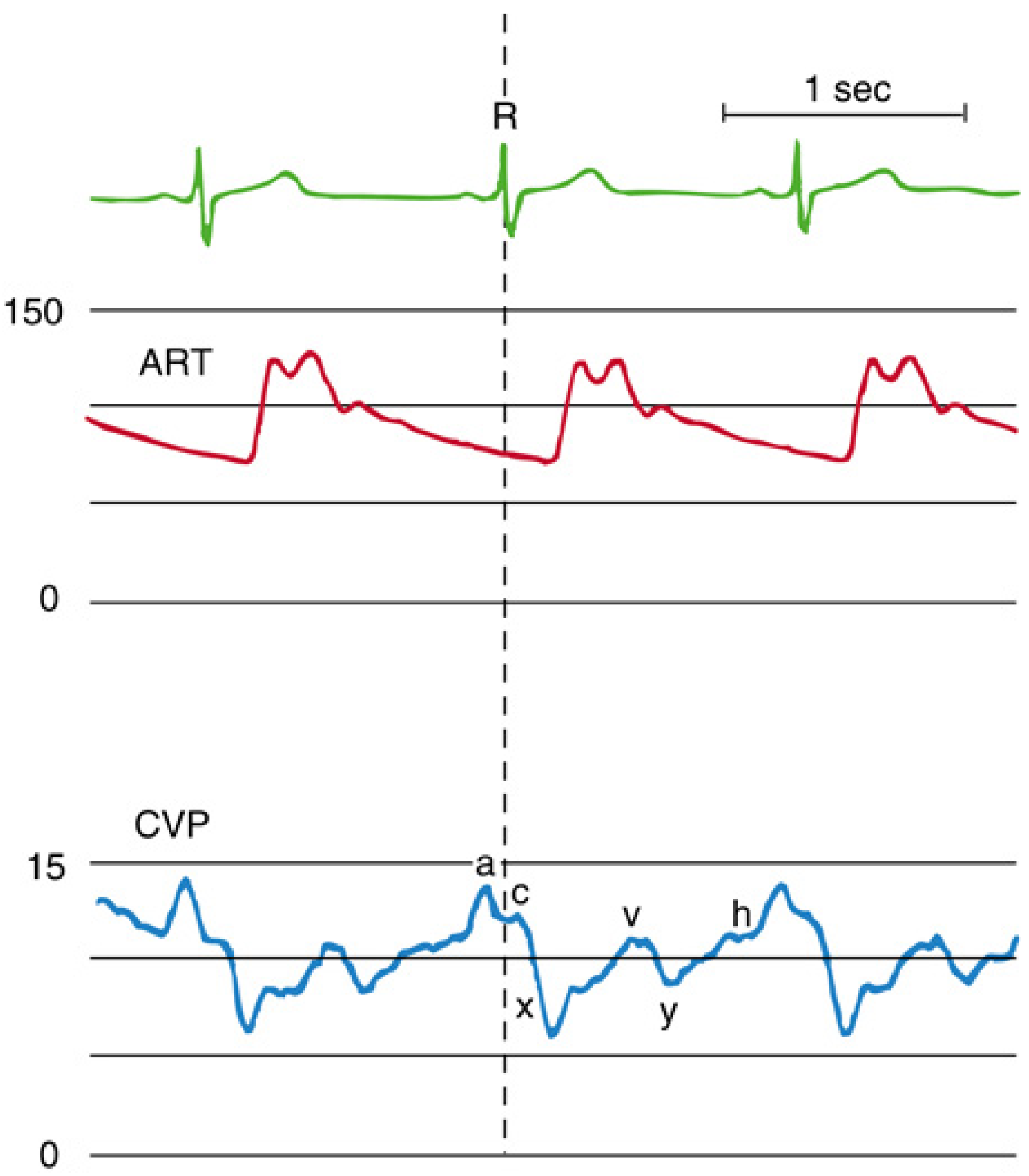
| Component | Phase of Cardiac Cycle | Mechanical Event | ECG Correlation |
|---|---|---|---|
| a wave | End-diastole | Atrial contraction → ↑RA pressure | After P wave |
| c wave | Early systole | TV closure + isovolumic RV contraction displacing TV toward RA | After R wave |
| x descent | Mid-systole | Atrial relaxation + tricuspid annulus drawn toward apex (systolic collapse) | Between R and T |
| v wave | Late systole | Venous filling of RA while TV is still closed | After T wave |
| y descent | Early diastole | TV opens → blood flows RA → RV (diastolic collapse) | After T wave |
| h wave | Mid-late diastole | Diastolic plateau; only seen in bradycardia + elevated CVP | - |
- a = Atrial contraction
- c = Closure of TV (isovolumic contraction)
- x = eXpulsion / atrial relaXation (systolic collapse)
- v = Venous filling of RA
- y = Yielding (TV opens, diastolic collapse)
Normal CVP Values
| Parameter | Value |
|---|---|
| Normal CVP | 2-8 mmHg |
| Typical healthy individual (Miller's) | 2-3 mmHg |
| MCFP - CVP gradient (drives venous return) | 6-8 mmHg (very small; hence small CVP changes = major haemodynamic effects) |
| CVP | Clinical State |
|---|---|
| <5 mmHg | Hypovolaemia |
| 5-12 mmHg | Normal |
| >12 mmHg | Fluid overload / RV failure / tamponade / PEEP / TR |
CVP Waveform Abnormalities (Miller's)
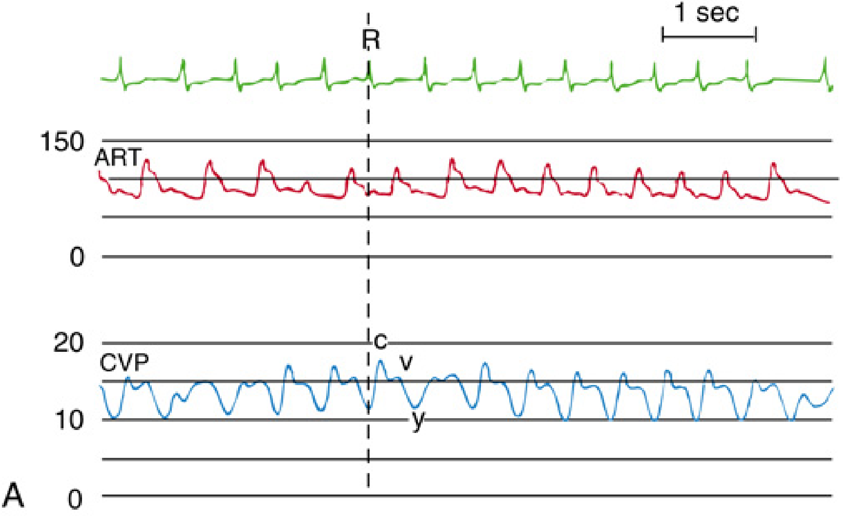
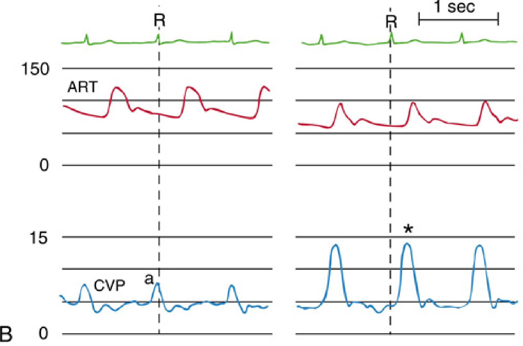
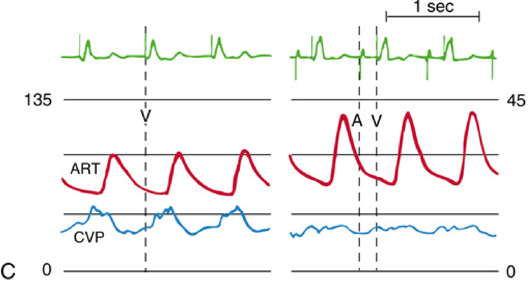
Full Waveform Abnormalities Table
| Condition | Waveform Change | Cause |
|---|---|---|
| Atrial fibrillation | Absent a wave | No organised atrial contraction |
| AV dissociation / junctional | Cannon a waves (giant intermittent) | Atrium contracts against closed TV |
| Tricuspid regurgitation | Tall c-v wave; x descent absent ("ventricularized") | Retrograde systolic filling of RA |
| Tricuspid stenosis | Prominent a wave; attenuated y descent | ↓ diastolic RA emptying |
| Cardiac tamponade | Absent y descent + ↑ mean CVP | Pericardial pressure prevents RV diastolic filling |
| Constrictive pericarditis | Prominent x AND y descents (W or M pattern) | Rapid early filling followed by abrupt restriction |
| RV ischaemia / pulmonary HTN | Prominent a wave; y descent preserved | ↓ RV compliance → stiff RV |
| Tachycardia | a and v waves merge; descents blend | Insufficient time between events |
| Bradycardia | All waves distinct; x and x' visible separately | More time between events; h wave may appear |
| PEEP / PPV | ↑ mean CVP (apparent); respiratory variation | Transmitted intrathoracic pressure |
CVP Monitoring - Technical Principles
→ Reference point: Phlebostatic axis
   = 4th ICS, mid-axillary line (= horizontal level of RA)
→ Zero transducer to atmosphere at this point before every reading
→ 1 cm error in zero reference = ~0.7 mmHg error in CVP
- Always at end-expiration (intrathoracic pressure closest to atmospheric)
- Spontaneous ventilation: end-expiration = highest CVP point on respiratory cycle
- IPPV: end-expiration = lowest CVP point on respiratory cycle
- Read from waveform, not mean digital display
Measured CVP rises with PEEP, but transmural CVP (true distending pressure) may not Correction: Transmural CVP ≈ Measured CVP - (PEEP × 0.5 to 0.75)
"CVP is NOT an accurate representation of cardiac preload and LV filling pressure"
- Same CVP = vastly different volumes on different portions of compliance curve
- LV function not reflected (CVP = right-sided pressure only)
- PEEP, tamponade, TR all distort CVP interpretation
- Dynamic indices (SVV, PPV) are superior predictors of fluid responsiveness
VII. COMPLICATIONS (Box 32.6 - Miller's)
Classification:
COMPLICATIONS OF CVP MONITORING
│
├── A. MECHANICAL
│     ├── Vascular injury: arterial, venous
│     ├── Cardiac tamponade (most catastrophic)
│     ├── Respiratory: pneumothorax, haemothorax, hydrothorax, chylothorax
│     ├── Airway compression (haematoma)
│     ├── Nerve injury
│     └── Arrhythmias
│
├── B. THROMBOEMBOLIC
│     ├── Venous thrombosis / DVT
│     ├── Pulmonary embolism
│     ├── Arterial thrombosis and embolism
│     ├── Catheter or guidewire embolism
│     └── Air embolism
│
└── C. INFECTIOUS
      ├── CLABSI (most common major late complication)
      ├── Insertion site infection
      ├── Catheter colonisation
      └── Endocarditis
Key Complication Data (Miller's):
| Complication | Incidence | Key Point |
|---|---|---|
| Arterial puncture | 1.9-15% | Most common acute mechanical complication |
| Pneumothorax - subclavian | 1.5% | Most common site for pneumothorax |
| Pneumothorax - IJV | 0.5% | Lower risk |
| Cardiac tamponade | Rare but catastrophic | Tip in RA/RV or abutting SVC wall at steep angle |
| DVT - femoral | Up to 21.5% | Highest thrombosis site |
| DVT - subclavian | 1.9% | Lowest thrombosis site |
| CLABSI | 3-5/1000 catheter days (pre-bundle) | Fell 50% (2008-2016), rose 91% during COVID |
Prevention Bundle (Miller's + CDC):
| Bundle Element | Detail |
|---|---|
| Hand hygiene | Alcohol-based rub |
| Maximal sterile barrier | Cap, mask, sterile gown + gloves, full drape |
| Chlorhexidine skin prep | >0.5% in alcohol; allow to dry |
| Optimal site | Subclavian preferred over femoral for infection |
| Daily line review | Remove when no longer needed |
| Antimicrobial catheter | When CLABSI rate >3.3/1000 catheter-days |
| No routine wire change | CDC: do NOT replace over guidewire on schedule |
VIII. CONFIRMATION OF CORRECT PLACEMENT
METHOD 1 - CXR (Mandatory after insertion)
→ Tip should lie at distal SVC / SVC-RA junction
→ Landmark: at or just above carina (carina = T4 level ≈ angle of Louis)
→ Danger sign: catheter tip curving and abutting SVC wall = risk of tamponade

METHOD 2 - Pressure waveform manometry
→ Transduced pressure shows low-pressure venous waveform (2-8 mmHg)
→ CVP waveform with a, c, v waves visible
→ If arterial trace (60-140 mmHg, pulsatile) → catheter in artery → remove

METHOD 3 - Blood gas from catheter
→ venous pO₂ and SpO₂ pattern confirms venous placement

METHOD 4 - Ultrasound
→ Real-time USS can confirm wire/catheter in vein before dilation
IX. VIVA QUICK-FIRE
| Q | A |
|---|---|
| Most preferred site for CVC in anaesthesia? | Right internal jugular vein |
| Why right IJV preferred over left? | Straighter path to SVC + RA; avoids thoracic duct |
| Insertion depth - right IJV? | 13-15 cm |
| Left-sided approach needs extra? | 3-5 cm more than right-sided |
| Tip position on CXR? | Distal SVC, at or just above carina |
| Most common acute mechanical complication? | Arterial puncture (1.9-15%) |
| Most catastrophic complication? | Cardiac tamponade |
| CXR sign warning of tamponade risk? | Catheter tip curving and abutting SVC wall at steep angle |
| Why Trendelenburg during insertion? | Distends veins; reduces air embolism risk |
| USS guidance - most strongly recommended for? | Internal jugular vein (Miller's) |
| Absent a wave on CVP? | Atrial fibrillation (no organised atrial contraction) |
| Cannon a waves? | AV dissociation / junctional rhythm - atrium against closed TV |
| Ventricularized CVP trace? | Tricuspid regurgitation (tall c-v wave, no x descent) |
| Prominent a + attenuated y descent? | Tricuspid stenosis |
| Absent y descent? | Cardiac tamponade |
| Prominent x AND y (W pattern)? | Constrictive pericarditis |
| Normal CVP value? | 2-8 mmHg (typically 2-3 mmHg in healthy - Miller's) |
| When to measure CVP? | End-expiration; patient supine; transducer at phlebostatic axis |
| Phlebostatic axis? | 4th ICS, mid-axillary line (= RA level) |
| Why is CVP poor predictor of preload? | Same CVP = different volumes at different compliance states; PEEP/TR/tamponade distort it |
| Highest infection risk site? | Femoral |
| Highest thrombosis site? | Femoral (21.5%) |
| Subclavian thrombosis rate? | 1.9% (lowest) |
| CLABSI trend 2008-2016? | Fell 50% with evidence-based bundles |
| CLABSI in COVID? | Rose 91%, especially in community hospitals |
| Position for VAE from CVC? | Left lateral decubitus + Trendelenburg (Durant's manoeuvre) |
| Guidewire - maximum advance length? | 15-20 cm (not beyond SVC-RA junction) |
| What if arrhythmia during wire insertion? | Withdraw wire 2-3 cm; almost always resolves |
| Protamine dose for heparin reversal? | 1 mg per 100 units heparin given |
Arterial cannulation - indications, contraindications, procedure ,complication as waveform