
Bhai mujhe ABG our ventilator settings ki detailed pdf chahiye
mkdir -p /home/daytona/workspace/abg-ventilator && ls /home/daytona/workspace/abg-ventilator/
~/abg-ventilator/generate_pdf.py
#!/usr/bin/env python3
"""
ABG & Ventilator Settings - Detailed Medical Reference PDF
Using ReportLab for professional PDF generation
"""

from reportlab.lib.pagesizes import A4
from reportlab.lib.styles import getSampleStyleSheet, ParagraphStyle
from reportlab.lib.units import cm, mm
from reportlab.lib.colors import (
    HexColor, white, black, Color
)
from reportlab.platypus import (
    SimpleDocTemplate, Paragraph, Spacer, Table, TableStyle,
    HRFlowable, PageBreak, KeepTogether
)
from reportlab.lib.enums import TA_CENTER, TA_LEFT, TA_JUSTIFY
from reportlab.lib import colors

# Color palette - medical/clinical theme
DARK_BLUE    = HexColor('#1A3C5E')
MED_BLUE     = HexColor('#2E6DA4')
LIGHT_BLUE   = HexColor('#D6E8F7')
ACCENT_GREEN = HexColor('#1E7E4B')
LIGHT_GREEN  = HexColor('#D4EDDA')
ACCENT_RED   = HexColor('#C0392B')
LIGHT_RED    = HexColor('#FDECEA')
ACCENT_AMBER = HexColor('#D68910')
LIGHT_AMBER  = HexColor('#FEF9E7')
ACCENT_PURPLE= HexColor('#6C3483')
LIGHT_PURPLE = HexColor('#EAD6F5')
DARK_GREY    = HexColor('#2C3E50')
MID_GREY     = HexColor('#566573')
LIGHT_GREY   = HexColor('#F2F3F4')
TABLE_ALT    = HexColor('#EBF5FB')

PAGE_W, PAGE_H = A4
MARGIN = 2 * cm

doc = SimpleDocTemplate(
    '/home/daytona/workspace/abg-ventilator/ABG_Ventilator_Settings.pdf',
    pagesize=A4,
    rightMargin=MARGIN, leftMargin=MARGIN,
    topMargin=2.5*cm, bottomMargin=2*cm,
    title='ABG & Ventilator Settings - Complete Medical Reference',
    author='Orris Medical AI'
)

styles = getSampleStyleSheet()

# ── Custom paragraph styles ────────────────────────────────────────────────
def make_style(name, parent='Normal', **kwargs):
    return ParagraphStyle(name, parent=styles[parent], **kwargs)

cover_title   = make_style('CoverTitle',  fontSize=28, textColor=white,
                            alignment=TA_CENTER, fontName='Helvetica-Bold',
                            spaceAfter=8)
cover_sub     = make_style('CoverSub',    fontSize=14, textColor=HexColor('#AED6F1'),
                            alignment=TA_CENTER, spaceAfter=4)
cover_note    = make_style('CoverNote',   fontSize=10, textColor=HexColor('#D5DBDB'),
                            alignment=TA_CENTER)
h1            = make_style('H1', fontSize=16, textColor=white,
                            fontName='Helvetica-Bold',
                            spaceBefore=6, spaceAfter=4, leading=20)
h2            = make_style('H2', fontSize=13, textColor=DARK_BLUE,
                            fontName='Helvetica-Bold',
                            spaceBefore=10, spaceAfter=4, leading=17)
h3            = make_style('H3', fontSize=11, textColor=MED_BLUE,
                            fontName='Helvetica-Bold',
                            spaceBefore=6, spaceAfter=3)
body          = make_style('Body', fontSize=9.5, leading=14,
                            textColor=DARK_GREY, spaceAfter=4,
                            alignment=TA_JUSTIFY)
body_sm       = make_style('BodySm', fontSize=8.5, leading=13,
                            textColor=DARK_GREY, spaceAfter=3)
bullet        = make_style('Bullet', fontSize=9.5, leading=14,
                            textColor=DARK_GREY, leftIndent=14,
                            bulletIndent=4, spaceAfter=2)
bullet_sm     = make_style('BulletSm', fontSize=8.5, leading=13,
                            textColor=DARK_GREY, leftIndent=18,
                            bulletIndent=6, spaceAfter=2)
box_title     = make_style('BoxTitle', fontSize=10, textColor=white,
                            fontName='Helvetica-Bold', spaceAfter=4)
box_body      = make_style('BoxBody', fontSize=9, leading=13,
                            textColor=DARK_GREY)
formula_style = make_style('Formula', fontSize=10, leading=14,
                            textColor=ACCENT_GREEN, fontName='Helvetica-Bold',
                            alignment=TA_CENTER, spaceBefore=4, spaceAfter=4)
warn_style    = make_style('Warn', fontSize=9, leading=13,
                            textColor=ACCENT_RED, fontName='Helvetica-Bold')
ref_style     = make_style('Ref', fontSize=7.5, textColor=MID_GREY,
                            leading=11, spaceAfter=2)

# ── Helper builders ────────────────────────────────────────────────────────

def section_header(title, color=DARK_BLUE, icon=''):
    """Colored section banner with title."""
    tbl = Table([[Paragraph(f'{icon}  {title}' if icon else title, h1)]],
                colWidths=[PAGE_W - 2*MARGIN])
    tbl.setStyle(TableStyle([
        ('BACKGROUND', (0,0), (-1,-1), color),
        ('ROUNDEDCORNERS', [6]),
        ('TOPPADDING',    (0,0), (-1,-1), 10),
        ('BOTTOMPADDING', (0,0), (-1,-1), 10),
        ('LEFTPADDING',   (0,0), (-1,-1), 14),
        ('RIGHTPADDING',  (0,0), (-1,-1), 14),
    ]))
    return tbl

def color_box(title, content_paras, bg=LIGHT_BLUE, border=MED_BLUE, title_bg=MED_BLUE):
    """Colored box with header strip and content."""
    rows = [[Paragraph(title, box_title)]]
    for p in content_paras:
        rows.append([p])
    tbl = Table(rows, colWidths=[PAGE_W - 2*MARGIN])
    style_cmds = [
        ('BACKGROUND', (0,0), (0,0), title_bg),
        ('BACKGROUND', (0,1), (-1,-1), bg),
        ('TOPPADDING',    (0,0), (-1,-1), 7),
        ('BOTTOMPADDING', (0,0), (-1,-1), 7),
        ('LEFTPADDING',   (0,0), (-1,-1), 12),
        ('RIGHTPADDING',  (0,0), (-1,-1), 12),
        ('BOX',           (0,0), (-1,-1), 1, border),
        ('GRID',          (0,0), (-1,-1), 0.3, HexColor('#AAAAAA')),
    ]
    tbl.setStyle(TableStyle(style_cmds))
    return tbl

def two_col_box(left_title, left_items, right_title, right_items,
                lbg=LIGHT_GREEN, lborder=ACCENT_GREEN,
                rbg=LIGHT_RED,   rborder=ACCENT_RED):
    """Side-by-side two-column colored boxes."""
    half = (PAGE_W - 2*MARGIN - 0.4*cm) / 2

    def build_cell_content(title, items, tbg, ibg, border_color):
        inner_rows = [[Paragraph(title, box_title)]]
        for it in items:
            inner_rows.append([Paragraph(f'• {it}', bullet_sm)])
        t = Table(inner_rows, colWidths=[half])
        t.setStyle(TableStyle([
            ('BACKGROUND', (0,0), (0,0), border_color),
            ('BACKGROUND', (0,1), (-1,-1), ibg),
            ('TOPPADDING',    (0,0), (-1,-1), 6),
            ('BOTTOMPADDING', (0,0), (-1,-1), 5),
            ('LEFTPADDING',   (0,0), (-1,-1), 10),
            ('RIGHTPADDING',  (0,0), (-1,-1), 10),
            ('BOX',           (0,0), (-1,-1), 1, border_color),
        ]))
        return t

    left_t  = build_cell_content(left_title,  left_items,  lborder, lbg, lborder)
    right_t = build_cell_content(right_title, right_items, rborder, rbg, rborder)
    outer = Table([[left_t, right_t]], colWidths=[half, half],
                  hAlign='LEFT')
    outer.setStyle(TableStyle([
        ('VALIGN',        (0,0), (-1,-1), 'TOP'),
        ('LEFTPADDING',   (0,0), (-1,-1), 0),
        ('RIGHTPADDING',  (0,0), (-1,-1), 0),
        ('TOPPADDING',    (0,0), (-1,-1), 0),
        ('BOTTOMPADDING', (0,0), (-1,-1), 0),
    ]))
    return outer

def data_table(headers, rows, col_widths=None, stripe_color=TABLE_ALT,
               header_color=DARK_BLUE):
    """Formatted data table with striped rows."""
    if col_widths is None:
        n = len(headers)
        col_widths = [(PAGE_W - 2*MARGIN) / n] * n
    header_row = [Paragraph(f'<b>{h}</b>', make_style(f'TH_{i}',
                  fontSize=9, textColor=white, fontName='Helvetica-Bold',
                  alignment=TA_CENTER)) for i, h in enumerate(headers)]
    all_rows = [header_row]
    for i, row in enumerate(rows):
        styled_row = [Paragraph(str(cell), make_style(f'TD_{i}_{j}',
                      fontSize=9, textColor=DARK_GREY,
                      alignment=TA_CENTER, leading=13))
                      for j, cell in enumerate(row)]
        all_rows.append(styled_row)

    tbl = Table(all_rows, colWidths=col_widths, repeatRows=1)
    style_cmds = [
        ('BACKGROUND',    (0,0), (-1,0),  header_color),
        ('TOPPADDING',    (0,0), (-1,-1), 6),
        ('BOTTOMPADDING', (0,0), (-1,-1), 6),
        ('LEFTPADDING',   (0,0), (-1,-1), 8),
        ('RIGHTPADDING',  (0,0), (-1,-1), 8),
        ('GRID',          (0,0), (-1,-1), 0.5, HexColor('#CCCCCC')),
        ('ROWBACKGROUNDS',(0,1), (-1,-1), [white, stripe_color]),
        ('VALIGN',        (0,0), (-1,-1), 'MIDDLE'),
    ]
    tbl.setStyle(TableStyle(style_cmds))
    return tbl

# ── Build story ────────────────────────────────────────────────────────────
story = []
S = Spacer

# ════════════════════════════════════════════════════════
# COVER PAGE
# ════════════════════════════════════════════════════════
cover_bg = Table(
    [[Paragraph('ABG & VENTILATOR SETTINGS', cover_title)],
     [Paragraph('Complete Medical Reference Guide', cover_sub)],
     [Spacer(1, 0.3*cm)],
     [Paragraph('Arterial Blood Gas Interpretation  •  Acid-Base Disorders  •  Mechanical Ventilation', cover_note)],
     [Spacer(1, 0.2*cm)],
     [Paragraph('ICU  |  Emergency Medicine  |  Pulmonology  |  Anaesthesia', cover_note)],
     [Spacer(1, 1*cm)],
     [Paragraph('Sources: Murray & Nadel\'s Respiratory Medicine  •  Barash Clinical Anaesthesia  •  Goldman-Cecil Medicine  •  Rosen\'s Emergency Medicine  •  Miller\'s Anaesthesia', ref_style)],
    ],
    colWidths=[PAGE_W - 2*MARGIN]
)
cover_bg.setStyle(TableStyle([
    ('BACKGROUND',    (0,0), (-1,-1), DARK_BLUE),
    ('TOPPADDING',    (0,0), (-1,-1), 30),
    ('BOTTOMPADDING', (0,0), (-1,-1), 10),
    ('LEFTPADDING',   (0,0), (-1,-1), 20),
    ('RIGHTPADDING',  (0,0), (-1,-1), 20),
    ('ROUNDEDCORNERS',[8]),
]))
story.append(Spacer(1, 1*cm))
story.append(cover_bg)
story.append(Spacer(1, 0.6*cm))

# Quick-Reference boxes on cover
qr_headers = ['Parameter', 'Normal Range']
qr_rows = [
    ['pH',          '7.35 – 7.45'],
    ['PaO₂',        '80 – 100 mmHg'],
    ['PaCO₂',       '35 – 45 mmHg'],
    ['HCO₃⁻',       '22 – 26 mEq/L'],
    ['BE',          '-2 to +2 mEq/L'],
    ['SaO₂',        '95 – 99%'],
    ['SpO₂ target', '≥ 94%'],
    ['P/F Ratio',   '> 300 (normal)'],
]
story.append(Paragraph('ABG Normal Reference Values', h2))
story.append(data_table(qr_headers, qr_rows,
                        col_widths=[9*cm, 8*cm]))

story.append(PageBreak())

# ════════════════════════════════════════════════════════
# SECTION 1 — ABG BASICS & NORMAL VALUES
# ════════════════════════════════════════════════════════
story.append(section_header('SECTION 1: ABG — Basics & Normal Values', DARK_BLUE))
story.append(S(1, 0.4*cm))

story.append(Paragraph('What is an ABG?', h2))
story.append(Paragraph(
    'Arterial Blood Gas (ABG) analysis measures pH, PaO₂, PaCO₂, HCO₃⁻, base excess, and oxygen saturation '
    'from an arterial blood sample. It is the <b>gold standard</b> for assessing oxygenation, ventilation, and '
    'acid-base status. ABG is superior to pulse oximetry because it directly measures PaO₂ and can detect '
    'abnormal haemoglobins (carboxyhemoglobin, methemoglobin) that falsely elevate SpO₂.',
    body))

story.append(Paragraph('Key Parameters', h2))

param_headers = ['Parameter', 'Full Name', 'Normal Range', 'Clinical Significance']
param_rows = [
    ['pH',       'Acidity/Alkalinity',         '7.35 – 7.45',    'Reflects overall acid-base balance. <7.2 = life-threatening acidaemia'],
    ['PaO₂',     'Partial Pressure of O₂',     '80 – 100 mmHg',  'Direct measure of oxygenation in arterial blood'],
    ['PaCO₂',    'Partial Pressure of CO₂',    '35 – 45 mmHg',   'Ventilatory parameter; raised = hypoventilation'],
    ['HCO₃⁻',    'Bicarbonate',                '22 – 26 mEq/L',  'Metabolic component; regulated by kidneys'],
    ['BE/BD',    'Base Excess / Deficit',       '-2 to +2 mEq/L', 'Positive = alkalosis; Negative = acidosis'],
    ['SaO₂',     'Arterial O₂ Saturation',     '95 – 99%',       'Haemoglobin saturation; SpO₂ is non-invasive estimate'],
    ['A-aDO₂',   'Alveolar-arterial O₂ diff.',  '<15 mmHg (<30 yr)','Elevated = V/Q mismatch, shunt, diffusion defect'],
    ['P/F Ratio','PaO₂/FiO₂ Ratio',            '>400 (ideal)',   '<300=mild ARDS; <200=moderate; <100=severe'],
]
story.append(data_table(param_headers, param_rows,
             col_widths=[2.2*cm, 4*cm, 3.5*cm, 7.5*cm]))

story.append(S(1, 0.4*cm))
story.append(Paragraph('Alveolar-Arterial (A-a) Gradient Formula', h3))
story.append(Paragraph(
    '<b>PAO₂ = (FiO₂ × [Patm - PH₂O]) - (PaCO₂ / RQ)</b><br/>'
    'On room air (sea level): <b>PAO₂ = 150 - (PaCO₂ / 0.8)</b><br/>'
    '<b>A-aDO₂ = PAO₂ - PaO₂</b>   |   Normal: <15 mmHg in young, increases with age (approx. age/4 + 4)',
    formula_style))

story.append(S(1, 0.4*cm))

# ════════════════════════════════════════════════════════
# SECTION 2 — STEPWISE ABG INTERPRETATION
# ════════════════════════════════════════════════════════
story.append(section_header('SECTION 2: Stepwise ABG Interpretation', MED_BLUE))
story.append(S(1, 0.4*cm))

story.append(Paragraph(
    'Use this 6-step systematic approach for every ABG (Barash Clinical Anaesthesia, 9e). '
    'Draw ABG and venous electrolytes simultaneously — calculated HCO₃⁻ on ABG should match '
    'measured serum HCO₃⁻ within 2–3 mEq/L; discrepancy = lab error or timing issue.',
    body))

steps_data = [
    ['STEP', 'ACTION', 'CRITERIA / DETAILS'],
    ['Step 1', 'Identify Acidaemia vs. Alkalemia',
     'pH < 7.35 = Acidaemia\npH > 7.45 = Alkalemia\npH 7.35–7.45 = Normal (may still have mixed disorder)'],
    ['Step 2', 'Determine Primary Disorder',
     'pH↓ + PaCO₂↑ = Respiratory Acidosis\npH↓ + HCO₃↓ = Metabolic Acidosis\npH↑ + PaCO₂↓ = Respiratory Alkalosis\npH↑ + HCO₃↑ = Metabolic Alkalosis'],
    ['Step 3', 'Assess Compensation\n(Acute vs. Chronic)',
     'Respiratory Acidosis — Acute: HCO₃ rises 1 per 10↑CO₂ | Chronic: rises 3.5 per 10↑CO₂\nRespiratory Alkalosis — Acute: HCO₃ falls 2 per 10↓CO₂ | Chronic: falls 5 per 10↓CO₂\nMetabolic Acidosis — Expected PaCO₂ = 1.5×HCO₃ + 8 ± 2 (Winter\'s formula)\nMetabolic Alkalosis — Expected PaCO₂ = 40 + 0.7×(HCO₃ - 24)'],
    ['Step 4', 'Calculate Anion Gap\n(Always — even if no met. acidosis)',
     'AG = Na⁺ - (Cl⁻ + HCO₃⁻)   Normal = 8–12 mEq/L (some sources ≤13)\nCorrect for albumin: Corrected AG = AG + 2.5 × (4 - albumin g/dL)\nHigh AG (>13) = MUDPILES causes\nNormal AG = hyperchloraemic acidosis'],
    ['Step 5', 'Assess Oxygenation',
     'PaO₂ < 60 mmHg = Hypoxaemia\nPaO₂/FiO₂ ratio — <300 mild ARDS, <200 moderate, <100 severe\nCalculate A-a gradient to identify shunt vs. hypoventilation'],
    ['Step 6', 'Delta-Delta (Δ/Δ) Ratio\n(If AG elevated)',
     'Δ/Δ = (AG - 12) / (24 - HCO₃)\nRatio 1–2 = Pure AG acidosis\nRatio < 1 = Concurrent non-AG metabolic acidosis\nRatio > 2 = Concurrent metabolic alkalosis or chronic resp. acidosis'],
]

step_tbl = Table(steps_data,
    colWidths=[2.2*cm, 4.5*cm, (PAGE_W - 2*MARGIN - 6.7*cm)])
step_tbl.setStyle(TableStyle([
    ('BACKGROUND',    (0,0), (-1,0),  DARK_BLUE),
    ('TEXTCOLOR',     (0,0), (-1,0),  white),
    ('FONTNAME',      (0,0), (-1,0),  'Helvetica-Bold'),
    ('FONTSIZE',      (0,0), (-1,-1), 9),
    ('ROWBACKGROUNDS',(0,1), (-1,-1), [white, LIGHT_BLUE]),
    ('BACKGROUND',    (0,1), (0,-1),  MED_BLUE),
    ('TEXTCOLOR',     (0,1), (0,-1),  white),
    ('FONTNAME',      (0,1), (0,-1),  'Helvetica-Bold'),
    ('ALIGN',         (0,0), (0,-1),  'CENTER'),
    ('VALIGN',        (0,0), (-1,-1), 'TOP'),
    ('TOPPADDING',    (0,0), (-1,-1), 7),
    ('BOTTOMPADDING', (0,0), (-1,-1), 7),
    ('LEFTPADDING',   (0,0), (-1,-1), 8),
    ('RIGHTPADDING',  (0,0), (-1,-1), 8),
    ('GRID',          (0,0), (-1,-1), 0.5, HexColor('#CCCCCC')),
]))
story.append(step_tbl)

story.append(PageBreak())

# ════════════════════════════════════════════════════════
# SECTION 3 — ACID-BASE DISORDERS
# ════════════════════════════════════════════════════════
story.append(section_header('SECTION 3: Acid-Base Disorders — Causes & Management', ACCENT_GREEN))
story.append(S(1, 0.3*cm))

story.append(Paragraph(
    'Acid-base disorders are found in 51–56% of hospitalised patients. Prevalence: Respiratory alkalosis '
    '(29–42%) > Metabolic alkalosis (16–28%) > Respiratory acidosis (26–27%) > Metabolic acidosis (10–12%). '
    'Mixed disorders account for ~6% (Barash Clinical Anaesthesia, 9e).',
    body))

story.append(S(1, 0.3*cm))

# 3a — Metabolic Acidosis
story.append(KeepTogether([
    Paragraph('3a. Metabolic Acidosis', h2),
    Paragraph(
        '<b>Definition:</b> pH↓, HCO₃⁻↓, PaCO₂↓ (respiratory compensation). '
        'Physiologic consequence: pH <7.2 causes impaired myocardial contractility, '
        'pulmonary hypertension, arrhythmia threshold↓, insulin resistance, hyperkalemia, '
        'decreased responsiveness to catecholamines.',
        body),
]))

ma_left  = ['Ketoacidosis (DKA, alcoholic, starvation)',
            'Lactic acidosis (sepsis, shock, tissue hypoxia)',
            'Uraemia / Advanced CKD',
            'Toxins: Methanol, Ethylene glycol, Salicylates',
            'Paraldehyde, Propylene glycol, Metformin']
ma_right = ['Renal Tubular Acidosis (type 1, 2, 4)',
            'Diarrhoea (GI HCO₃⁻ loss)',
            'Carbonic anhydrase inhibitors (acetazolamide)',
            '0.9% Saline (hyperchloraemic)',
            'Ureteroenteric diversion, Biliary fistula']
story.append(two_col_box('HIGH AG Causes (AG > 13) — MUDPILES', ma_left,
                         'NORMAL AG Causes (Hyperchloraemic)', ma_right,
                         lbg=LIGHT_AMBER, lborder=ACCENT_AMBER,
                         rbg=LIGHT_BLUE, rborder=MED_BLUE))
story.append(S(1, 0.3*cm))
story.append(Paragraph(
    '<b>Management:</b> Treat the underlying cause. Ventilatory compensation must be maintained if '
    'patient is on MV. No strong evidence for routine NaHCO₃ in critically ill patients unless pH <7.2 '
    'in septic/hypovolemic shock (BICAR-ICU trial — no mortality benefit at 28 days with bicarbonate '
    'vs. placebo in mixed organ failure). For patients on MV, adjust ventilator to maintain compensatory '
    'hyperventilation; correct shock, hypoxia, and metabolic causes.',
    body))

story.append(S(1, 0.3*cm))

# 3b — Metabolic Alkalosis
story.append(Paragraph('3b. Metabolic Alkalosis', h2))
story.append(Paragraph(
    '<b>Definition:</b> pH↑, HCO₃⁻↑, PaCO₂↑ (respiratory compensation, hypoventilation). '
    'Expected PaCO₂ = 40 + 0.7 × (HCO₃measured − 24). '
    'Compensation is limited by hypoxic drive; PaCO₂ rarely exceeds 55 mmHg.',
    body))

malk_left  = ['Vomiting / NG suction (Cl⁻ and H⁺ loss)',
              'Diuretics (loop/thiazide — K⁺ and Cl⁻ loss)',
              'Contraction alkalosis',
              'Post-hypercapnic alkalosis']
malk_right = ['Primary hyperaldosteronism / Cushing\'s',
              'Excess NaHCO₃ or antacid ingestion',
              'Milk-alkali syndrome',
              'Hypomagnesaemia, Hypokalaemia']
story.append(two_col_box('Saline-Responsive (UCl⁻ < 20 mEq/L)', malk_left,
                         'Saline-Resistant (UCl⁻ > 20 mEq/L)', malk_right,
                         lbg=LIGHT_GREEN, lborder=ACCENT_GREEN,
                         rbg=LIGHT_PURPLE, rborder=ACCENT_PURPLE))

story.append(S(1, 0.3*cm))

# 3c — Respiratory Acidosis
story.append(Paragraph('3c. Respiratory Acidosis', h2))
story.append(Paragraph(
    '<b>Definition:</b> pH↓, PaCO₂↑ (>45 mmHg), HCO₃⁻↑ (compensation). '
    '<b>Cause = alveolar hypoventilation.</b> '
    'Acute: HCO₃ +1 per 10↑PaCO₂. Chronic: HCO₃ +3.5 per 10↑PaCO₂.',
    body))

story.append(two_col_box('Central Causes', [
    'CNS depression (opioids, benzos, sedatives)',
    'Brainstem lesion / CVA',
    'Obesity hypoventilation (OHS)',
    'Central sleep apnoea'],
    'Peripheral / Mechanical Causes', [
    'COPD exacerbation, severe asthma',
    'Neuromuscular disease (GBS, MG, SCI)',
    'Pneumothorax, Haemothorax, ARDS',
    'Chest wall deformity, Massive obesity'],
    lbg=LIGHT_RED, lborder=ACCENT_RED,
    rbg=LIGHT_AMBER, rborder=ACCENT_AMBER))
story.append(S(1, 0.3*cm))
story.append(Paragraph(
    '<b>Management:</b> Treat underlying cause. If patient cannot compensate, NIV (BiPAP) is first-line '
    'for COPD (reduces mortality, avoids intubation). Intubate if: pH <7.25, declining mentation, '
    'haemodynamic instability, mask intolerance, failure of NIV in 30–120 min.',
    body))

story.append(S(1, 0.3*cm))

# 3d — Respiratory Alkalosis
story.append(Paragraph('3d. Respiratory Alkalosis', h2))
story.append(Paragraph(
    '<b>Definition:</b> pH↑, PaCO₂↓ (<35 mmHg), HCO₃⁻↓ (compensation). Most common acid-base '
    'disorder in hospitalised patients. Acute: HCO₃ -2 per 10↓PaCO₂. Chronic: HCO₃ -5 per 10↓PaCO₂.',
    body))

ra_causes = ['Anxiety / Pain / Psychogenic hyperventilation',
             'Fever, Sepsis (early), Pregnancy',
             'Pulmonary embolism (V/Q mismatch triggers hyperventilation)',
             'Liver failure (central hyperventilation)',
             'Salicylate toxicity (early phase)',
             'Hypoxia-driven hyperventilation (altitude, pneumonia)',
             'Iatrogenic (excessive MV settings)']
story.append(color_box('Common Causes of Respiratory Alkalosis',
    [Paragraph(f'• {c}', bullet_sm) for c in ra_causes],
    bg=LIGHT_BLUE, border=MED_BLUE, title_bg=MED_BLUE))

story.append(PageBreak())

# ════════════════════════════════════════════════════════
# SECTION 4 — OXYGENATION & HYPOXAEMIA
# ════════════════════════════════════════════════════════
story.append(section_header('SECTION 4: Oxygenation & Hypoxaemia', ACCENT_PURPLE))
story.append(S(1, 0.4*cm))

story.append(Paragraph('Causes of Hypoxaemia — 5 Mechanisms', h2))

hypox_headers = ['Mechanism', 'A-aDO₂', 'PaCO₂', 'Response to O₂', 'Examples']
hypox_rows = [
    ['Hypoventilation',      'Normal',   '↑',      'Good',      'Sedation, OHS, NMD'],
    ['V/Q Mismatch',         '↑',        'N or ↓', 'Good',      'COPD, asthma, pneumonia, PE'],
    ['Diffusion Defect',     '↑',        'N or ↓', 'Good',      'ILD, emphysema, early ARDS'],
    ['Right-to-Left Shunt',  '↑',        'N or ↓', 'Poor (<10% response)', 'ARDS, ASD/VSD, hepatopulmonary'],
    ['Low FiO₂ / Altitude',  'Normal',   'N or ↓', 'Good',      'High altitude, confined space'],
]
story.append(data_table(hypox_headers, hypox_rows,
    col_widths=[3.5*cm, 2.5*cm, 2*cm, 4.5*cm, 4.7*cm],
    header_color=ACCENT_PURPLE))

story.append(S(1, 0.4*cm))
story.append(Paragraph('PaO₂/FiO₂ Ratio (P/F Ratio) — ARDS Classification (Berlin 2012)', h2))

pf_headers = ['P/F Ratio (mmHg)', 'ARDS Category', 'PEEP Requirement', 'Mortality']
pf_rows = [
    ['>400',       'Normal',            '—',       '—'],
    ['300–400',    'Borderline / Early ALI', '≥5 cmH₂O', 'Low'],
    ['200–300',    'Mild ARDS',         '≥5 cmH₂O', '27%'],
    ['100–200',    'Moderate ARDS',     '≥5 cmH₂O', '32%'],
    ['<100',       'Severe ARDS',       '≥5 cmH₂O', '45%'],
]
story.append(data_table(pf_headers, pf_rows,
    col_widths=[4*cm, 5*cm, 4.5*cm, 3.7*cm],
    header_color=ACCENT_RED))

story.append(S(1, 0.4*cm))

story.append(color_box('SpO₂ vs. ABG — Key Limitation',
    [Paragraph(
        'Pulse oximetry CANNOT detect hypoventilation in patients on supplemental oxygen. '
        'The sigmoid shape of the O₂-Hb dissociation curve means PaO₂ can fall significantly '
        'before SpO₂ drops. PaCO₂ can rise to dangerous levels while SpO₂ appears normal. '
        '<b>Always use ABG or capnography when hypercarbia is suspected</b> — especially in patients '
        'receiving supplemental O₂. Errors also occur with carboxyhemoglobin, methemoglobin, '
        'dark nail polish, poor perfusion, or jaundice (Murray & Nadel, 2022).',
        body_sm)],
    bg=LIGHT_AMBER, border=ACCENT_AMBER, title_bg=ACCENT_AMBER))

story.append(PageBreak())

# ════════════════════════════════════════════════════════
# SECTION 5 — MECHANICAL VENTILATION MODES
# ════════════════════════════════════════════════════════
story.append(section_header('SECTION 5: Mechanical Ventilation — Modes', DARK_GREY))
story.append(S(1, 0.4*cm))

story.append(Paragraph(
    'Mechanical ventilation replaces or supplements the work of breathing. Understanding '
    'ventilator modes is essential for appropriate management of critically ill patients.',
    body))

story.append(Paragraph('Ventilator Modes Overview', h2))

modes_headers = ['Mode', 'Abbreviation', 'Trigger', 'Control Variable', 'Clinical Use']
modes_rows = [
    ['Volume Control – Assist Control',  'VC-AC / A/C', 'Patient or time', 'Fixed tidal volume', 'Standard first-line mode in ICU; guarantees VT'],
    ['Pressure Control – Assist Control','PC-AC',        'Patient or time', 'Fixed PIP',          'ARDS (limits barotrauma); variable VT'],
    ['Synchronised Intermittent Mandatory Ventilation', 'SIMV', 'Patient or time', 'Volume or Pressure', 'Weaning (controversial — now less used)'],
    ['Pressure Support Ventilation',     'PSV / PS',     'Patient',         'Pressure support level', 'Spontaneous breathing trials; weaning'],
    ['High-Frequency Oscillatory Ventilation', 'HFOV',   'Machine',         'Pressure amplitude', 'Severe ARDS rescue; paediatric'],
    ['Airway Pressure Release Ventilation', 'APRV',      'Patient',         'CPAP + release time', 'ARDS — open-lung approach'],
    ['Continuous Positive Airway Pressure', 'CPAP',      'Patient fully',   'Constant pressure', 'Spontaneous breathing + PEEP only; NIV/post-extubation'],
    ['BiLevel Positive Airway Pressure',  'BiPAP',       'Patient / timed', 'Two pressure levels (IPAP/EPAP)', 'NIV for COPD, OHS, CHF; avoids intubation'],
]
story.append(data_table(modes_headers, modes_rows,
    col_widths=[3.5*cm, 2.2*cm, 2.8*cm, 3.2*cm, 5.5*cm],
    header_color=DARK_GREY))

story.append(S(1, 0.4*cm))

story.append(Paragraph('Volume Control vs. Pressure Control — Key Differences', h3))

vpc_headers = ['Feature', 'Volume Control (VC)', 'Pressure Control (PC)']
vpc_rows = [
    ['VT delivery',    'Fixed, guaranteed',          'Variable (depends on compliance & resistance)'],
    ['Peak pressure',  'Variable — can be dangerously high', 'Fixed — safer from barotrauma perspective'],
    ['Flow pattern',   'Square (decelerating optional)', 'Decelerating (more physiological)'],
    ['Risk',           'Volutrauma/barotrauma if compliance↓', 'Inadequate VT if compliance changes'],
    ['Alarm priority', 'Monitor peak pressure',      'Monitor tidal volume'],
    ['Best for',       'Obstructive (asthma, COPD)', 'ARDS, restrictive lung disease'],
]
story.append(data_table(vpc_headers, vpc_rows,
    col_widths=[3.5*cm, 6.5*cm, 7.2*cm],
    header_color=MED_BLUE))

story.append(PageBreak())

# ════════════════════════════════════════════════════════
# SECTION 6 — VENTILATOR SETTINGS
# ════════════════════════════════════════════════════════
story.append(section_header('SECTION 6: Initial Ventilator Settings', MED_BLUE))
story.append(S(1, 0.4*cm))

story.append(Paragraph(
    'Settings below are for a typical adult patient on Volume Assist-Control. Adjust for clinical '
    'context (ARDS, COPD, asthma, neuromuscular disease, post-op). Always reassess within 30–60 '
    'minutes with ABG and clinical response.',
    body))

settings_headers = ['Parameter', 'Standard Setting', 'ARDS Protocol', 'COPD/Asthma', 'Rationale']
settings_rows = [
    ['FiO₂', 'Start 1.0 (100%)\ntitrate to SpO₂ 94–98%',
     'Titrate to SpO₂ 88–95%\n(permissive hypoxaemia)',
     'Titrate to SpO₂ ≥92%',
     'Avoid O₂ toxicity; target minimum FiO₂ to achieve adequate SpO₂'],
    ['Tidal Volume (VT)', '6–8 mL/kg IBW\n(max 10 mL/kg)',
     '<b>6 mL/kg IBW</b> (lung-protective; ARDSnet)',
     '6–8 mL/kg IBW',
     'Low VT reduces VILI (ventilator-induced lung injury). IBW = ideal body weight'],
    ['Respiratory Rate', '12–16 breaths/min',
     '16–25 (adjust to pH ≥7.25)',
     '<b>≤10 breaths/min</b> (allow expiration time)',
     'Low RR in obstructive disease prevents air trapping (auto-PEEP)'],
    ['PEEP', '5 cmH₂O',
     '<b>Higher PEEP ≥8–15 cmH₂O</b>\n(per ARDSnet PEEP/FiO₂ table)',
     'Low: 0–5 cmH₂O\n(match to intrinsic PEEP)',
     'Recruits atelectatic alveoli; prevents cyclical collapse. Risk: barotrauma if excessive'],
    ['Peak Inspiratory Pressure (PIP)', '<35 cmH₂O preferred',
     '<b><30 cmH₂O</b>\n(limit plateau P)',
     '<40 cmH₂O acceptable in severe obstruction',
     'High PIP = barotrauma risk. Difference (PIP – Plateau P) = airway resistance'],
    ['Plateau Pressure (Pplat)', 'Monitor; <30 cmH₂O ideal',
     '<b>≤30 cmH₂O</b> (ARDSnet protocol)',
     'May be elevated due to air trapping; true Pplat may be lower',
     'Reflects alveolar distension. Calculate: pause inspiratory hold for 0.5 sec'],
    ['I:E Ratio', '1:2 (default)',
     '1:2 to 1:3',
     '<b>1:3 to 1:4</b> (prolonged expiration to prevent air trapping)',
     'Obstructive disease needs long expiratory time. Inverse I:E ratio used in ARDS rescue'],
    ['Inspiratory Flow Rate', '40–60 L/min',
     '40–60 L/min',
     '<b>>60 L/min</b> (allows more expiratory time)',
     'High flow rate in COPD/asthma shortens inspiratory time, prolongs expiration'],
    ['Trigger Sensitivity', '-1 to -2 cmH₂O (pressure)\nor 1–2 L/min (flow)',
     '-1 to -2 cmH₂O',
     '-1 to -2 cmH₂O',
     'Too sensitive = auto-triggering. Too insensitive = patient-ventilator dyssynchrony'],
]

story.append(data_table(settings_headers, settings_rows,
    col_widths=[3.2*cm, 3.8*cm, 3.8*cm, 3.8*cm, 5.6*cm],
    header_color=MED_BLUE))

story.append(S(1, 0.4*cm))

story.append(Paragraph('Ideal Body Weight (IBW) Calculator', h3))
story.append(Paragraph(
    '<b>Males:</b>   IBW (kg) = 50 + 2.3 × (height in inches – 60)<br/>'
    '<b>Females:</b> IBW (kg) = 45.5 + 2.3 × (height in inches – 60)<br/>'
    'Or: <b>Males:</b> 50 + 0.91 × (height cm – 152.4)   |   <b>Females:</b> 45.5 + 0.91 × (height cm – 152.4)',
    formula_style))

story.append(PageBreak())

# ════════════════════════════════════════════════════════
# SECTION 7 — LUNG-PROTECTIVE VENTILATION (ARDSnet)
# ════════════════════════════════════════════════════════
story.append(section_header('SECTION 7: Lung-Protective Ventilation & ARDS Protocol', ACCENT_RED))
story.append(S(1, 0.4*cm))

story.append(Paragraph(
    'The ARDSNet ARMA trial (2000) demonstrated a 22% relative reduction in mortality '
    'using low-tidal-volume ventilation (6 mL/kg IBW vs. 12 mL/kg IBW). '
    'Goldman-Cecil Medicine describes ARDS as diffuse lung injury with severe hypoxaemia (P/F ratio-based), '
    'bilateral infiltrates, not explained by cardiac failure.',
    body))

story.append(Paragraph('ARDSnet Low-Tidal-Volume Protocol — Step by Step', h2))

ardsnet = [
    '1. Set VT = 8 mL/kg IBW initially, then reduce by 1 mL/kg every 2 hours to target 6 mL/kg IBW',
    '2. Set initial RR to maintain minute ventilation; adjust to pH target (not CO₂)',
    '3. Maintain Plateau Pressure ≤30 cmH₂O (measure every 4 hours via inspiratory hold)',
    '4. Use PEEP/FiO₂ table to optimise oxygenation (higher PEEP in moderate-severe ARDS)',
    '5. Target SpO₂ 88–95% or PaO₂ 55–80 mmHg (permissive hypoxaemia acceptable)',
    '6. Target pH 7.25–7.45. If pH <7.25 despite max RR (35/min): consider NaHCO₃ infusion',
    '7. If plateau pressure >30 cmH₂O: reduce VT by 1 mL/kg (minimum 4 mL/kg IBW)',
]
for step in ardsnet:
    story.append(Paragraph(step, bullet))

story.append(S(1, 0.3*cm))

story.append(Paragraph('PEEP / FiO₂ Table (ARDSnet — Higher PEEP Strategy)', h3))
peep_headers = ['FiO₂', '0.3', '0.4', '0.5', '0.6', '0.7', '0.8', '0.9', '1.0']
peep_lower   = ['Lower PEEP', '5',  '5–8',  '8–10', '10',  '10–12', '14',  '14–18', '18–24']
peep_higher  = ['Higher PEEP', '5–14', '14–16', '16–18', '20', '20',  '20–22', '22', '22–24']

pf_tbl = Table([peep_headers, peep_lower, peep_higher],
               colWidths=[(PAGE_W - 2*MARGIN)/9] * 9)
pf_tbl.setStyle(TableStyle([
    ('BACKGROUND',    (0,0), (-1,0), DARK_BLUE),
    ('TEXTCOLOR',     (0,0), (-1,0), white),
    ('FONTNAME',      (0,0), (-1,-1), 'Helvetica-Bold'),
    ('FONTSIZE',      (0,0), (-1,-1), 8.5),
    ('ROWBACKGROUNDS',(0,1), (-1,-1), [LIGHT_BLUE, LIGHT_GREEN]),
    ('ALIGN',         (0,0), (-1,-1), 'CENTER'),
    ('VALIGN',        (0,0), (-1,-1), 'MIDDLE'),
    ('GRID',          (0,0), (-1,-1), 0.5, HexColor('#CCCCCC')),
    ('TOPPADDING',    (0,0), (-1,-1), 6),
    ('BOTTOMPADDING', (0,0), (-1,-1), 6),
]))
story.append(pf_tbl)
story.append(S(1, 0.3*cm))

story.append(Paragraph('Rescue Therapies in Severe ARDS', h3))
rescue_therapies = [
    'Prone positioning (≥16 hrs/day) — reduces mortality in severe ARDS (PROSEVA trial, P/F <150)',
    'Neuromuscular blockade (cisatracurium 48 hrs) — considered in P/F <150 (ACURASYS trial); ROSE trial showed no benefit at 90 days — use selectively',
    'Inhaled pulmonary vasodilators (NO, prostacyclin) — improves oxygenation; no mortality benefit',
    'High-frequency oscillatory ventilation (HFOV) — rescue only (OSCAR/OSCILLATE trials showed no benefit)',
    'Extracorporeal membrane oxygenation (ECMO) — for refractory ARDS unresponsive to all measures',
    'Recruitment manoeuvres — sustained inflation (40 cmH₂O × 40 sec) or stepwise; use cautiously',
]
for th in rescue_therapies:
    story.append(Paragraph(f'• {th}', bullet))

story.append(PageBreak())

# ════════════════════════════════════════════════════════
# SECTION 8 — VENT SETTINGS FOR SPECIFIC CONDITIONS
# ════════════════════════════════════════════════════════
story.append(section_header('SECTION 8: Disease-Specific Ventilator Strategies', ACCENT_AMBER))
story.append(S(1, 0.4*cm))

# COPD
story.append(Paragraph('COPD Exacerbation', h2))
story.append(color_box('COPD — Ventilation Principles',
    [Paragraph(p, body_sm) for p in [
        '• NIV (BiPAP) is PREFERRED over invasive MV for acute COPD exacerbation when patient is cooperative, haemodynamically stable, '
        'and SpO₂ responsive. Intubate if pH <7.25 after 30–120 min NIV trial, deteriorating mentation, or haemodynamic instability '
        '(Goldman-Cecil Medicine).',
        '• Goal: Prevent auto-PEEP (breath stacking). Set low RR (8–10/min), high inspiratory flow (>60 L/min), prolonged I:E ratio (1:3 to 1:4).',
        '• PEEP: Match extrinsic PEEP to ~80% of intrinsic (auto) PEEP measured on expiratory hold. This reduces work of breathing without '
        'causing overdistension.',
        '• Permissive hypercapnia is acceptable — pH target ≥7.25. Do not normalise CO₂ rapidly (risks post-hypercapnic alkalosis).',
        '• Oxygen target SpO₂ 88–92% to prevent suppression of hypoxic drive in chronic CO₂ retainers.',
    ]],
    bg=LIGHT_BLUE, border=MED_BLUE, title_bg=DARK_BLUE))

story.append(S(1, 0.3*cm))

# Asthma
story.append(Paragraph('Severe Asthma (Status Asthmaticus)', h2))
story.append(color_box('Asthma — Ventilation Principles',
    [Paragraph(p, body_sm) for p in [
        '• Intubation indications: Coma, cardiac/respiratory arrest, paradoxical breathing, refractory hypoxaemia, failed NIV. '
        'Use RSI with Ketamine (1–2 mg/kg preferred — bronchodilatory) or Propofol (1.5–2 mg/kg — caution: hypotension). '
        'Succinylcholine 1.5 mg/kg or Rocuronium 1 mg/kg for paralysis (Rosen\'s EM).',
        '• ETT size ≥8.0 mm (facilitates suctioning, mucous plug removal, bronchoscopy).',
        '• Permissive hypercapnia strategy: Decrease hyperinflation is priority, not normalising CO₂. '
        'PaCO₂ can rise but avoid >100 mmHg (cerebral vasodilation risk, max CBF at PaCO₂ 120 mmHg).',
        '• VT: 6–8 mL/kg IBW. RR: <10/min. High inspiratory flow >60 L/min. Low PEEP initially (0–5 cmH₂O).',
        '• Extrinsic PEEP may be titrated to match intrinsic PEEP to improve triggering and reduce WOB — do not exceed intrinsic PEEP.',
        '• Target SpO₂ >92%. Inline bronchodilators, IV corticosteroids, IV magnesium, aggressive sedation (avoid histamine-releasing agents like morphine — use fentanyl/remifentanil).',
        '• Monitor: Auto-PEEP (expiratory hold), PIP, plateau pressure, capnography continuously.',
    ]],
    bg=LIGHT_GREEN, border=ACCENT_GREEN, title_bg=ACCENT_GREEN))

story.append(S(1, 0.3*cm))

# Post-op
story.append(Paragraph('Post-Operative Ventilation', h2))
post_op_items = [
    'Standard: VC-AC, VT 6–8 mL/kg, RR 12–14, PEEP 5, FiO₂ to wean as tolerated',
    'Wean FiO₂ to ≤0.40 as SpO₂ allows; then wean to PSV',
    'Extubate when: Alert, follows commands, intact cough/gag, SpO₂ ≥95% on FiO₂ ≤0.40, PEEP ≤5',
    'Spontaneous Breathing Trial (SBT): T-piece or PSV 5–8 + PEEP 5 for 30–120 min',
    'Rapid Shallow Breathing Index (RSBI = RR/VT) <105 breaths/min/L predicts extubation success',
]
for it in post_op_items:
    story.append(Paragraph(f'• {it}', bullet))

story.append(PageBreak())

# ════════════════════════════════════════════════════════
# SECTION 9 — COMPLICATIONS & TROUBLESHOOTING
# ════════════════════════════════════════════════════════
story.append(section_header('SECTION 9: Complications & Troubleshooting on MV', ACCENT_RED))
story.append(S(1, 0.4*cm))

story.append(Paragraph('Ventilator Alarms — Common Causes & Actions', h2))

alarm_headers = ['Alarm', 'Likely Causes', 'Immediate Action']
alarm_rows = [
    ['High Peak Pressure',
     'Secretions/mucous plug, Bronchospasm, Kinking of ETT, Pneumothorax, Biting on tube',
     'Suction airway; check ETT position; listen for bilateral BS; check for auto-PEEP; stat CXR'],
    ['High Plateau Pressure\n(>30 cmH₂O)',
     'Decreased compliance (ARDS, pulmonary oedema, pneumothorax), Auto-PEEP, Pneumothorax',
     'Reduce VT; check for auto-PEEP; inspiratory hold to measure plateau; consider needle decompression if PTX suspected'],
    ['Low Tidal Volume',
     'Patient-vent dyssynchrony, Leak (circuit, ETT cuff), Worsening compliance, Sedation inadequate',
     'Check circuit connections; check ETT cuff pressure (target 20–30 cmH₂O); increase sedation if dysynchrony'],
    ['Low SpO₂ / Desaturation',
     'ETT displacement, Mucus plug, Pneumothorax, ARDS worsening, Sputum plugging',
     'Manual ventilate with 100% O₂ first; suction; verify ETT position; auscultate; urgent CXR'],
    ['Apnoea Alarm',
     'Apnoea (patient asleep/sedated), ETT disconnection, Trigger threshold too insensitive',
     'Check patient and connections; adjust trigger sensitivity; increase mandatory breath rate if apnoeic'],
    ['Auto-PEEP\n(Intrinsic PEEP)',
     'Obstructive disease (COPD/asthma), High RR, Short expiratory time, High minute ventilation',
     'Decrease RR; decrease VT; increase I:E ratio (more expiratory time); disconnect and allow full exhalation'],
]
story.append(data_table(alarm_headers, alarm_rows,
    col_widths=[3.5*cm, 6.5*cm, 7.2*cm],
    header_color=ACCENT_RED))

story.append(S(1, 0.4*cm))

story.append(Paragraph('Ventilator-Associated Complications', h2))

complic_left = [
    'Ventilator-Induced Lung Injury (VILI)',
    'Barotrauma: pneumothorax, pneumomediastinum',
    'Volutrauma: alveolar overdistension',
    'Atelectrauma: cyclical opening/closing',
    'Biotrauma: inflammatory mediator release',
]
complic_right = [
    'Ventilator-Associated Pneumonia (VAP)',
    'Haemodynamic compromise (↓ preload)',
    'Diaphragm atrophy (ventilator-induced)',
    'Oxygen toxicity (FiO₂ >0.6 prolonged)',
    'Tracheal injury from ETT/suctioning',
]
story.append(two_col_box('Lung Complications', complic_left,
                         'Systemic Complications', complic_right,
                         lbg=LIGHT_RED, lborder=ACCENT_RED,
                         rbg=LIGHT_AMBER, rborder=ACCENT_AMBER))

story.append(S(1, 0.4*cm))

story.append(color_box('VAP Prevention Bundle (Standard ICU Protocol)',
    [Paragraph(f'• {item}', bullet_sm) for item in [
        'Head of bed elevation 30–45°',
        'Daily sedation interruption + spontaneous breathing trial',
        'Oral care with chlorhexidine every 6–8 hours',
        'Subglottic secretion drainage (if available)',
        'Minimise unnecessary circuit changes',
        'DVT prophylaxis and stress ulcer prophylaxis',
    ]],
    bg=LIGHT_GREEN, border=ACCENT_GREEN, title_bg=ACCENT_GREEN))

story.append(PageBreak())

# ════════════════════════════════════════════════════════
# SECTION 10 — WEANING FROM MV
# ════════════════════════════════════════════════════════
story.append(section_header('SECTION 10: Weaning from Mechanical Ventilation', MED_BLUE))
story.append(S(1, 0.4*cm))

story.append(Paragraph('Readiness Criteria for Weaning', h2))

readiness = [
    'Underlying cause of respiratory failure is resolving or resolved',
    'Patient is awake, alert, and able to follow simple commands',
    'Haemodynamically stable (vasopressors low-dose or off)',
    'FiO₂ ≤0.40–0.50 and SpO₂ ≥92–95% on those settings',
    'PEEP ≤5–8 cmH₂O',
    'Adequate cough reflex (for secretion clearance)',
    'Absence of excessive secretions',
    'No unresolved pneumothorax or significant fluid overload',
]
for r in readiness:
    story.append(Paragraph(f'• {r}', bullet))

story.append(S(1, 0.3*cm))

story.append(Paragraph('Weaning Predictors', h2))
wean_headers = ['Predictor', 'Formula / Threshold', 'Interpretation']
wean_rows = [
    ['RSBI (Rapid Shallow Breathing Index)',
     'RSBI = RR / VT (L)\nMeasure on minimal support for 1 min',
     '<105 = likely to succeed\n>105 = likely to fail'],
    ['Negative Inspiratory Force (NIF / MIP)',
     'Maximum inspiratory pressure\n(20-sec valve occlusion)',
     'More negative than -20 to -30 cmH₂O = adequate\n<-20 cmH₂O = weak; poor prognosis'],
    ['Minute Ventilation (Ve)',
     'VT × RR\nMeasure on no support',
     '<10–12 L/min suggests adequate ventilatory reserve'],
    ['P0.1 (Airway Occlusion Pressure)',
     'Inspiratory pressure generated in first 0.1 sec against occluded airway',
     '<6 cmH₂O = low drive (easy wean)\n>6 cmH₂O = high drive (difficult wean)'],
    ['Spontaneous Breathing Trial (SBT)',
     '30–120 min on T-piece or\nPSV 5–7 + PEEP 5',
     'Pass = extubate if no signs of failure (RR >35, SpO₂ <90%, use of accessory muscles, agitation, diaphoresis)'],
]
story.append(data_table(wean_headers, wean_rows,
    col_widths=[4.5*cm, 5*cm, 7.7*cm]))

story.append(S(1, 0.4*cm))

story.append(Paragraph('Post-Extubation Management', h3))
post_ext = [
    'High-flow nasal cannula (HFNC) — preferred for hypoxaemic patients post-extubation (reduces reintubation risk)',
    'NIV (BiPAP) — particularly useful in post-extubation respiratory failure in COPD, cardiac patients',
    'Criteria for reintubation: RR >35, SpO₂ <88% on max O₂, PaCO₂ rising + pH falling, haemodynamic instability, GCS decline',
    'Reintubation within 48 hours associated with higher ICU and hospital mortality — extubate carefully',
]
for item in post_ext:
    story.append(Paragraph(f'• {item}', bullet))

story.append(PageBreak())

# ════════════════════════════════════════════════════════
# SECTION 11 — QUICK REFERENCE SUMMARY
# ════════════════════════════════════════════════════════
story.append(section_header('SECTION 11: Quick Reference Summary Cards', DARK_BLUE))
story.append(S(1, 0.3*cm))

# ABG quick card
abg_qr = [
    ['pH',        '7.35–7.45',   '<7.35 = Acidaemia',             '>7.45 = Alkalemia'],
    ['PaCO₂',     '35–45 mmHg',  '↑ = Resp Acidosis / Met Comp',  '↓ = Resp Alkalosis / Met Comp'],
    ['HCO₃⁻',     '22–26 mEq/L', '↓ = Met Acidosis / Resp Comp',  '↑ = Met Alkalosis / Resp Comp'],
    ['BE',        '-2 to +2',    '< -2 = Base Deficit (acidosis)', '> +2 = Base Excess (alkalosis)'],
    ['PaO₂',      '80–100 mmHg', '<60 = Significant hypoxaemia',  '<40 = Severe/life-threatening'],
    ['P/F ratio', '>300',        '<200 = Moderate ARDS',          '<100 = Severe ARDS'],
]
qr_tbl = Table([['Parameter','Normal','Low Value Means','High Value Means']] + abg_qr,
               colWidths=[3*cm, 3.5*cm, 5.5*cm, 5.2*cm])
qr_tbl.setStyle(TableStyle([
    ('BACKGROUND',    (0,0), (-1,0),  DARK_BLUE),
    ('TEXTCOLOR',     (0,0), (-1,0),  white),
    ('FONTNAME',      (0,0), (-1,0),  'Helvetica-Bold'),
    ('FONTSIZE',      (0,0), (-1,-1), 9),
    ('ROWBACKGROUNDS',(0,1), (-1,-1), [white, LIGHT_BLUE]),
    ('GRID',          (0,0), (-1,-1), 0.5, HexColor('#CCCCCC')),
    ('TOPPADDING',    (0,0), (-1,-1), 6),
    ('BOTTOMPADDING', (0,0), (-1,-1), 6),
    ('LEFTPADDING',   (0,0), (-1,-1), 8),
    ('VALIGN',        (0,0), (-1,-1), 'MIDDLE'),
]))
story.append(Paragraph('ABG Parameters at a Glance', h2))
story.append(qr_tbl)

story.append(S(1, 0.4*cm))

story.append(Paragraph('Compensation Formulas — Quick Reference', h2))
comp_rows = [
    ['Metabolic Acidosis',   'PaCO₂ = 1.5 × HCO₃ + 8 ± 2   (Winter\'s formula)'],
    ['Metabolic Alkalosis',  'PaCO₂ = 40 + 0.7 × (HCO₃ − 24)'],
    ['Resp. Acidosis Acute',   'HCO₃ ↑ 1 mEq/L per 10 mmHg ↑ PaCO₂'],
    ['Resp. Acidosis Chronic', 'HCO₃ ↑ 3.5 mEq/L per 10 mmHg ↑ PaCO₂'],
    ['Resp. Alkalosis Acute',  'HCO₃ ↓ 2 mEq/L per 10 mmHg ↓ PaCO₂'],
    ['Resp. Alkalosis Chronic','HCO₃ ↓ 5 mEq/L per 10 mmHg ↓ PaCO₂'],
]
comp_tbl = Table([['Primary Disorder','Expected Compensation']] + comp_rows,
    colWidths=[6*cm, (PAGE_W - 2*MARGIN - 6*cm)])
comp_tbl.setStyle(TableStyle([
    ('BACKGROUND',    (0,0), (-1,0),  ACCENT_GREEN),
    ('TEXTCOLOR',     (0,0), (-1,0),  white),
    ('FONTNAME',      (0,0), (-1,0),  'Helvetica-Bold'),
    ('FONTSIZE',      (0,0), (-1,-1), 9.5),
    ('ROWBACKGROUNDS',(0,1), (-1,-1), [LIGHT_GREEN, white]),
    ('GRID',          (0,0), (-1,-1), 0.5, HexColor('#CCCCCC')),
    ('FONTNAME',      (0,1), (-1,-1), 'Helvetica-Bold'),
    ('TEXTCOLOR',     (0,1), (0,-1),  ACCENT_GREEN),
    ('TOPPADDING',    (0,0), (-1,-1), 7),
    ('BOTTOMPADDING', (0,0), (-1,-1), 7),
    ('LEFTPADDING',   (0,0), (-1,-1), 10),
]))
story.append(comp_tbl)

story.append(S(1, 0.4*cm))

# Initial vent settings quick-ref
story.append(Paragraph('Ventilator Initial Settings — Quick Card', h2))
vent_qr = [
    ['FiO₂',          '1.0 (100%)',          'Titrate SpO₂ 94–98%',      '≤0.40 goal to avoid O₂ toxicity'],
    ['VT',             '6–8 mL/kg IBW',       '6 mL/kg (ARDS)',           'Never >10 mL/kg'],
    ['RR',             '12–16 /min',          '≤10 (COPD/asthma)',        '16–25 (ARDS)'],
    ['PEEP',           '5 cmH₂O',            '8–15+ (ARDS)',             '0–5 (obstructive disease)'],
    ['I:E',            '1:2',                 '1:3 to 1:4 (COPD)',        'Inverse 2:1 in ARDS rescue'],
    ['Flow Rate',      '40–60 L/min',         '>60 L/min (COPD/asthma)', 'Standard for most others'],
    ['PIP target',     '<35 cmH₂O',           '<40 acceptable (asthma)', 'Alarm at 40 cmH₂O'],
    ['Plateau P',      '<30 cmH₂O',           'Strict <30 in ARDS',      'Measure via inspiratory hold'],
    ['Trigger',        '-1 to -2 cmH₂O',      'Flow trigger 1–2 L/min',  'Avoid auto-triggering'],
]
story.append(data_table(['Parameter','Standard','COPD/Asthma','ARDS / Note'],
                        vent_qr,
                        col_widths=[3.5*cm, 4*cm, 4.5*cm, 5.2*cm]))

story.append(S(1, 0.4*cm))

# Footer references
story.append(HRFlowable(width='100%', thickness=1, color=DARK_BLUE))
story.append(S(1, 0.2*cm))
refs = [
    'Murray & Nadel\'s Textbook of Respiratory Medicine (2022) — Oxygenation detection, ABG, pulmonary oedema',
    'Barash, Cullen & Stoelting\'s Clinical Anaesthesia, 9th Ed — Acid-base interpretation, ABG stepwise approach',
    'Goldman-Cecil Medicine, International Ed — COPD acute respiratory failure, ARDS definition & management',
    'Rosen\'s Emergency Medicine, 9th Ed — Mechanical ventilation in asthma, RSI, permissive hypercapnia',
    'Miller\'s Anaesthesia, 10th Ed — Ventilator modes, pressure support, PEEP titration',
    'ARDSNet ARMA Trial (2000) — Low tidal volume ventilation protocol',
]
story.append(Paragraph('References & Sources', h3))
for r in refs:
    story.append(Paragraph(f'• {r}', ref_style))

# Build PDF
doc.build(story)
print("PDF generated successfully!")
print("File: /home/daytona/workspace/abg-ventilator/ABG_Ventilator_Settings.pdf")
cd /home/daytona/workspace/abg-ventilator && python3 generate_pdf.py 2>&1
ls -lh /home/daytona/workspace/abg-ventilator/ABG_Ventilator_Settings.pdf
ABG Ventilator Settings
PDF Document · PDF
PDF mein kya-kya hai — 11 Sections:
| Section | Content |
|---|---|
| Cover + Normal Values | pH, PaO₂, PaCO₂, HCO₃, BE, SaO₂, P/F ratio — sabke normal ranges ek jagah |
| Section 1 | ABG basics — A-a gradient formula, key parameters explained |
| Section 2 | 6-Step Stepwise ABG Interpretation — systematic approach (Winter's formula, delta-delta, anion gap) |
| Section 3 | Acid-base disorders — Metabolic Acidosis (MUDPILES), Metabolic Alkalosis, Resp. Acidosis, Resp. Alkalosis — causes + management |
| Section 4 | Oxygenation & Hypoxaemia — 5 mechanisms, P/F ratio table, SpO₂ vs ABG limitations |
| Section 5 | Ventilator Modes — VC-AC, PC-AC, SIMV, PSV, APRV, HFOV, CPAP, BiPAP |
| Section 6 | Initial Ventilator Settings — FiO₂, VT, RR, PEEP, PIP, Plateau P, I:E ratio, flow rate, trigger sensitivity (Standard vs ARDS vs COPD/Asthma) |
| Section 7 | ARDSnet Lung-Protective Protocol — Step-by-step, PEEP/FiO₂ table, rescue therapies (prone, ECMO, NO) |
| Section 8 | Disease-specific ventilation — COPD, Status Asthmaticus (RSI agents, permissive hypercapnia), Post-op |
| Section 9 | Complications & troubleshooting — Alarm causes + actions, VAP prevention bundle |
| Section 10 | Weaning criteria, RSBI, NIF, SBT, post-extubation management |
| Section 11 | Quick Reference Summary Cards — ABG at a glance, compensation formulas, initial vent settings |
ABG our ventilator settings detailed me bata
🩸 ARTERIAL BLOOD GAS (ABG)
Normal Values
| Parameter | Full Name | Normal Range |
|---|---|---|
| pH | Acidity/Alkalinity | 7.35 – 7.45 |
| PaO₂ | Partial pressure of O₂ | 80 – 100 mmHg |
| PaCO₂ | Partial pressure of CO₂ | 35 – 45 mmHg |
| HCO₃⁻ | Bicarbonate | 22 – 26 mEq/L |
| BE/BD | Base Excess / Deficit | -2 to +2 mEq/L |
| SaO₂ | Arterial O₂ saturation | 95 – 99% |
| P/F Ratio | PaO₂ / FiO₂ | > 300 (normal) |
ABG Interpretation — 6-Step Method
Step 1 — pH dekho (Acidaemia ya Alkalemia?)
- pH < 7.35 = Acidaemia
- pH > 7.45 = Alkalemia
- pH normal bhi ho sakta hai mixed disorder mein
Step 2 — Primary disorder kya hai?
| pH | PaCO₂ | HCO₃ | Diagnosis |
|---|---|---|---|
| ↓ | ↑ | Normal/↑ | Respiratory Acidosis |
| ↓ | ↓ | ↓ | Metabolic Acidosis |
| ↑ | ↓ | Normal/↓ | Respiratory Alkalosis |
| ↑ | ↑ | ↑ | Metabolic Alkalosis |
Step 3 — Compensation appropriate hai?
| Primary Disorder | Expected Compensation Formula |
|---|---|
| Metabolic Acidosis | PaCO₂ = 1.5 × HCO₃ + 8 ± 2 (Winter's formula) |
| Metabolic Alkalosis | PaCO₂ = 40 + 0.7 × (HCO₃ − 24) |
| Resp. Acidosis Acute | HCO₃ ↑ 1 mEq/L per 10 mmHg ↑ CO₂ |
| Resp. Acidosis Chronic | HCO₃ ↑ 3.5 mEq/L per 10 mmHg ↑ CO₂ |
| Resp. Alkalosis Acute | HCO₃ ↓ 2 mEq/L per 10 mmHg ↓ CO₂ |
| Resp. Alkalosis Chronic | HCO₃ ↓ 5 mEq/L per 10 mmHg ↓ CO₂ |
Agar compensation expected formula se match nahi karta → Mixed disorder hai
Step 4 — Anion Gap calculate karo (hamesha!)
- Normal AG = 8–12 mEq/L
- Albumin correct karo: Corrected AG = AG + 2.5 × (4 − albumin)
| AG | Diagnosis | Causes (MUDPILES) |
|---|---|---|
| High AG (>13) | AGMA | Methanol, Uraemia, DKA, Paraldehyde, Isoniazid/Iron, Lactic acidosis, Ethylene glycol, Salicylates |
| Normal AG | NAGMA (Hyperchloraemic) | Diarrhoea, RTA, Saline infusion, Carbonic anhydrase inhibitors |
Step 5 — Oxygenation assess karo
- PAO₂ = 150 − (PaCO₂ / 0.8) (room air, sea level)
- Normal A-aDO₂ < 15 mmHg (youth mein), age ke saath badhta hai
| Mechanism | A-a Gradient | PaCO₂ | O₂ Response |
|---|---|---|---|
| Hypoventilation | Normal | ↑ | Good |
| V/Q Mismatch | ↑ | N/↓ | Good |
| Diffusion defect | ↑ | N/↓ | Good |
| Right-to-left shunt | ↑ | N/↓ | Poor |
| Low FiO₂ | Normal | N/↓ | Good |
Step 6 — Delta-Delta Ratio (agar AG high hai)
| Ratio | Meaning |
|---|---|
| 1 – 2 | Pure AG metabolic acidosis |
| < 1 | Concurrent non-AG metabolic acidosis bhi hai |
| > 2 | Concurrent metabolic alkalosis ya chronic resp. acidosis |
4 Acid-Base Disorders — Detail
🔴 Metabolic Acidosis
- Physiologic consequences (pH <7.2): Myocardial contractility ↓, pulmonary HTN, arrhythmia threshold ↓, insulin resistance, hyperkalemia, catecholamine response ↓
- Management: Underlying cause treat karo. NaHCO₃ routinely nahi dete — BICAR-ICU trial mein no mortality benefit. pH <7.2 in septic shock mein selectively use karo.
🟡 Metabolic Alkalosis
- Saline-responsive (UCl⁻ <20): Vomiting, NG suction, diuretics, contraction alkalosis
- Saline-resistant (UCl⁻ >20): Hyperaldosteronism, Cushing's, exogenous steroids
🟠 Respiratory Acidosis
- Cause = alveolar hypoventilation
- CNS: Opioids, sedatives, brainstem lesion, OHS
- Peripheral: COPD, asthma, NMD (GBS, MG), pneumothorax
- Management: NIV (BiPAP) first-line COPD mein. Intubate if pH <7.25 after 30–120 min NIV trial.
🔵 Respiratory Alkalosis
- Most common acid-base disorder in hospitalised patients
- Causes: Anxiety, fever, sepsis (early), PE, liver failure, salicylate toxicity, iatrogenic (excess MV)
🫁 MECHANICAL VENTILATION SETTINGS
Ventilator Modes
| Mode | Abbreviation | Key Feature | Use Case |
|---|---|---|---|
| Volume Control - Assist Control | VC-AC | Fixed VT guaranteed | Standard ICU first-line |
| Pressure Control - Assist Control | PC-AC | Fixed PIP, variable VT | ARDS — barotrauma limit |
| Synchronised IMV | SIMV | Mandatory + spontaneous | Weaning (controversial) |
| Pressure Support Ventilation | PSV | Patient-triggered, pressure-limited | Weaning, SBT |
| BiPAP | BiPAP | IPAP + EPAP, non-invasive | COPD, OHS, CHF — avoid intubation |
| CPAP | CPAP | Constant pressure, fully spontaneous | Post-extubation, mild hypoxia |
| APRV | APRV | High CPAP + brief releases | Severe ARDS — open lung |
| HFOV | HFOV | Very high RR, tiny VT | Rescue severe ARDS, paeds |
Initial Ventilator Settings (VC-AC — Standard Adult)
1. FiO₂ (Fraction of Inspired Oxygen)
- Start: 1.0 (100%)
- Titrate: SpO₂ 94–98% ke liye minimum FiO₂ use karo
- Goal: FiO₂ ≤0.40 jald se jald (O₂ toxicity prevent karne ke liye — prolonged >0.60 = toxic)
- ARDS mein: Target SpO₂ 88–95% (permissive hypoxaemia acceptable)
2. Tidal Volume (VT)
- Standard: 6–8 mL/kg IBW
- ARDS: 6 mL/kg IBW (ARDSnet protocol — 22% mortality reduction)
- Maximum: Never exceed 10 mL/kg
- IBW formula:
- Male: 50 + 0.91 × (height cm − 152.4)
- Female: 45.5 + 0.91 × (height cm − 152.4)
⚠️ IBW use karo, actual weight nahi — especially obese patients mein
3. Respiratory Rate (RR)
- Standard: 12–16 breaths/min
- ARDS: 16–25/min (minute ventilation maintain karne ke liye, pH target)
- COPD/Asthma: ≤10/min — expiration ke liye time chahiye, auto-PEEP prevent karo
- Adjust based on: pH (not PaCO₂) — pH 7.25–7.45 target
4. PEEP (Positive End-Expiratory Pressure)
- Standard: 5 cmH₂O
- ARDS: 8–15+ cmH₂O (ARDSnet PEEP/FiO₂ table)
- COPD/Asthma: 0–5 cmH₂O (match to intrinsic PEEP)
- Purpose: Alveoli ko collapse hone se bachata hai; oxygenation improve karta hai
- Risk: Barotrauma if excessive; haemodynamic compromise (↓ venous return)
| FiO₂ | 0.3 | 0.4 | 0.5 | 0.6 | 0.7 | 0.8 | 0.9 | 1.0 |
|---|---|---|---|---|---|---|---|---|
| Higher PEEP | 5–14 | 14–16 | 16–18 | 20 | 20 | 20–22 | 22 | 22–24 |
5. Peak Inspiratory Pressure (PIP) & Plateau Pressure (Pplat)
- PIP target: < 35 cmH₂O (alarm at 40)
- Plateau Pressure target: < 30 cmH₂O (strict in ARDS)
- Measure Pplat: Inspiratory hold 0.5 seconds
- PIP − Pplat = airway resistance (high = secretions/bronchospasm)
- Pplat − PEEP = driving pressure — keep < 15 cmH₂O in ARDS
6. I:E Ratio (Inspiratory : Expiratory)
- Standard: 1:2
- COPD/Asthma: 1:3 to 1:4 (expiration time badhao, auto-PEEP bachao)
- ARDS rescue: Inverse ratio 2:1 (mean airway pressure badhaata hai, oxygenation improve)
7. Inspiratory Flow Rate
- Standard: 40–60 L/min
- COPD/Asthma: > 60 L/min (inspiratory time kam karo, expiratory time badhao)
8. Trigger Sensitivity
- Pressure trigger: -1 to -2 cmH₂O
- Flow trigger: 1–2 L/min
- Too sensitive → Auto-triggering
- Too insensitive → Patient-ventilator dyssynchrony
Disease-Specific Settings Summary
| Parameter | Standard | ARDS | COPD | Asthma |
|---|---|---|---|---|
| FiO₂ | Titrate 94–98% | 88–95% SpO₂ | 88–92% SpO₂ | ≥92% SpO₂ |
| VT | 6–8 mL/kg | 6 mL/kg | 6–8 mL/kg | 6–8 mL/kg |
| RR | 12–16 | 16–25 | ≤10 | ≤10 |
| PEEP | 5 | 8–15+ | 0–5 (match intrinsic) | 0–5 (match intrinsic) |
| I:E | 1:2 | 1:2 | 1:3–1:4 | 1:3–1:4 |
| Flow | 40–60 L/min | 40–60 | >60 | >60 |
| Strategy | Standard | Low VT lung-protective | NIV prefer, permissive hypercapnia | Ketamine RSI, permissive hypercapnia |
Auto-PEEP (Intrinsic PEEP) — Important Concept
- Occurs in COPD, asthma, high RR, low I:E ratio
- Detect: Expiratory hold manoeuvre on ventilator
- Consequences: Barotrauma, haemodynamic instability, patient-vent dyssynchrony
- Fix: ↓ RR, ↓ VT, ↑ expiratory time, ↑ flow rate, sometimes briefly disconnect patient
Ventilator Alarms — Troubleshooting
| Alarm | Common Causes | Action |
|---|---|---|
| High PIP | Secretions, bronchospasm, kinked ETT, pneumothorax | Suction → check ETT → auscultate → CXR |
| High Pplat (>30) | ↓ Compliance, ARDS, pneumothorax, auto-PEEP | Reduce VT, check auto-PEEP, rule out pneumothorax |
| Low VT | Circuit leak, ETT cuff leak, dyssynchrony | Check cuff pressure (target 20–30 cmH₂O), circuit |
| Desaturation | ETT displacement, mucus plug, PTX, ARDS ↑ | Manual bag 100% O₂ → suction → verify ETT position |
| Auto-PEEP | Obstructive disease, high RR | ↓ RR, ↑ I:E, disconnect briefly |
Weaning se Ventilator
- Underlying cause resolve ho rahi ho
- Alert, follows commands
- Haemodynamically stable
- FiO₂ ≤0.40–0.50, SpO₂ ≥92–95%
- PEEP ≤5–8 cmH₂O
RSBI = RR / VT(L)
- < 105 = Extubation likely to succeed
- > 105 = Likely to fail
P/F Ratio — ARDS Severity
| P/F Ratio | Category | Mortality |
|---|---|---|
| > 400 | Normal | — |
| 300–400 | Mild ALI | Low |
| 200–300 | Mild ARDS | ~27% |
| 100–200 | Moderate ARDS | ~32% |
| < 100 | Severe ARDS | ~45% |
- Prone positioning ≥16 hrs/day — mortality ↓ in P/F <150 (PROSEVA trial)
- Neuromuscular blockade (cisatracurium) — selective use
- Inhaled NO / Prostacyclin — oxygenation ↑, no mortality benefit
- ECMO — last resort
I want to learn CT brain, diagnosis and anatomy according to CT brain in hindi english
CT scan brain hemorrhage stroke diagnosis
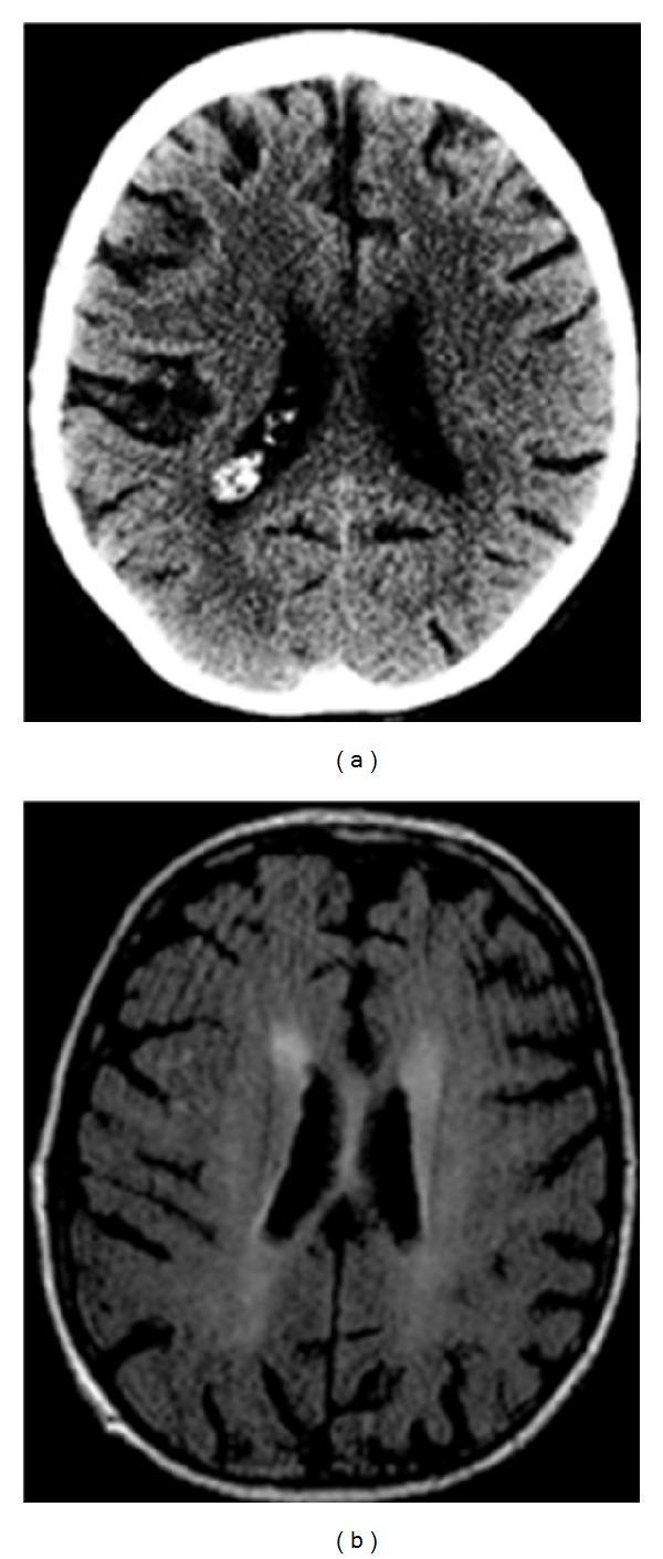
This composite figure presents two axial cross-sections of the brain for clinical diagnosis. Image (a) is an unenhanced Computed Tomography (CT) scan at the level of the lateral ventricles, showing a localized area of hyperattenuation within the left central sulcus, consistent with a focal subarachnoid hemorrhage. Notably, there is an absence of significant mass effect or midline shift. Image (b) is a Magnetic Resonance (MR) FLAIR sequence showing the same anatomical level. It reveals bilateral, ill-defined areas of hyperintensity in the periventricular and deep white matter, representing non-specific leukoaraiosis or chronic microvascular changes. These combined findings—isolated sulcal hemorrhage and white matter disease—are key radiologic markers in the assessment of Cerebral Amyloid Angiopathy (CAA). The target audience includes radiology residents and neurology specialists focusing on stroke and neurodegenerative pathologies.
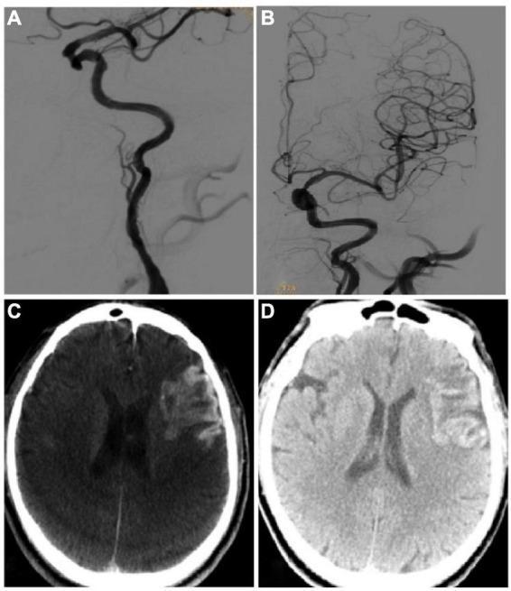
This composite image presents a clinical case of endovascular treatment for stroke and its subsequent complications. Panels A and B are Digital Subtraction Angiography (DSA) images of the left internal carotid artery (ICA). Panel A demonstrates a long-segment occlusion of the ICA characterized by a abrupt cessation of contrast flow. Panel B shows the same vessel post-recanalization after the deployment of two endovascular stents, revealing restored patency and distal intracranial filling. Panels C and D are axial CT scans of the brain at the level of the lateral ventricles. Panel C (Xper CT) was obtained immediately post-procedure and shows a large, sheet-like hyperdense area in the left hemisphere, occupying over 30% of the infarcted territory, consistent with a Type III hemorrhagic transformation or contrast extravasation. Panel D is a non-contrast CT plain scan from the following day, showing the persistence of this hyperdense lesion with a slight reduction in density, confirming the diagnosis of hemorrhagic transformation within the ischemic stroke zone. The image illustrates the successful technical outcome of endovascular recanalization and the serious potential complication of reperfusion-related hemorrhage.
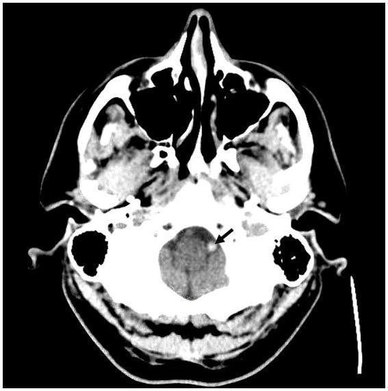
This diagnostic image is an axial non-contrast computed tomography (CT) scan of the head at the level of the posterior fossa. The image displays the skull base, facial bones, and intracranial contents. The cerebellum and brainstem are visible with normal gray-matter density. A key finding is indicated by a black arrow pointing to the left intracranial vertebral artery, which exhibits a distinct 'hyperdense vessel sign'. This hyperdensity, appearing brighter (more opaque) than typical intravascular blood and the surrounding brain parenchyma, is a radiographic marker often associated with acute thromboembolic occlusion. No obvious acute intracranial hemorrhage or midline shift is observed at this level. This visual evidence is clinically significant for the early diagnosis of acute ischemic stroke within the posterior circulation, specifically involving the vertebral artery.
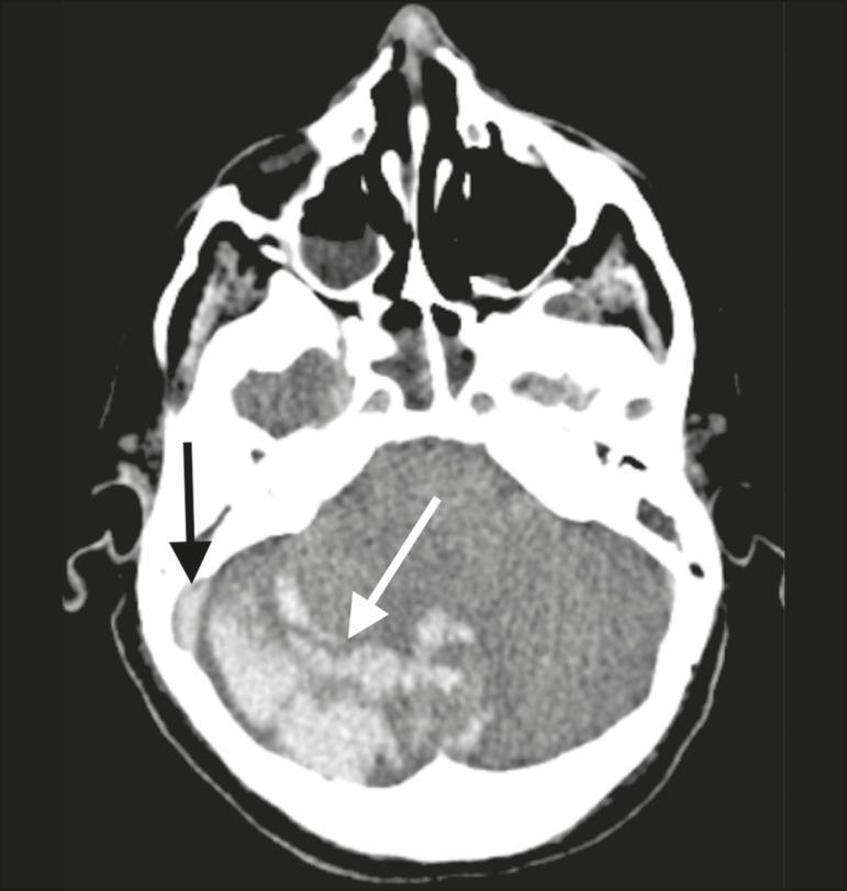
Non-contrast axial brain CT scan focusing on the posterior fossa. The image demonstrates a heterogeneous hyperdense area indicative of a hematoma (white arrow) located within the cerebellar vermis and the right cerebellar hemisphere. Furthermore, a focal hyperdensity is observed within the right sigmoid and transverse sinuses (black arrow), characteristic of the 'dense sinus sign.' This finding is highly suggestive of cerebral venous sinus thrombosis (CVST) resulting in secondary venous hemorrhagic infarction within the cerebellum. The image illustrates the radiological correlation between dural venous sinus occlusion and subsequent parenchymal hemorrhage. Clinical relevance includes the diagnosis of stroke complications in the context of hypercoagulable states, such as those observed in systemic inflammatory or viral conditions like COVID-19. This diagnostic image is intended for neurology and radiology students to identify signs of dural sinus thrombosis and associated hemorrhagic changes.
CT brain normal anatomy ventricles basal ganglia
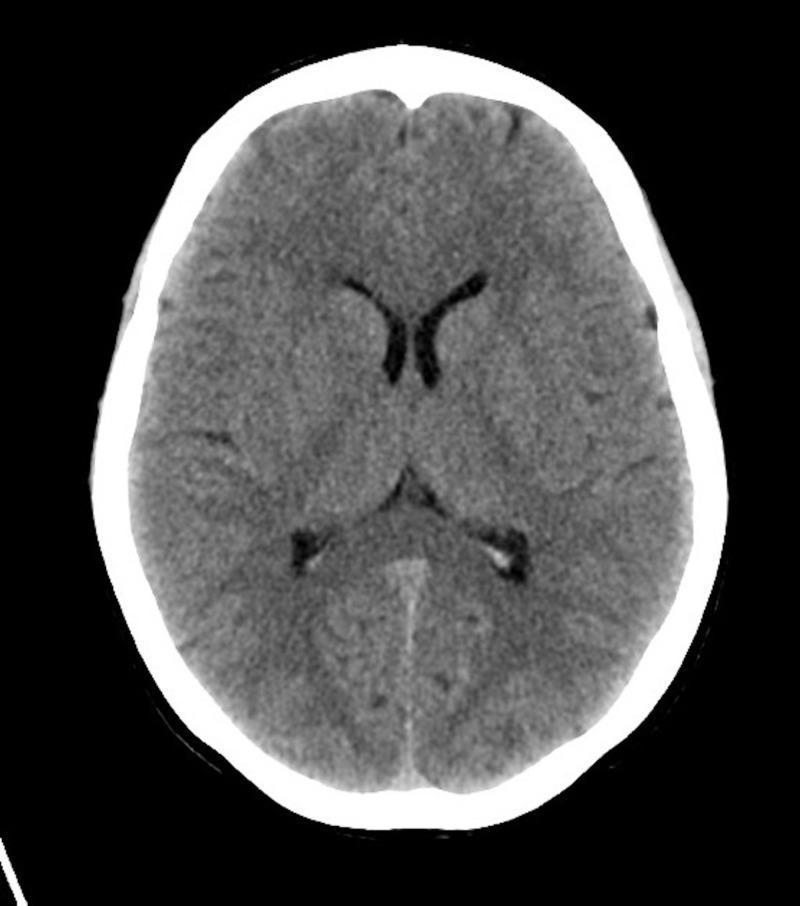
This diagnostic image is an axial non-contrast Computed Tomography (CT) scan of the brain, demonstrating normal anatomy at the level of the basal ganglia and lateral ventricles. The brain parenchyma shows appropriate attenuation with preserved gray-white matter differentiation. Key anatomical structures visible include the frontal horns of the lateral ventricles, which are symmetric and non-dilated, and the midline falx cerebri. The basal ganglia and thalamus appear unremarkable, without signs of hemorrhage, mass effect, or focal ischemia. The sulci and subarachnoid spaces are normal for age, and the overlying calvarium is intact without fractures. This imaging is used in clinical education to demonstrate a normal neurological study in patients presenting with altered mental status (AMS), specifically to rule out intracranial pathology such as stroke, trauma, or space-occupying lesions, thereby pointing toward metabolic etiologies like alkalosis.
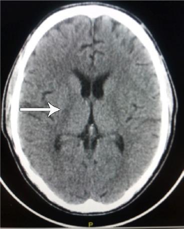
This diagnostic image is a non-contrast axial (transverse) computed tomography (CT) scan of the human brain. The scan displays the supratentorial anatomy, including the frontal, parietal, and occipital lobes, with visible lateral ventricles and the third ventricle along the midline. The brain parenchyma shows generally preserved symmetry with normal appearing sulci and gyri, indicating an absence of significant global atrophy or mass effect. A white arrow points to a focal, subtle hyperdensity located in the region of the basal ganglia, specifically involving the right putamen or globus pallidus area. This finding is clinically significant in the context of movement disorders, such as Parkinson's Disease (PD) or secondary parkinsonism, where vascular changes or calcifications in the basal ganglia may be relevant. The image serves as an educational example of neuroradiological assessment for focal density changes in patients presenting with progressive neurological symptoms.
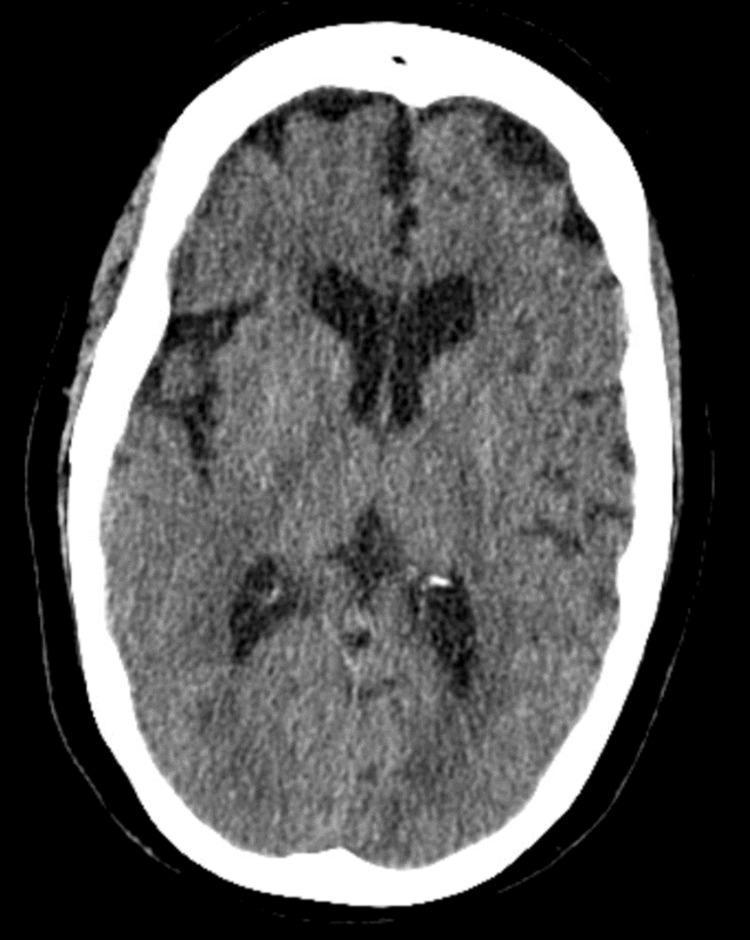
This diagnostic image is an axial non-contrast computed tomography (CT) scan of the head at the level of the lateral ventricles and basal ganglia. The image demonstrates normal intracranial anatomy for educational comparison. The brain parenchyma shows appropriate differentiation between gray and white matter with no evidence of acute hemorrhage, mass effect, or focal infarcts. The ventricular system, including the frontal horns of the lateral ventricles and the third ventricle, is centrally located and of normal size (hypodense appearance due to cerebrospinal fluid). The midline is unremarkable, with a centrally aligned falx cerebri and no evidence of midline shift. Cortical sulci and sylvian fissures are visible and of normal prominence for age. The surrounding calvarium is intact and typically hyperdense (bright white) on this soft tissue window setting. This scan serves as a clinical reference for a negative or 'normal' head CT in an emergency or neurological assessment context.
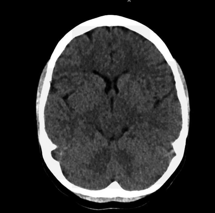
This diagnostic image is a non-contrast Computed Tomography (CT) scan of the head in an axial plane. The scan demonstrates the intracranial anatomy at the level of the basal ganglia and lateral ventricles. The brain parenchyma shows normal, homogeneous attenuation without evidence of acute intracranial hemorrhage, midline shift, or mass effect. Key anatomical structures visible include the frontal and occipital lobes, the Sylvian fissures, and the anterior horns of the lateral ventricles, which appear as hypodense (dark), fluid-filled spaces. The skull is depicted as a hyperdense (bright white) peripheral ring, indicating normal calcification of the cranial vault. This image is representative of a routine screening for neurological symptoms, such as suspected subarachnoid hemorrhage, where the initial radiological finding is unremarkable. The scan is clinically significant for students and practitioners to understand the baseline appearance of a 'normal' head CT in an emergency or diagnostic triage context.
subarachnoid hemorrhage epidural subdural hematoma CT brain
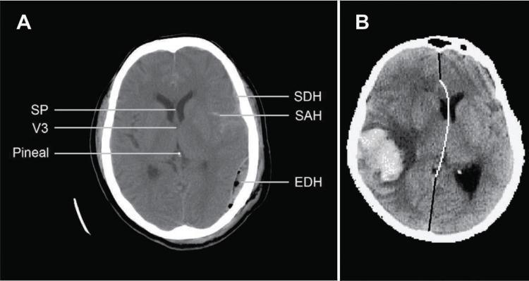
This educational composite contains two axial non-contrast computed tomography (CT) scans of the brain demonstrating various intracranial hemorrhages and mass effect. Image A illustrates traumatic brain injury findings with multiple hemorrhage types: a crescentic subdural hematoma (SDH), subarachnoid hemorrhage (SAH) within the sulci, and a biconvex/lens-shaped epidural hematoma (EDH) on the patient's right side. Key anatomical landmarks annotated include the septum pellucidum (SP), third ventricle (V3), and the pineal gland. Image B depicts a large, hyperdense, oval-shaped intracerebral hematoma (ICH) in the right hemisphere. This lesion exerts significant mass effect, resulting in a prominent midline shift (MLS), visualized by a curved white line showing the deviation of the brain's midline structures toward the left. The content is designed for neurosurgical and radiological education, focusing on identifying different hemorrhage patterns and quantifying midline shift in acute clinical settings.
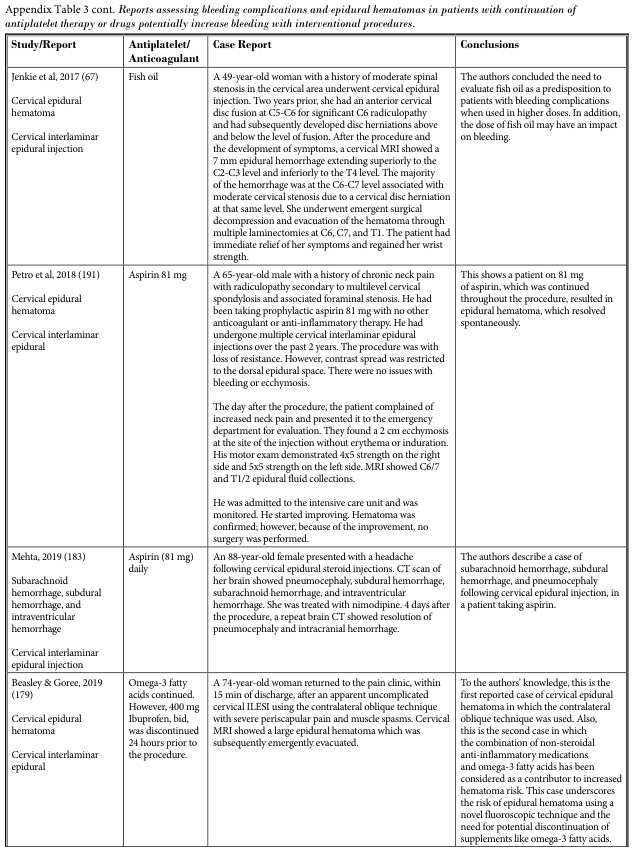
Appendix Table 3 cont. Reports assessing bleeding complications and epidural hematomas in patients with continuation of antiplatelet therapy or drugs potentially increase bleeding with interventional procedure.
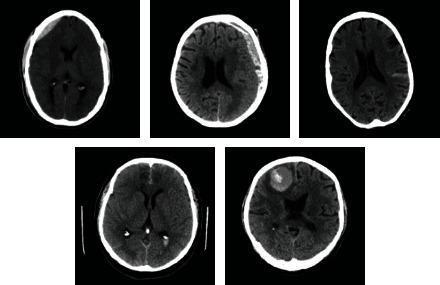
This composite diagnostic image features five axial non-contrast Computed Tomography (CT) scans of the human brain, illustrating various presentations of intracranial hemorrhage. The scans demonstrate distinct pathological features: 1) a hyperdense, biconvex (lens-shaped) collection consistent with an epidural hematoma in the left frontal region; 2) a crescent-shaped hyperdense collection along the cerebral hemisphere characteristic of a subdural hematoma; 3) a scan showing relatively normal brain parenchyma and ventricular system for comparison; 4) a scan exhibiting subtle hyperdensity within the cortical sulci indicative of subarachnoid hemorrhage; and 5) a focal, well-defined hyperdense intraparenchymal lesion in the right frontal lobe surrounded by a thin rim of hypodensity, representing an intraparenchymal hemorrhage with associated vasogenic edema. These images serve as educational examples for identifying different hemorrhage subtypes based on location, morphology, and density. The collection is highly relevant for neuroradiology training and the development of automated detection algorithms.
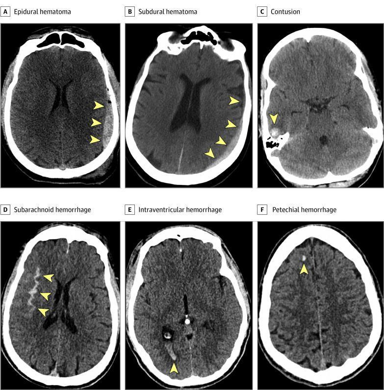
This composite educational graphic presents six axial noncontrast computed tomography (CT) scans of the brain, demonstrating common subtypes of acute traumatic intracranial hemorrhage (ICH) as defined by the NINDS Common Data Elements. Panel A shows an Epidural Hematoma (EDH) characterized by a high-density, biconvex (lens-shaped) collection between the skull and dura mater. Panel B illustrates a Subdural Hematoma (SDH) as a crescent-shaped hyperdensity along the brain surface. Panel C demonstrates a Cerebral Contusion, appearing as a heterogeneous intraparenchymal lesion with mixed densities. Panel D shows a Subarachnoid Hemorrhage (SAH) with hyperdense blood filling the cortical sulci and fissures. Panel E displays an Intraventricular Hemorrhage (IVH) with high-density blood layering within the posterior horns of the lateral ventricles. Panel F identifies a Petechial Hemorrhage, seen as a small, punctate focus of high density within the white matter, often associated with diffuse axonal injury. Yellow arrowheads highlight the specific pathologies in each diagnostic image, emphasizing the distinct morphology and anatomical compartmentalization of each bleed type.
ischemic stroke CT brain hypodense infarct MCA territory
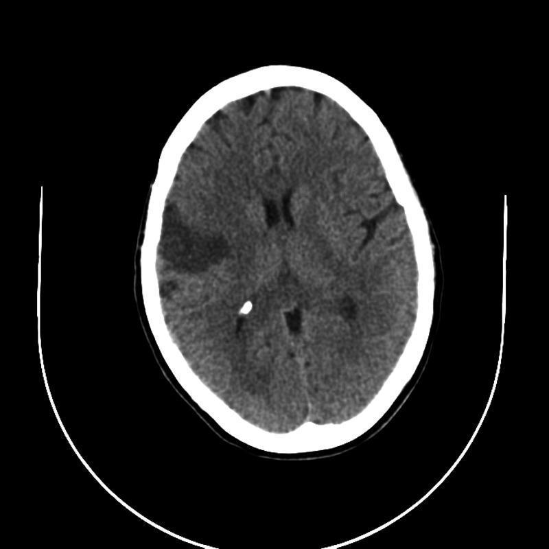
This diagnostic image is an axial non-contrast CT scan of the brain, rather than MRI as some tags suggest, displaying acute to subacute ischemic changes. The primary finding is a moderately sized, wedge-shaped area of hypodensity in the right cerebral hemisphere, corresponding to the vascular territory of the middle cerebral artery (MCA). This hypodense region involves the cortical ribbon and subcortical white matter, indicative of a cytotoxic edema and infarction. A secondary, smaller, and more subtle area of hypodensity is observed in the left MCA territory, suggesting bilateral involvement. Key anatomical landmarks include the hyperdense skull, cerebral sulci, and the ventricular system, which shows slight compression or effacement adjacent to the larger infarct site. The image illustrates the classic radiological presentation of ischemic stroke in the MCA distribution, serving as a critical educational example for identifying vascular territory infarcts and associated tissue density changes in neuroimaging.
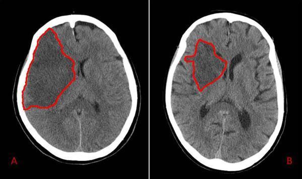
This diagnostic image displays two side-by-side axial non-contrast CT scans of the brain, labeled A and B, illustrating varying extents of ischemic stroke in the right middle cerebral artery (MCA) territory. In both panels, the infarcted areas are demarcated by red outlines. Panel A demonstrates a large, confluent hypodense region involving the right frontal and temporal lobes, extending to the cortical surface, consistent with a total MCA occlusion. There is a visible mass effect characterized by effacement of the right lateral ventricle and a subtle midline shift to the left. Panel B shows a smaller, more localized hypodensity primarily affecting the deeper parenchyma and subcortical structures of the right hemisphere, sparing the peripheral cortex, typical of a partial MCA territory infarct. In panel B, the ventricular system and midline structures remain relatively preserved compared to panel A. This comparison highlights the radiological presentation of different stroke severities and their secondary effects on brain morphology 48 hours post-admission.
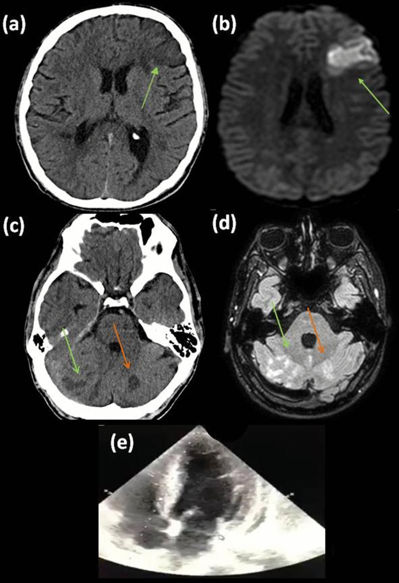
This composite figure illustrates a case of multi-territory embolic ischemic strokes and cardiac imaging. (a) Non-contrast axial CT scan of the brain shows a faint hypodense area in the left frontal lobe. (b) Corresponding Diffusion-Weighted Imaging (DWI) MRI confirms an acute infarct in the left middle cerebral artery (MCA) superior trunk territory, appearing as a bright, hyperintense signal indicating restricted diffusion. (c) A more caudal axial CT slice reveals multiple small hypodense lesions in the right cerebellar hemisphere and vermis (green arrows), alongside an older, well-defined hypodense chronic infarct in the left cerebellar hemisphere (orange arrow). (d) Fluid-Attenuated Inversion Recovery (FLAIR) MRI at the same level as (c) shows these acute/subacute cerebellar lesions as hyperintense signals (green arrows) and the chronic left-sided infarct (orange arrow). (e) Transthoracic echocardiogram (TTE), apical four-chamber view, demonstrating the resolution (lysis) of a previously documented left ventricular thrombus. The series highlights the importance of multimodal imaging (CT, MRI-DWI/FLAIR) and cardiac assessment in the evaluation of cardioembolic stroke.
brain tumor CT scan mass effect midline shift
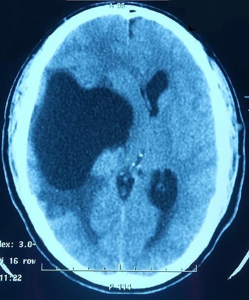
Axial non-contrast computed tomography (CT) scan of the brain demonstrating a large, well-circumscribed, hypodense lesion in the right temporal and frontoparietal regions. The lesion exhibits significant mass effect, evidenced by a marked midline shift toward the left and subfalcine herniation. The right lateral ventricle is severely compressed and effaced, while the contralateral left lateral ventricle shows compensatory enlargement or distortion. Peripheral vasogenic edema is visible as ill-defined hypodensity in the adjacent white matter. These findings are consistent with a space-occupying intracranial lesion, such as a large cyst, tumor, or abscess. Clinical context suggests this specific case involves neurosarcoidosis presenting as a granulomatous mass, initially mimicking cerebral tuberculosis. This image is educational for illustrating intracranial mass effect, ventricular compression, and midline shift in neuro-radiology.
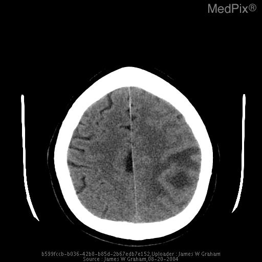
**Medical Image Description**
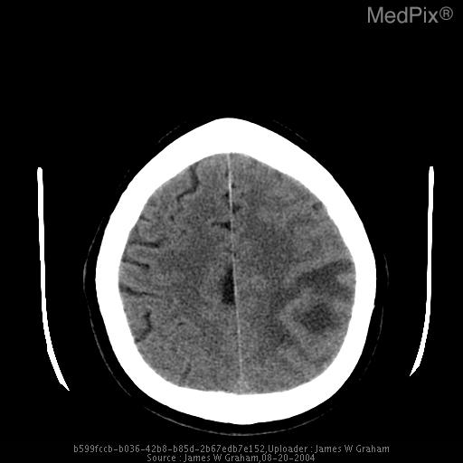
**Imaging Modality:** Non-contrast axial CT scan of the head.
🧠 CT BRAIN — Complete Guide (Hindi-English)
CT Scan Kya Hota Hai? / What is CT Scan?
| Density | CT mein dikhna | Matlab |
|---|---|---|
| Hyperdense (Bright/White) | Roshan/Safed | Blood (acute), Bone, Calcification, Contrast |
| Isodense | Grey | Normal brain tissue |
| Hypodense (Dark/Black) | Kaala/Dark | CSF, Water, Oedema, Old infarct, Air |
Trick: "Fresh blood = WHITE, Old blood/water/CSF = BLACK"
PART 1 — CT BRAIN ANATOMY (Normal Structures)
CT Brain ke Levels — Upar se Neeche
🔵 Level 1 — Skull Base / Posterior Fossa Level
- Cerebellum (bilateral, posterior) — grey matter, smooth
- Brainstem (Pons/Medulla) — midline mein
- 4th Ventricle — dark (CSF), midline
- Temporal bones — bright (bone)
- Mastoid air cells — dark (air)
- Orbits — eyes dikhte hain
- Ethmoid sinuses
🟢 Level 2 — Suprasellar / Basal Cisterns Level
- Basal cisterns — star-shaped dark area (CSF)
- Suprasellar cistern ("5-pointed star")
- Interpeduncular cistern
- Ambient cistern
- Midbrain — midline mein
- Temporal lobes — lateral
- Circle of Willis area
- 3rd Ventricle — thin midline dark line
⚠️ SAH mein yeh cisterns bright (hyperdense) ho jaati hain — normal star shape nahi dikhta
🟡 Level 3 — Basal Ganglia / Lateral Ventricle Level (MOST IMPORTANT)
| Structure | Location | Appearance |
|---|---|---|
| Lateral Ventricles (frontal horns) | Bilateral, midline ke paas | Hypodense (dark) — CSF |
| 3rd Ventricle | Midline, thin | Hypodense |
| Caudate Nucleus | Anteromedial to lateral ventricle | Isodense (grey) |
| Putamen | Lateral | Isodense |
| Globus Pallidus | Medial to putamen | Slightly hyperdense (can calcify) |
| Internal Capsule | Between basal ganglia & thalamus | White matter (hypodense) |
| Thalamus | Bilateral, central | Isodense |
| External Capsule | Lateral | White matter |
| Insula | Lateral cortex | Grey matter |
| Sylvian Fissure | Lateral | Dark (CSF) |
| Falx Cerebri | Midline | Bright (dura) |
🟠 Level 4 — Body of Lateral Ventricles Level
- Body of Lateral Ventricles — bilateral dark spaces
- Septum Pellucidum — thin midline structure between ventricles
- Corpus Callosum — connects both hemispheres (hypodense/white matter)
- Caudate (body)
- Choroid Plexus — in ventricles, may calcify (normally bright dots)
- Parietal lobes — bilateral
🔴 Level 5 — High Convexity / Vertex Level
- Cerebral sulci — dark (CSF)
- Gyri — grey matter (isodense)
- Cortex — grey (darker) vs White matter (lighter)
- Falx Cerebri — midline bright line
- Superior sagittal sinus — midline posterior, slightly bright
Normal CT Brain — Kya Dekhna Hai
Normal CT Brain mein Dekhne ka Checklist (ABCDE)
| Letter | Check | Details |
|---|---|---|
| A | Asymmetry / Midline shift | Midline centered hai? Falx cerebri midline par hai? Septum pellucidum shifted nahi? |
| B | Blood / Bone | Koi hyperdense (white) area hai? Skull fracture? |
| C | CSF spaces | Ventricles size normal? Sulci visible? Cisterns open? |
| D | Density changes | Hypodense areas (oedema/infarct)? Hyperdense areas (blood)? |
| E | Extra-axial spaces | Subdural/Epidural space mein kuch hai? |
PART 2 — CT BRAIN DIAGNOSES (Pathological Findings)
🔴 1. INTRACRANIAL HEMORRHAGE — Types
🅰️ Epidural Hematoma (EDH)
| Feature | Details |
|---|---|
| Shape | Biconvex / Lens-shaped (dono taraf bulging) |
| CT appearance | Hyperdense (bright white) |
| Location | Temporal area most common (middle meningeal artery injury) |
| Cause | Head trauma, temporal bone fracture |
| Crossing sutures? | NO — sutures cross nahi karta |
| Classic history | Lucid interval — conscious → unconscious |
| Emergency | Surgical evacuation needed |
Trick: EDH = Egg-shaped = biconvex lens
🅱️ Subdural Hematoma (SDH)
| Feature | Details |
|---|---|
| Shape | Crescent-shaped (brain surface ko follow karta hai) |
| CT appearance | Acute = Hyperdense |
| Location | Brain ki surface par, falx ke saath bhi |
| Cause | Bridging vein injury, elderly falls, anticoagulants |
| Crossing sutures? | YES — sutures cross kar sakta hai |
| Midline shift | Common — brain ko push karta hai |
| SDH Age | CT Density |
|---|---|
| Acute (0–3 days) | Hyperdense (White) |
| Subacute (3 days–3 weeks) | Isodense (Grey — miss hone ka risk!) |
| Chronic (>3 weeks) | Hypodense (Dark) |

🅲️ Subarachnoid Hemorrhage (SAH)
| Feature | Details |
|---|---|
| CT appearance | Hyperdense (white) blood in sulci, cisterns, fissures |
| Classic finding | Star-shaped basal cisterns bright ho jaati hain |
| Cause | Berry aneurysm rupture (most common), trauma |
| Classic symptom | "Thunderclap headache" — worst headache of life |
| CT sensitivity | 90–95% within first 12 hours; decreases with time |
| If CT negative | LP (Lumbar puncture) karo — xanthochromia check |
🅳️ Intracerebral Hemorrhage (ICH)
| Feature | Details |
|---|---|
| CT appearance | Round/oval hyperdense (bright) lesion within brain parenchyma |
| Oedema | Surrounding hypodense ring (vasogenic oedema) |
| Cause | HTN (most common — basal ganglia, thalamus, pons), AVM, coagulopathy, amyloid angiopathy |
| HTN bleed location | Putamen > Thalamus > Pons > Cerebellum > Caudate |
| Mass effect | Ventricles compress, midline shift |
Putamen (35%) → Thalamus (15%) → Pons (10%) → Cerebellum (10%) → Caudate (5%)
🅴️ Intraventricular Hemorrhage (IVH)
| Feature | Details |
|---|---|
| CT appearance | Bright blood within ventricles |
| Pattern | Blood fills and expands ventricles |
| Cause | Extension from ICH, trauma, AVM, premature neonates |
| Complication | Hydrocephalus (blood blocks CSF flow) |
🔵 2. ISCHEMIC STROKE (Infarction)
| Feature | Details |
|---|---|
| CT appearance | Hypodense (dark) area in vascular territory |
| Early (0–6 hrs) | CT almost normal ho sakta hai — MRI better hai |
| Early signs | Loss of grey-white differentiation, sulcal effacement, dense MCA sign |
| Late (>24 hrs) | Clearly hypodense wedge-shaped area |
| Territory | MCA, ACA, PCA territory follow karta hai |
Acute Stroke Early CT Signs:
- Dense MCA sign — MCA vessel bright white dikhti hai (thrombus)
- Loss of insular ribbon — insula ka grey-white differentiation khatam
- Sulcal effacement — sulci nahi dikhte (oedema)
- Obscuration of basal ganglia
MCA vs ACA vs PCA Territory
| Artery | Area Supplied | Deficit |
|---|---|---|
| MCA | Frontal + Parietal + Temporal lateral | Contralateral hemiplegia (face + arm > leg), aphasia (dominant) |
| ACA | Frontal lobe medial | Contralateral leg weakness |
| PCA | Occipital lobe | Homonymous hemianopia |
| PICA | Cerebellum (lateral medulla) | Wallenberg syndrome |
| Basilar artery | Brainstem + Bilateral cerebellum | Locked-in syndrome, coma |
Dense MCA Sign & Hyperdense Vessel Sign:
🟢 3. CEREBRAL VENOUS SINUS THROMBOSIS (CVST)
| CT Finding | Details |
|---|---|
| Dense sinus sign | Involved sinus hyperdense (bright) — thrombus |
| Empty delta sign | Contrast CT mein — sinus walls enhance but centre dark (filling defect) |
| Venous infarct | Unusual location, haemorrhagic, doesn't follow arterial territory |
| Bilateral involvement | Both hemispheres affected |
| Causes | OCP, pregnancy, dehydration, hypercoagulable states, COVID-19 |
🟡 4. HYDROCEPHALUS
| Type | Cause | CT Finding |
|---|---|---|
| Obstructive (Non-communicating) | Tumour blocking CSF path | Only some ventricles dilated (proximal to block) |
| Communicating | Impaired reabsorption (post-SAH, meningitis) | All 4 ventricles dilated |
| Normal Pressure Hydrocephalus (NPH) | Unknown | Ventricles large, sulci small (tight), periventricular lucency |
🟠 5. BRAIN TUMOURS
| Feature | Details |
|---|---|
| Mass effect | Brain ko push karta hai, sulci compress hote hain |
| Midline shift | Falx cerebri aur septum pellucidum shift hote hain |
| Oedema | Surrounding hypodensity (vasogenic oedema) — "white matter finger-like" |
| Calcification | Some tumours calcify (meningioma, oligodendroglioma) |
| Enhancement | Contrast CT par ring enhancement ya solid enhancement |
| Tumour | CT Features |
|---|---|
| Glioblastoma (GBM) | Irregular ring-enhancing lesion, central necrosis, massive oedema |
| Meningioma | Hyperdense, calcified, attached to dura, homogeneous enhancement |
| Metastasis | Multiple lesions, ring-enhancing, at grey-white junction |
| Abscess | Ring-enhancing, thin smooth wall, restricted diffusion on MRI |
| Primary CNS Lymphoma | Periventricular, homogeneous enhancement |
🔵 6. TRAUMA — Head Injury CT (Marshall Scale)
Traumatic Brain Injury CT Findings:
| Finding | Description |
|---|---|
| Cerebral contusion | Heterogeneous (mixed density) intraparenchymal lesion |
| Diffuse Axonal Injury (DAI) | CT mostly normal/subtle petechiae at grey-white junction; MRI better |
| Skull fracture | Cortical break in bone window |
| Pneumocephalus | Air (hypodense black) inside skull — open fracture sign |
| Midline shift | Measure at level of septum pellucidum |
| Herniation | Uncal (temporal lobe herniates into tentorial notch), Subfalcine, Tonsillar |
Marshall CT Classification (TBI Severity):
| Grade | CT Finding | Mortality |
|---|---|---|
| I | No visible pathology | 9.6% |
| II | Cisterns present, shift <5mm, no clot >25mL | 13.5% |
| III | Cisterns compressed/absent, shift <5mm | 34.0% |
| IV | Shift >5mm, no clot >25mL | 56.2% |
| V | Any evacuated mass lesion | — |
| VI | Non-evacuated clot >25mL | — |
🔴 7. BRAIN ATROPHY
| Finding | Description |
|---|---|
| Sulcal widening | Sulci wider than normal for age |
| Ventricle enlargement | Ventricles bigger — "ex vacuo" dilatation |
| Decreased brain volume | Grey and white matter reduced |
| Causes | Alzheimer's, ageing, chronic alcoholism, post-trauma |
PART 3 — COMPARISON TABLE (CT vs MRI)
| Feature | CT Brain | MRI Brain |
|---|---|---|
| Speed | Fast (5–10 min) | Slow (30–60 min) |
| Acute hemorrhage | Excellent (hyperdense) | Good |
| Acute ischemia (<6 hrs) | Poor | Excellent (DWI) |
| Bone/fracture | Excellent | Poor |
| Posterior fossa | Limited (bone artifact) | Excellent |
| Brainstem lesions | Poor | Excellent |
| Availability/Cost | Wide/Cheap | Limited/Expensive |
| Radiation | Yes | No |
| Metal implants | OK | Contraindicated |
PART 4 — CT BRAIN WINDOWS
| Window | Centre / Width | Use |
|---|---|---|
| Brain/Soft tissue window | C: 35, W: 80 | Brain parenchyma (grey-white, blood) |
| Blood window | C: 55, W: 160 | Hemorrhage detection |
| Bone window | C: 350, W: 2000 | Skull fracture, calvarium |
| Subdural window | C: 70, W: 200 | Isodense SDH detect karna |
| Lung window | Low centre | Pneumocephalus |
PART 5 — QUICK DIAGNOSIS SUMMARY (Cheat Sheet)
| CT Finding | Diagnosis |
|---|---|
| Biconvex bright lesion | Epidural Hematoma (EDH) |
| Crescent-shaped bright lesion | Acute Subdural Hematoma |
| Crescent-shaped dark lesion | Chronic Subdural Hematoma |
| Bright blood in cisterns/sulci | Subarachnoid Hemorrhage (SAH) |
| Round bright lesion in basal ganglia | Hypertensive ICH |
| Wedge-shaped dark area | Ischemic Infarct |
| Ventricles all dilated | Communicating Hydrocephalus |
| Ring-enhancing lesion | Abscess / GBM / Metastasis |
| Periventricular bright lesion | CNS Lymphoma |
| Dense MCA sign | Acute MCA stroke |
| Dense sinus | CVST |
| Multiple bright calcified lesions | Neurocysticercosis |
| Sulci widened + ventricles big | Cerebral Atrophy |
| Bright falx / tentorium | SAH (blood tracking) |
| Midline shift | Mass effect — tumour/hematoma/large infarct |
PART 6 — CT BRAIN PADHNE KA SYSTEMATIC APPROACH
Step 1: BONE WINDOW
        → Skull fracture? Calvarium intact?

Step 2: MIDLINE
        → Falx cerebri midline par hai?
        → Septum pellucidum shift nahi?
        → Midline shift measure karo (>5mm = significant)

Step 3: VENTRICLES
        → Size: Dilated? Compressed?
        → Symmetry: Bilateral equal size?
        → Blood in ventricles (IVH)?
        → Hydrocephalus?

Step 4: CISTERNS
        → Basal cisterns (star shape) patent?
        → Compressed cisterns = raised ICP
        → Blood in cisterns = SAH

Step 5: PARENCHYMA (Brain tissue)
        → Any hyperdense (bright) areas = fresh blood
        → Any hypodense (dark) areas = infarct/oedema/tumour
        → Grey-white differentiation normal?
        → Sulci visible?

Step 6: EXTRA-AXIAL SPACES
        → Epidural space mein kuch hai?
        → Subdural space clean?

Step 7: POSTERIOR FOSSA
        → Cerebellum symmetric?
        → 4th ventricle midline?
        → Brainstem normal?
Bhai LFT ek dam simple me samjhao yr ke kaise our kya diagnosis banaye
liver function tests LFT interpretation bilirubin ALT AST alkaline phosphatase
Create a comparison table PDF for LFT patterns
Bhai intubation ke pure explained notes bana de hinglish me with photos kuch bhi nahi chhutna chahiye