
Variant types of thyroid malignancy, pathology, management, staging follow up
"thyroid cancer" AND "management"
Thyroid Malignancy: Variants, Pathology, Management, Staging & Follow-up
Overview
| Type | % of cases | Cell of origin |
|---|---|---|
| Papillary Thyroid Carcinoma (PTC) | ~84% | Follicular epithelium |
| Follicular Thyroid Carcinoma (FTC) | ~11% | Follicular epithelium |
| Oncocytic (Hürthle cell) Carcinoma (OCA) | ~3–4% | Follicular epithelium |
| Poorly Differentiated TC (PDTC) | ~1–2% | Follicular epithelium |
| Medullary Thyroid Carcinoma (MTC) | ~2–5% | Parafollicular C cells |
| Anaplastic Thyroid Carcinoma (ATC) | ~1% | Follicular epithelium (dedifferentiated) |
| Primary Thyroid Lymphoma | Rare | Lymphoid tissue |
1. Papillary Thyroid Carcinoma (PTC)
Pathology
- Branching papillae with fibrovascular stalks covered by cuboidal epithelium
- Ground-glass (Orphan Annie) nuclei - optically clear appearance due to finely dispersed chromatin
- Intranuclear pseudoinclusions and nuclear grooves (cytoplasmic invaginations)
- Psammoma bodies - concentric calcifications in papillary cores; almost never in follicular or medullary carcinomas
- Lymphatic invasion common; blood vessel invasion uncommon in smaller lesions
- Cervical lymph node metastases in up to 50% of cases
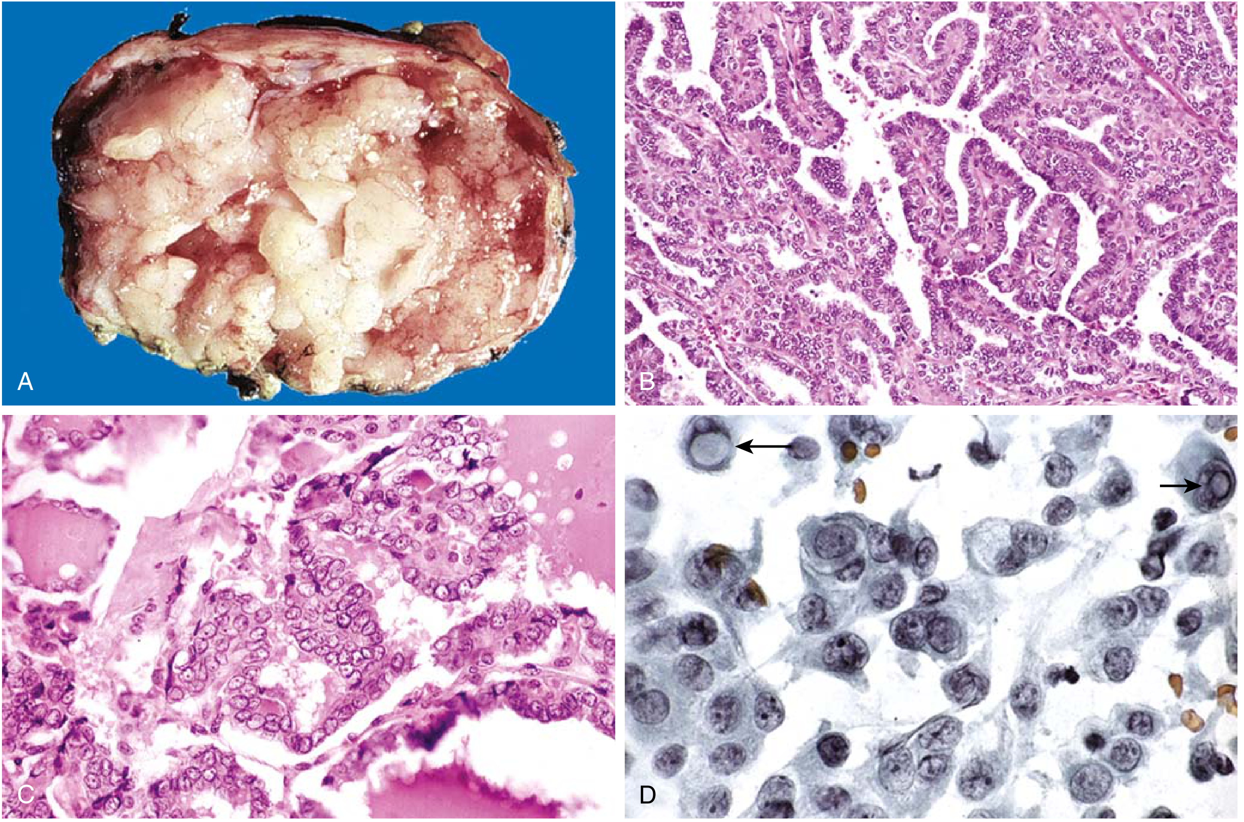
- BRAF V600E - most common point mutation (~45% of PTC), activates MAPK pathway, associated with aggressive features and RAI resistance
- RAS mutations - 20-30%, more common in follicular variant PTC; activates both MAPK and PI3K-AKT
- RET/PTC rearrangements - 5-10%, characteristic of radiation-induced PTC; multifocal, more extrathyroidal extension
- TERT promoter mutations - up to 27%, especially co-occurring with BRAF; associated with RAI resistance and high recurrence risk
- PAX8-PPARG - seen in follicular variant PTC
- Classical/conventional
- Follicular variant (encapsulated vs infiltrative) - most common variant; almost totally follicular architecture but PTC nuclear features
- Tall cell variant - aggressive, occurs in older patients
- Columnar cell variant - aggressive
- Diffuse sclerosing variant
- Solid/trabecular variant
- Hobnail variant - high recurrence risk
- Noninvasive follicular thyroid neoplasm with papillary-like nuclear features (NIFTP) - reclassified as non-malignant
2. Follicular Thyroid Carcinoma (FTC)
Pathology
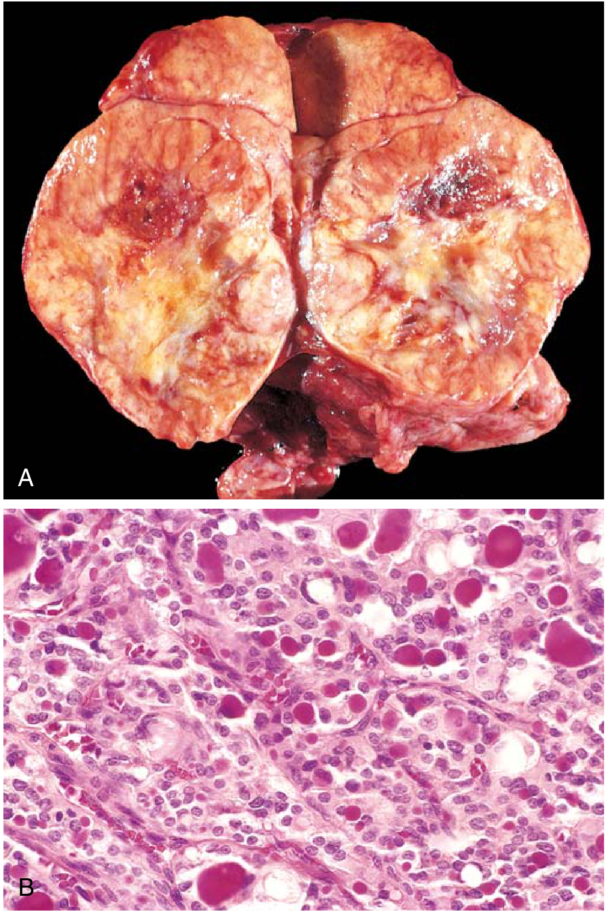
- Minimally invasive - capsular invasion only; very low metastatic risk; <10% die within 10 years
- Widely invasive/angioinvasive - blood vessel invasion; metastases to lung, bone, liver; up to 50% die within 10 years
3. Oncocytic (Hürthle Cell) Carcinoma (OCA)
4. Medullary Thyroid Carcinoma (MTC)
Pathology
- RET proto-oncogene (chromosome 10q11.2) - germline mutations in virtually all hereditary MTC; somatic (acquired) RET mutations in ~50% of sporadic MTC
- Codon C634 mutations are most common in hereditary MTC
- Sporadic (~75%) - unifocal; usually presents 4th-6th decade; up to 50% have palpable neck mass; cervical nodal metastases in >70% with palpable tumor; distant mets (liver, mediastinum, lungs, bone) in 10-15%
- Familial (~25%):
- MEN 2A (Sipple syndrome) - MTC + pheochromocytoma + primary hyperparathyroidism; codon 634 RET mutation; presents 3rd decade
- MEN 2B - MTC + pheochromocytoma + marfanoid habitus + mucosal neuromas + intestinal ganglioneuromatosis; codon M918T mutation; most aggressive; presents before 2nd decade
- Familial MTC - MTC without other MEN features
- Highest risk (MEN 2B, M918T mutation) - prophylactic thyroidectomy within first 6 months of life
- High risk (C634F/G/R/S/W/Y, C609, C611, etc.) - thyroidectomy before age 5 years
- Moderate risk - thyroidectomy timing guided by calcitonin levels/imaging
5. Anaplastic Thyroid Carcinoma (ATC)
Pathology
6. TNM Staging (AJCC 8th Edition)
Primary Tumor (T)
| Stage | Description |
|---|---|
| T1a | ≤1 cm, limited to thyroid |
| T1b | >1 cm, ≤2 cm, limited to thyroid |
| T2 | >2 cm, ≤4 cm, limited to thyroid |
| T3a | >4 cm, limited to thyroid |
| T3b | Gross extrathyroidal extension into strap muscles only (any size) |
| T4a | Gross ETE into subcutaneous tissue, larynx, trachea, esophagus, or recurrent laryngeal nerve |
| T4b | Gross ETE into prevertebral fascia, or encasing carotid artery or mediastinal vessels |
Regional Lymph Nodes (N)
| Stage | Description |
|---|---|
| N1a | Metastasis to level VI or VII (pretracheal, paratracheal, prelaryngeal/Delphian, upper mediastinal) |
| N1b | Metastasis to lateral neck (levels I-V) or retropharyngeal nodes |
Stage Grouping - Differentiated TC (PTC/FTC/OCA/PDTC)
| Stage | TNM |
|---|---|
| I | Any T, Any N, M0 |
| II | Any T, Any N, M1 |
| Stage | TNM |
|---|---|
| I | T1-T2, N0, M0 |
| II | T1-T2 N1 M0, or T3a/T3b any N M0 |
| III | T4a, any N, M0 |
| IVA | T4b, any N, M0 |
| IVB | Any T, any N, M1 |
Additional Risk-Stratification Systems
- MACIS (Mayo Clinic): Score based on Metastasis + Age + Completeness of resection + Invasion + Size. Score <6.0 = 99% 20-year survival
- AMES: Age, Metastases, Extent of tumor, Size - divides into low-risk and high-risk groups
- ATA Dynamic Risk Stratification - assigns response-to-therapy categories after treatment:
- Excellent response - no clinical, biochemical, or structural evidence of disease (1-4% recurrence risk)
- Biochemical incomplete response - abnormal Tg or rising anti-Tg antibodies, no structural disease (50% achieve NED spontaneously or with Rx)
- Structural incomplete response - persistent locoregional or distant disease (mortality up to 50% with distant mets)
- Indeterminate response - nonspecific findings (15-20% develop structural disease)
7. Management
Differentiated Thyroid Cancer (PTC, FTC, OCA)
-
Thyroid lobectomy - acceptable for low-risk, unilateral DTC 1-4 cm, no ETE, no metastatic disease; recommended for <1 cm DTC (after active surveillance discussion)
-
Total thyroidectomy - preferred for (Sabiston, p. 1515):
- Tumor ≥4 cm
- Clinically evident nodal metastases
- Gross extrathyroidal extension
- Evidence of distant metastatic disease
- Radiation-induced DTC
- Familial non-medullary TC
- Multifocal bilateral DTC
-
Active surveillance - an option for small (<1.5 cm) intrathyroidal micropapillary cancers without cervical LN mets, ETE, or proximity to the recurrent laryngeal nerve
- Therapeutic compartment-based dissection for clinically/radiographically evident nodal disease
- Central compartment (level VI ± VII) dissection for ipsilateral central nodal involvement
- Lateral neck dissection (levels II-V) for proven lateral neck metastases
- Prophylactic central neck dissection remains controversial
- Not indicated for T1 tumors ≤2 cm confined to thyroid (no recurrence/survival benefit)
- No benefit demonstrated for larger (>2 cm but <4 cm) low-risk tumors
- Indicated for higher risk patients: larger tumors, aggressive variants, vascular invasion, ETE, large-volume lymph node metastases, older patients
- Low-dose ¹³¹I (1100 MBq / 30 mCi) for remnant ablation; high-dose (up to 5500 MBq / 150 mCi) for adjuvant treatment of residual/metastatic disease
- Requires TSH stimulation (TSH >25 mIU/L) - achieved by either thyroid hormone withdrawal or recombinant human TSH (rhTSH, thyrotropin alfa)
- Preceded by low-iodine diet for 1-2 weeks
- Levothyroxine to suppress TSH - rationale: differentiated TCs express TSH-R and remain responsive to TSH stimulation
- Degree of suppression tailored to risk: high-risk patients aim for TSH <0.1 mIU/L; low-risk patients can maintain low-normal TSH
- Lenvatinib and sorafenib (multi-kinase inhibitors) - approved for RAI-refractory progressive differentiated TC
- BRAF inhibitors (dabrafenib + trametinib) for BRAF V600E-mutant RAI-refractory DTC
Medullary Thyroid Carcinoma
- Surgery is the mainstay - total thyroidectomy with central neck dissection; lateral neck dissection if lateral nodal disease
- Exclude pheochromocytoma first in all RET mutation carriers before surgery (risk of hypertensive crisis)
- No RAI - MTC does not take up radioiodine
- External beam radiation - palliative for unresectable or metastatic disease
- Vandetanib and cabozantinib - approved kinase inhibitors (targeting RET, VEGFR, EGFR) for advanced/metastatic MTC; provide improved progression-free survival
- Selpercatinib - selective RET inhibitor with high response rates in RET-mutant MTC (2020 FDA approval); pralsetinib is an alternative
- Genetic testing - all patients with MTC should be tested for RET germline mutations; positive results trigger cascade testing of first-degree relatives
Anaplastic Thyroid Carcinoma
- Prognosis is dismal - median survival 3-6 months; nearly 100% mortality (Harrison's, p. 3092)
- RAI is usually ineffective (loss of iodine uptake)
- Multimodal approach:
- Surgery (if resectable) - debulking may relieve airway obstruction
- External beam radiation therapy - can be continued if tumors are responsive; often combined with chemotherapy
- Chemotherapy - anthracyclines (doxorubicin), paclitaxel, cisplatin; generally ineffective as monotherapy
- BRAF V600E + MEK inhibitor - dabrafenib + trametinib received FDA approval (2018) for BRAF V600E-mutant ATC; response rates ~69%, some durable responses; the first effective regimen for ATC
- Clinical trial enrollment is strongly encouraged
Thyroid Lymphoma
- Most arise in background of Hashimoto's thyroiditis
- Most common type: diffuse large B-cell lymphoma (DLBCL)
- Highly sensitive to external beam radiation when localized
- Surgery should be avoided as initial therapy (may spread localized disease)
- For disseminated disease: systemic lymphoma protocols (R-CHOP)
8. Follow-up
Differentiated Thyroid Cancer
- Sensitive marker for residual or recurrent disease after total thyroidectomy + RAI ablation
- Modern assays with functional sensitivity ~0.1 ng/mL
- Anti-Tg antibodies must be measured simultaneously (interfere with Tg assay)
- Rising Tg or anti-Tg antibodies after initial ablation indicate recurrence
- Performed approximately 6 months after thyroid ablation; more sensitive than whole-body scan for detecting cervical LN recurrence
- Continued at regular intervals based on risk
- Post-treatment WBS after ¹³¹I therapy confirms uptake and identifies metastatic disease
- Stimulated WBS (with TSH elevation) used in high-risk or biochemical-incomplete-response patients
- Excellent response - 1-4% recurrence risk; follow-up can be relaxed
- Biochemical incomplete - 50% achieve NED with time or additional therapy
- Structural incomplete - disease-specific mortality up to 11% (locoregional) to 50% (distant)
- Indeterminate - 15-20% develop structural disease; continued monitoring
- Years 1-2: Tg every 6-12 months + neck ultrasound at ~6 months then annually
- Years 3-5: Annual Tg + ultrasound (longer intervals if excellent response)
- Long-term: Low-risk patients with undetectable Tg after lobectomy only require TSH + Tg annually
Medullary Thyroid Carcinoma
- Serum calcitonin - primary marker for residual/recurrent disease; measured at 2-3 months post-op, then every 6 months
- CEA - concurrent monitoring; rapid CEA rise without calcitonin rise suggests dedifferentiation
- Neck ultrasound - every 6 months
- CT chest/abdomen, bone scan, MRI liver - for elevated calcitonin or suspected distant metastases
- RET mutation-positive family members - annual calcitonin screening; prophylactic thyroidectomy timing based on ATA risk category
Anaplastic Thyroid Carcinoma
- Imaging (CT neck/chest/abdomen) every 2-3 months given the aggressive disease course
- Follow-up for treatment response and palliative needs
Detailed notes on minimally invasive procedures for thyroid surgery and list out recent advances in thyroid surgery (malignant and non malignant)
"thyroid surgery" AND "minimally invasive" AND "robotic"
recent advances minimally invasive thyroid surgery 2024 2025 techniques robotic endoscopic
PMID: 41173534
PMID: 41182840
"thermal ablation" AND "thyroid" AND "radiofrequency"
PMID: 37604507
https://pmc.ncbi.nlm.nih.gov/articles/PMC12385003
"fluorescence" AND "parathyroid" AND "thyroidectomy"
https://pmc.ncbi.nlm.nih.gov/articles/PMC12799353
"active surveillance" AND "thyroid microcarcinoma"
Minimally Invasive Thyroid Surgery: Detailed Notes & Recent Advances
PART 1: MINIMALLY INVASIVE PROCEDURES FOR THYROID SURGERY
Historical Background
- Cervical minimally invasive - smaller incision in standard location
- Remote access - incision placed away from the neck entirely
A. Cervical Minimally Invasive Approaches
1. Mini-Open / Small-Incision Thyroidectomy
- Cervicotomy reduced from 6-8 cm to 3-4 cm using standard instruments
- With endoscopic assistance: 1.5-2.5 cm incision
- Terminology: minimally invasive, video-assisted, videoscopic/endoscopic, mini-open
- All demonstrate reasonable feasibility and safety in carefully selected patients
2. Minimally Invasive Video-Assisted Thyroidectomy (MIVAT)
- Introduced by Miccoli (Italy) in 1999
- Incision: 1.5-2 cm suprasternal central cervical incision
- Uses 5 mm 30-degree endoscope + conventional instruments (no gas insufflation needed)
- Patient selection: Thyroid nodule ≤3.5 cm, total gland volume ≤25 mL, no previous neck surgery/irradiation, no thyroiditis
- Advantages: Direct access, no remote flap dissection, preserves cosmetic benefit of small scar
- The endoscope provides excellent magnified visualization of RLN, superior laryngeal nerve, and parathyroid glands (see illustration below)
B. Remote Access Approaches
- Transaxillary
- Bilateral Axillo-Breast Approach (BABA)
- Retroauricular (facelift/hairline)
- Transoral (TOETVA)
1. Transaxillary Thyroidectomy
- Single 5-6 cm incision in the axilla (or hairline of axilla)
- Gasless technique: subcutaneous tunnel created with long custom retractors
- CO2-assisted variant also used
- Robotic arms + endoscope traverse tunnel from axilla to thyroid
- Da Vinci system provides 3D visualization, 540° of wristed instrumentation, motion scaling
- Conversion to open thyroidectomy: 1 case
- Permanent RLN injury: 1.3%
- Permanent hypoparathyroidism: 1.1%
- Neck hematoma: 0.3%
- Approach-specific complication (arm lymphedema): 1 case, resolved conservatively
- Zero cancer recurrences among 133 patients with histologic malignancy (Sabiston, p. 1529)
- Benign or malignant thyroid disease without ETE or lateral nodal metastases
- Thyroid volume not excessively large (varies by guideline, typically <10 cm in dominant dimension)
- No prior neck surgery
- Patient preference for cosmetic outcome
2. Bilateral Axillo-Breast Approach (BABA)
- Three or four small port incisions: one in each axilla + one or two periareolar incisions
- Creates working space over anterior chest/subcutaneous tissue with CO2 insufflation or gasless retraction
- Advantage: Bilateral thyroid access possible from symmetric ports; symmetric cosmetic incisions hidden in natural creases
- Disadvantage: Larger skin flap area than transaxillary; longer dissection tunnel
- Widely adopted in East Asia; growing use with robotic assistance (robotic BABA)
- Has been used for total thyroidectomy + central neck dissection
3. Retroauricular / Facelift Approach
- Single incision in the retroauricular hairline (facelift incision)
- Tunnel dissection over the sternocleidomastoid to the thyroid
- Robotic assistance (Terris 2012 series, n=15): demonstrated shorter operative time vs transaxillary; amenable to outpatient setting
- Incision hidden in hairline - not visible frontally
- Shorter operative tunnel than transaxillary
- Can be performed ambulatory/outpatient
- Unilateral access; contralateral lobe access more limited
- Risk of greater auricular nerve injury
- Requires familiarity with facelift dissection planes
4. Transoral Endoscopic Thyroidectomy Vestibular Approach (TOETVA)
- Three port incisions in the oral vestibule (inside the lower lip) - not through oral mucosa
- Uses CO2 gas insufflation
- Central 10 mm trocar + two 5 mm lateral trocars
- Standard laparoscopic instruments or robotic adaptation
- Mental nerve at risk during lateral trocar placement (must stay medial to canine teeth)
- Permanent RLN injury: 0%
- Permanent hypoparathyroidism: 0%
- Neck hematoma requiring open thyroidectomy: 1 case
- Intraoperative conversion: 3 cases
- Transient mental nerve palsy: 0.7% (3 patients)
- Postoperative infection: 0% (Sabiston, p. 1530)
Comparison of Remote Access Approaches
| Feature | Transaxillary | BABA | Retroauricular | TOETVA |
|---|---|---|---|---|
| Skin incision | Axilla | Axilla + areola | Hairline | None |
| Gas/gasless | Both | CO2 or gasless | Both | CO2 |
| Bilateral access | Limited | Yes | Limited | Yes |
| Cosmesis | Excellent | Excellent | Excellent | Best (no scar) |
| Approach complications | Brachial plexus, lymphedema | Skin flap seroma | Greater auricular nerve | Mental nerve palsy |
| Central neck dissection | Yes (with robotic) | Yes | Limited | Yes |
| Learning curve | High | High | Moderate | High |
| Adoption | US, Korea | Korea, China | US (niche) | Thailand, Asia, growing globally |
C. Non-Surgical Ablative Procedures (Percutaneous Minimally Invasive)
1. Ethanol (Alcohol) Ablation
- 95-99% dehydrated ethanol injected under US guidance
- Mechanism: coagulative necrosis (cellular dehydration) + ischemic necrosis (small vessel thrombosis)
- Best indication: predominantly cystic thyroid nodules (recurrence after aspiration alone is common)
- Very effective for cystic lesions; less effective for solid nodules
- Low cost; minimal equipment
2. Radiofrequency Ablation (RFA)
- Symptomatic benign nonfunctional thyroid nodules with ATA very low to low suspicion (ACR TI-RADS 1-3) or intermediate suspicion (TI-RADS 4) with benign cytology - two benign biopsies required
- Autonomous functioning nodules (toxic adenoma) with contraindications to RAI or surgery (resolution of hyperthyroidism is less predictable)
- Nodule volume reduction: 65% at 6 months, 77% at 12 months
- Improvement in compressive symptoms and cosmesis
- Better preservation of thyroid function vs surgery
- Better health-related quality of life vs surgery (Sabiston, p. 1530)
- Local recurrence: 2% (RFA), 2% (microwave), 3% (laser)
- Lymph node metastasis: 1% (RFA, lowest)
- No distant metastasis events
- RFA showed superior outcomes in most measures for T1N0M0 PTC
- Not recommended for indeterminate nodules (no histology, risk of tumor progression with incomplete treatment)
3. Microwave Ablation (MWA)
- Electromagnetic field (900-2500 MHz) oscillates polar water molecules to generate heat
- Achieves higher temperatures and larger ablation zones faster than RFA
- Advantage: Less susceptible to heat-sink effect near blood vessels
- Complication rate higher than RFA (13% minor complications in meta-analysis)
- Complete disappearance rate: 71% vs 93% for laser
4. Laser Ablation (LA)
- Single or multiple optical fibers deliver focused laser energy causing thermal coagulation
- Higher complete disappearance rate (93%) than RFA or MWA in meta-analysis
- Local recurrence: 3%
- Minor complication rate 6%
5. High-Intensity Focused Ultrasound (HIFU)
- Ultrasound waves focused to generate heat at target location
- Microbubble formation via cavitation causes hemorrhagic necrosis
- Non-invasive - no needle/probe insertion; entirely external device
- Limited penetration depth; precise targeting required
- Approved in Europe; growing evidence base
PART 2: RECENT ADVANCES IN THYROID SURGERY (2023-2026)
A. Surgical Technique Advances
1. Single-Port (SP) Robotic Thyroidectomy (da Vinci SP System)
- SP Transaxillary - most studied; STAR-RND (Single-port Transaxillary Radical Neck Dissection) and GOSTA-RND approaches allow radical neck dissection with lymph node yields of 25-30, operative times 180-220 minutes
- SP Areolar (SPRA) - periareolar single-port approach
- SP Hairline (retroauricular) - single-port variant
- SP Transoral (TORT) - under development
2. 2025 International Consensus Statement on Remote-Access Surgery
- Minimum surgeon volume requirements for safe practice
- Structured training pathways including cadaveric dissection and proctoring
- Patient eligibility criteria based on disease characteristics
- Oncologic outcomes comparable to open surgery with experienced surgeons
- Improved cosmetic satisfaction
- Development of high-volume centers of excellence recommended
3. 2025 ATA Guidelines Update - Surgical Management Changes
- Lobectomy increasingly preferred for low-risk DTC, expanding beyond prior 2015 thresholds
- Active surveillance recommendations for very low-risk papillary microcarcinoma - avoidance of immediate surgery for select patients
- Detailed guidance on intraoperative parathyroid management (Recommendation 24) - devascularized glands should undergo autotransplantation with frozen section confirmation
- Therapeutic central neck dissection (level VI + upper VII) for clinically involved central nodes (strong recommendation, moderate certainty)
B. Intraoperative Technology Advances
4. Near-Infrared Autofluorescence (NIRAF) for Parathyroid Identification
- Parathyroid tissue emits autofluorescence at 820 nm when excited with near-infrared laser (discovered ~2008; now FDA-approved devices)
- FDA-approved products: PTeye (probe-based; AiBiomed) and Fluobeam-800 (image-based; Fluoptics)
- Sensitivity and accuracy >90% for parathyroid detection
- Limitation: Cannot differentiate healthy vs diseased parathyroid; cannot assess viability; penetration limited to ~5 mm (only useful once gland is exposed)
- Replaces intraoperative frozen section/aspiration for PTH in many centers, potentially reducing operative time
- NOT yet routine but gaining adoption (Fischer's Mastery of Surgery, p. 1172; Schwartz's, p. 1686)
5. Indocyanine Green (ICG) Perfusion Angiography
- IV injection of ICG used with near-infrared camera to generate intraoperative perfusion map of parathyroid glands and their vascular pedicles
- Predicts which parathyroid glands will remain viable after thyroidectomy
- Helps decide which devascularized glands require autotransplantation
- Evidence: reduces permanent hypoparathyroidism rates post-thyroidectomy
- Meta-analysis (PMID 40225464): Image-based NIRAF showed superior sensitivity vs probe-based for parathyroid identification
6. Continuous Intraoperative Neuromonitoring (CIONM)
- Evolution from intermittent to continuous RLN monitoring during thyroidectomy
- Intermittent: periodic direct stimulation and laryngeal palpation
- CIONM: EMG-based continuous system via endotracheal tube electrodes provides real-time warning before impending RLN injury
- Allows the surgeon to pause and reposition before permanent injury occurs
- Current evidence: Multiple studies confirm feasibility; however, no large RCT has proven superiority over intermittent monitoring in reducing injury rates when RLN is routinely visualized (Schwartz's, p. 1686)
- Most useful in reoperative thyroid surgery, large goiters, and thyroid malignancy with ETE
C. Non-Surgical Minimally Invasive Advances (Ablation for Malignancy)
7. Thermal Ablation for Low-Risk Papillary Thyroid Microcarcinoma (PTmC)
- Systematic review and meta-analysis (PMID 37604507, n=36 studies): RFA, MWA, and laser ablation all reliable for T1N0M0 PTC; RFA shows best overall outcomes
- 3-year follow-up RFA meta-analysis for PTmC (PMID 38870871): Low local recurrence (~2%), no distant metastases
- 2026 Ontario Health Technology Assessment (PMID 42311839): Ablative technologies for thyroid nodules formally assessed; supports use for benign disease; evidence for malignancy still maturing
8. Thermal Ablation for Indeterminate Nodules (Bethesda III/IV)
- 2025 systematic review (PMID 40658618): Thermal ablation for indeterminate nodules reduces nodule volume and symptoms
- Major caveat: absence of histology means malignancy cannot be fully excluded; close follow-up mandatory
- Being studied as a bridge or alternative to diagnostic lobectomy in selected Bethesda III cases
9. Quality of Life Data for Benign Disease
- 2025 systematic review (PMID 41213637): Thermal ablation of benign nodules results in significant QoL improvement; comparable or superior to surgical outcomes for symptom control
D. Advances in Management Strategy
10. Active Surveillance for Papillary Thyroid Microcarcinoma
- Japanese Kuma Hospital and Cancer Institute Hospital data (20+ years) established that most PTmC (<1 cm) grow very slowly and rarely progress
- Now incorporated into ATA 2025 guidelines as an accepted alternative to immediate surgery for selected low-risk PTmC
- Criteria: unifocal, intrathyroidal, no LN/distant mets, not adjacent to RLN or trachea, no aggressive histologic features
- A 2026 meta-analysis (PMID 41452620, JAMA Otolaryngol) comparing patient-reported outcomes across treatment strategies (active surveillance, ablation, surgery) for PTmC supports active surveillance as having excellent outcomes with lower treatment burden
11. Active Surveillance for Locoregional Recurrent DTC
- 2026 systematic review and meta-analysis (PMID 41791888, Thyroid): Active surveillance is viable for selected patients with locoregional recurrent DTC, particularly small (<1 cm) cervical nodal metastases with low growth kinetics
12. Lobectomy vs Total Thyroidectomy - Expanding Lobectomy Indications
- Large observational data (SEER, NCDB) confirmed equivalent survival between lobectomy and total thyroidectomy for DTC 1-4 cm without high-risk features
- ATA 2025 guidelines expand lobectomy indications; total thyroidectomy reserved for high-risk disease
- 2025 systematic review (PMID 40915491) on lobectomy vs total thyroidectomy specifically in pediatric DTC shows comparable oncologic outcomes
E. Molecular and Technological Advances
13. Molecular Profiling to Guide Surgery
- ThyroSeq v3, Afirma GSC, ThyGenX/ThyraMIR - gene expression classifiers for indeterminate (Bethesda III/IV) FNA cytology
- Allows "rule out" malignancy → avoidance of diagnostic lobectomy for benign nodules
- Allows "rule in" high-risk mutations (BRAF, TERT, RET fusions) → guide extent of surgery (lobectomy vs total thyroidectomy)
- High-risk molecular profiles (BRAF + TERT co-mutation) favor total thyroidectomy even for smaller tumors
14. Fluorescence-Guided Sentinel Lymph Node Biopsy
- ICG-based sentinel node mapping for thyroid cancer being explored
- May reduce extent of central neck dissection in low-risk PTC by accurately identifying sentinel nodes
- Still investigational; not yet standard of care
15. AI-Assisted Ultrasound and Surgical Navigation
- AI-based ultrasound risk stratification systems increasingly validated
- Computer vision tools for intraoperative RLN recognition being developed
- Augmented reality overlay systems for thyroid surgery orientation under investigation
F. Energy Devices for Hemostasis
16. Advanced Energy Devices
- Harmonic scalpel (ultrasonic coagulation) and LigaSure (vessel sealing) now standard in most thyroid centers
- Reduce operative time, decrease blood loss, and reduce need for suture ligation of inferior thyroid artery branches
- Meta-analyses confirm reduced operative time by ~20-30 minutes and lower drain output compared to traditional cold-knife/suture technique
- Thermal spread is the key safety concern near RLN - energy devices must be kept >3 mm from the nerve
Summary Table: Recent Advances at a Glance
| Domain | Advance | Year / Status |
|---|---|---|
| Robotic surgery | Single-port da Vinci SP thyroidectomy | 2020s - actively studied |
| Remote access | 2025 International Consensus Statement | 2025 - published |
| Guidelines | 2025 ATA DTC Management Guidelines | 2025 - major update |
| Parathyroid preservation | NIRAF + ICG angiography | FDA approved; growing adoption |
| Nerve protection | Continuous intraoperative RLN monitoring | Widely available; evidence accumulating |
| Ablation - benign | RFA, MWA, laser, HIFU for benign nodules | Established; guideline-endorsed |
| Ablation - malignant | RFA/MWA for PTmC | Promising; not yet first-line per ATA |
| Ablation - indeterminate | Thermal ablation for Bethesda III/IV | Under investigation |
| Strategy - benign | Expanded active surveillance for low-risk PTC | 2025 ATA guidelines |
| Strategy - malignant | Lobectomy preferred for low-risk DTC 1-4 cm | Expanded in 2025 ATA |
| Molecular | Gene expression classifiers guiding surgery | Established (ThyroSeq v3, Afirma GSC) |
| Oncology | Dabrafenib + trametinib for BRAF+ ATC | FDA approved 2018; changing ATC outcomes |
- Sabiston Textbook of Surgery, pp. 1529-1530
- Scott-Brown's Otorhinolaryngology (Vol 3), p. 521
- Schwartz's Principles of Surgery 11E, p. 1686
- Fischer's Mastery of Surgery 8E, p. 1172
- Cummings Otolaryngology, p. 2391
- 2025 International Consensus on Remote-Access Thyroidectomy (PMID 41173534)
- Thermal ablation for T1N0M0 PTC - Meta-analysis (PMID 37604507)
- RFA for PTmC 3-year follow-up (PMID 38870871)
- Patient-reported outcomes in PTmC treatment (PMID 41452620)
- Single-port robotic thyroidectomy review 2025 (PMC12799353)
I didn’t understand the follow up part. When required each tests and if patient underwent thyroidectomy then what all follow up should be taken in each variant ?
Post-Thyroidectomy Follow-Up: A Complete Guide by Variant
- Which type of thyroid cancer - each variant has different markers and risks
- What surgery was done - lobectomy vs total thyroidectomy vs total thyroidectomy + RAI
- How the patient responds to initial treatment (dynamic risk re-stratification)
UNDERSTANDING THE FOUNDATION: WHY FOLLOW-UP DIFFERS
The Two Pillars of Follow-Up Assessment
- Low risk (~3% recurrence) - T1/T2, intrathyroidal, no aggressive histology, no vascular invasion, no/minimal nodal micrometastases
- Intermediate risk (~21%) - microscopic ETE, aggressive histology, vascular invasion, >5 LN mets all <3 cm
- High risk (~68%) - gross ETE, incomplete resection, distant metastases, gross nodal mets ≥3 cm
| Response | Definition | What it means |
|---|---|---|
| Excellent | Suppressed Tg <0.2 ng/mL + normal neck ultrasound + no structural disease | 1-4% future recurrence risk; loosen follow-up |
| Indeterminate | Suppressed Tg <1 µg/L but stimulated Tg 1-10 µg/L; nonspecific imaging | 15-20% will develop structural disease; maintain monitoring |
| Biochemical incomplete | Abnormal Tg OR rising anti-Tg antibodies, no locatable structural disease | 50% eventually achieve NED; 20% develop structural disease |
| Structural incomplete | Persistent locoregional or distant disease on imaging | Mortality 11% (locoregional) to 50% (distant mets) |
1. PAPILLARY THYROID CARCINOMA (PTC) FOLLOW-UP
Scenario A: Total Thyroidectomy + RAI Ablation (most common for intermediate/high risk)
| Test | What it detects | Why it matters |
|---|---|---|
| Serum Thyroglobulin (Tg) | Residual or recurrent thyroid tissue/cancer | Produced only by thyroid follicular cells; after total thyroidectomy + RAI, any Tg = residual cancer |
| Anti-Tg Antibodies (TgAb) | Presence of antibodies that interfere with Tg assay | Must be measured every time you measure Tg; rising TgAb is itself a surrogate marker for recurrence when Tg is unreliable |
| Neck Ultrasound (US) | Thyroid bed recurrence, cervical lymph node mets | More sensitive than whole-body scan for cervical recurrence |
| Whole-Body RAI Scan (WBS) | Iodine-avid distant metastases | Only for specific indications (not routine) |
| TSH | Adequate TSH suppression | Levothyroxine dose titration |
| CT chest / FDG-PET | Distant mets when Tg elevated but US negative | Secondary imaging when Tg >10 ng/mL but neck US is normal |
- Serum calcium + PTH - detect post-op hypoparathyroidism
- TSH - start levothyroxine; titrate dose
- High-risk patients: TSH target <0.1 mIU/L (suppressive)
- Low-risk patients: TSH target 0.5-2.0 mIU/L (low-normal, not suppressive)
- RLN function - indirect laryngoscopy if voice changes present
- TSH stimulation (>25 mIU/L) - achieved by either:
- Thyroid hormone withdrawal x 2-4 weeks, OR
- rhTSH (Thyrogen) injections x 2 days
- Low-iodine diet x 1-2 weeks before RAI
- Pre-ablation scan with ¹²³I or ¹³¹I (optional) - to assess remnant size
- Baseline serum Tg under TSH stimulation
- Neck ultrasound (performed by a skilled thyroid-focused ultrasonographer - not a routine scan)
- Stimulated serum Tg - either under TSH withdrawal or rhTSH
- Classify the patient into one of the 4 dynamic response categories (table above)
- Stimulated Tg <1 µg/L + normal US = excellent/indeterminate response
- Stimulated Tg ≥10 µg/L = structural incomplete; further imaging required
- Rising Tg on any two consecutive measurements = recurrence until proven otherwise
- ¹³¹I WBS to locate iodine-avid disease
- CT chest if Tg >10 ng/mL with normal neck US (mediastinal, hilar, or pulmonary mets)
- FDG-PET CT if Tg elevated and ¹³¹I WBS is negative (dedifferentiated, RAI-refractory disease)
| Time Point | Tests Required |
|---|---|
| Every 6-12 months, Years 1-2 | Serum Tg + TgAb + TSH; Neck US at 6 months (then only if clinically indicated) |
| Annually, Years 3-5 | Serum Tg + TgAb + TSH; Neck US annually |
| After Year 5 (excellent response) | Annual Tg + TSH only; ultrasound can be every 2-3 years or on clinical indication |
- Risk of structural recurrence <3% at 5 years (Harrison's, p. 3091)
- Can transition to annual Tg + TSH without stimulation tests
- Neck ultrasound frequency can be reduced to every 2-3 years
Scenario B: Lobectomy Only (for low-risk T1b-T2, no RAI given)
- Tg is NOT a reliable marker (residual normal thyroid still produces Tg)
- Instead use:
- Annual TSH + Tg - monitor for Tg trend (rising trend is suspicious even if absolute value elevated)
- Neck ultrasound - focus on contralateral lobe, thyroid bed, central and lateral neck nodes; at 6 months then annually
- No RAI, no WBS needed
- TSH target: low-normal range (0.5-2 mIU/L) - mild suppression or low-normal; full suppression not needed for low-risk disease after lobectomy
When to Order Each Imaging Test - PTC Summary
| Test | When to Order |
|---|---|
| Neck ultrasound | 6 months post-ablation (routine); then annually for 3-5 years; ALWAYS when Tg rises |
| Stimulated Tg (rhTSH or withdrawal) | 9-12 months post-ablation (first response assessment); if Tg on LT4 rises |
| ¹³¹I Whole-body scan | Only if: known iodine-avid mets, elevated Tg with negative US, or post-treatment verification of ablation |
| CT chest/neck | Tg >10 ng/mL with normal neck US; suspicion of mediastinal or pulmonary mets |
| FDG-PET CT | Elevated Tg (>10 ng/mL) + negative RAI scan = RAI-refractory dedifferentiated disease |
| MRI spine/bone scan | Symptomatic bone pain + elevated Tg; known distant mets |
2. FOLLICULAR THYROID CARCINOMA (FTC) FOLLOW-UP
- Same tools: Tg + TgAb, neck US, TSH
- Same dynamic risk stratification system applies
- Tg is equally reliable as a tumor marker after total thyroidectomy + RAI
| Feature | PTC | FTC |
|---|---|---|
| Spread pattern | Lymphatic (neck nodes) | Hematogenous (lung, bone, liver) |
| Where recurrences appear | Cervical lymph nodes (80%) | Distant sites more common |
| Neck US sensitivity | High (catches most recurrences) | Less reliable alone - distant mets more frequent |
| RAI uptake in mets | Variable | Often retained in well-differentiated mets (useful therapeutically) |
- Neck ultrasound is still done but cross-sectional imaging and WBS are more important for FTC surveillance
- For widely invasive FTC or angioinvasive FTC: periodic CT chest even in the absence of elevated Tg (lung mets may be RAI-avid but slow-growing)
- Bone scan or MRI skeleton if bone mets suspected (FTC metastasizes to bone more than PTC)
| Time | Tests |
|---|---|
| 3-6 months post-op | TSH, Tg, TgAb; neck US; calcium if total thyroidectomy |
| 9-12 months | Stimulated Tg + neck US; post-RAI WBS review → dynamic risk classification |
| Annually (Years 1-5) | TSH + Tg + TgAb + neck US; chest CT if high-risk/Tg rising |
| Long-term (excellent response) | Annual TSH + Tg only; imaging on clinical indication |
- Young age, male gender, large tumors, distant metastases (Cummings, p. 2394)
- Angioinvasive FTC (>4 foci of vascular invasion) - high-risk, requires aggressive surveillance
3. ONCOCYTIC (HÜRTHLE CELL) CARCINOMA (OCA) FOLLOW-UP
- RAI uptake is often poor (Hürthle cells lose iodine-concentrating ability) → WBS is less useful
- Tg is still produced and used as a marker
- FDG-PET CT is more useful than RAI scan for surveillance when Tg is elevated
- Hematogenous spread (like FTC) → CT chest + bones important
- Otherwise follows the same schedule as FTC above, but with FDG-PET preferred over WBS
4. MEDULLARY THYROID CARCINOMA (MTC) FOLLOW-UP
- No Tg - C cells do not produce thyroglobulin
- No RAI - C cells do not take up iodine
- No TSH suppression needed - MTC is not TSH-responsive
| Marker | Role |
|---|---|
| Serum Calcitonin | Primary marker of residual/recurrent MTC; very sensitive; produced by C cells |
| Serum CEA (carcinoembryonic antigen) | Secondary marker; rapid CEA rise without calcitonin rise = dedifferentiation (worse prognosis) |
Post-Thyroidectomy MTC Follow-Up Schedule
- Serum calcitonin - to assess whether biochemical cure was achieved
- Serum CEA
- RET mutation status (if not already done) - for family screening
| Post-op calcitonin | Action |
|---|---|
| Undetectable (normal) | Biochemical cure achieved; follow every 6 months with calcitonin + CEA |
| Low but detectable (elevated but <150 pg/mL) | Perform neck ultrasound; residual microscopic disease likely |
| Elevated ≥150 pg/mL | Full metastatic workup (see below) |
| Interval | Tests |
|---|---|
| Every 6 months (all patients) | Serum calcitonin + CEA |
| Annually | Neck ultrasound (if calcitonin is undetectable or stable low-level) |
| If calcitonin ≥150 pg/mL or rising | Full metastatic workup: CT neck + chest, CT or MRI abdomen/liver, axial MRI or bone scintigraphy |
- Calcitonin: highly sensitive for MTC; correlates with tumor burden
- CEA: a rising CEA with a stable or falling calcitonin suggests the tumor is dedifferentiating (losing calcitonin production) - this is a poor prognostic sign
- Calcitonin doubling time: if calcitonin doubles in <6 months, poor prognosis; if >2 years, favorable
- Standard levothyroxine replacement only (not suppressive dosing)
- TSH suppression has no benefit in MTC (Goldman-Cecil, p. 2446)
- All first-degree relatives of RET mutation-positive patients: annual calcitonin + genetic testing
- Prophylactic thyroidectomy timing based on specific RET mutation risk category (ATA 2015)
5. ANAPLASTIC THYROID CARCINOMA (ATC) FOLLOW-UP
- CT neck + chest + abdomen every 2-3 months - given aggressive disease course and rapid progression
- Assess airway status at every visit (ATC compresses trachea rapidly)
- Monitor for treatment response to dabrafenib + trametinib (in BRAF V600E+ cases)
- If BRAF V600E mutation-positive: PET-CT can monitor metabolic response to targeted therapy
- Focus shifts to symptom control, airway management, and palliative care planning
6. THYROID LYMPHOMA FOLLOW-UP
- Follows lymphoma surveillance protocols (not thyroid-specific)
- CT neck/chest/abdomen/pelvis every 3-6 months initially
- Annual surveillance imaging after complete remission
- No Tg or calcitonin monitoring needed
- Monitor thyroid function (LT4 replacement) if thyroid was irradiated or removed
COMPLETE FOLLOW-UP SUMMARY TABLE
| Variant | Marker 1 | Marker 2 | RAI Scan | Neck US | Other Imaging | TSH Target |
|---|---|---|---|---|---|---|
| PTC (total thyroidectomy + RAI) | Tg (+ TgAb) | Anti-Tg Ab | Yes (selective) | Yes - 6 months, then annual | CT chest / FDG-PET if Tg elevated + WBS negative | Suppressive (<0.1) high-risk; Low-normal (0.5-2) low-risk |
| PTC (lobectomy only) | Tg trend | TgAb | No | Yes - 6 months, then annual | CT if clinically indicated | Low-normal (0.5-2) |
| FTC | Tg (+ TgAb) | Anti-Tg Ab | Yes (selective; may have less uptake) | Yes - annual | CT chest + bone survey (angioinvasive) | Suppressive (high-risk); Low-normal (low-risk) |
| OCA (Hürthle cell) | Tg | TgAb | Poor uptake; limited use | Yes | FDG-PET preferred over WBS | Suppressive (high-risk) |
| MTC | Calcitonin | CEA | None (no iodine uptake) | Yes (if calcitonin elevated) | CT neck/chest, MRI liver/abdomen, bone scan (if calcitonin ≥150) | Normal (no suppression needed) |
| ATC | None specific | - | No | - | CT every 2-3 months | Replacement only |
| Lymphoma | Lymphoma markers | - | No | - | CT body (lymphoma protocol) | Replacement only |
THE DYNAMIC FOLLOW-UP DECISION TREE (SIMPLIFIED)
After total thyroidectomy + RAI (DTC patients):
|
9-12 months: Neck US + Stimulated Tg
|
┌───────────────┼───────────────┐
| | |
EXCELLENT INDETERMINATE INCOMPLETE
(Tg <1 + US (Tg 1-10 or (Tg ≥10 or
normal) non-specific) structural disease)
| | |
Annual Tg Keep annual Further workup:
+ TSH, US Tg + US; repeat WBS → CT → PET
every 2-3 yr stimulated Tg → Re-treat (RAI /
in 12-18 months kinase inhibitors)
KEY NUMBERS TO REMEMBER
| Threshold | Clinical meaning |
|---|---|
| Tg <0.2 ng/mL on LT4 + normal US | Excellent response; risk of recurrence <3% at 5 years |
| Tg >1 µg/L on LT4 | Requires stimulated Tg + further imaging |
| Tg >10 ng/mL + normal neck US | Order CT chest + FDG-PET CT |
| Tg sensitivity: 97% sensitivity, 100% specificity for recurrence | Most reliable tumor marker in thyroid oncology |
| Calcitonin <150 pg/mL post-MTC surgery | Neck US only |
| Calcitonin ≥150 pg/mL post-MTC surgery | Full metastatic workup |
| Calcitonin doubling time <6 months | Poor prognosis MTC |
- Harrison's Principles of Internal Medicine 22E, pp. 3090-3091
- Goldman-Cecil Medicine, pp. 2445-2446
- Scott-Brown's Otorhinolaryngology (Vol 1), pp. 831-832
- Cummings Otolaryngology, pp. 2393-2394
- Sabiston Textbook of Surgery, pp. 1520-1522