
Right side pain and TMj arthralgia what could be the casue of dizziness
temporomandibular joint dizziness vertigo
TMJ arthralgia dizziness vertigo mechanism causes right side pain
TMJ temporomandibular joint anatomy ear proximity diagram
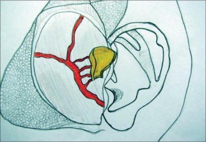
This medical illustration depicts the surgical anatomy and harvesting of a vascularized conchal cartilage flap from the ear, frequently used for temporomandibular joint (TMJ) reconstruction. The diagram shows the external ear with a highlighted yellow segment representing the harvested auricular cartilage. A critical component shown is the superficial temporal artery (STA) and its branching vascular network, rendered in red, which provides the arterial supply to the soft tissue cuff (temporoparietal fascia or TPF) attached to the cartilage. The illustration demonstrates the spatial relationship between the donor site at the cymba conchae/helix root and the arterial pedicle, highlighting the importance of preserving vascularity for the successful creation of an autologous, vascularized interpositional flap. This content is intended for surgical planning and education in plastic surgery and maxillofacial procedures, illustrating the technique of harvesting a composite tissue graft with its associated blood supply to ensure graft viability.
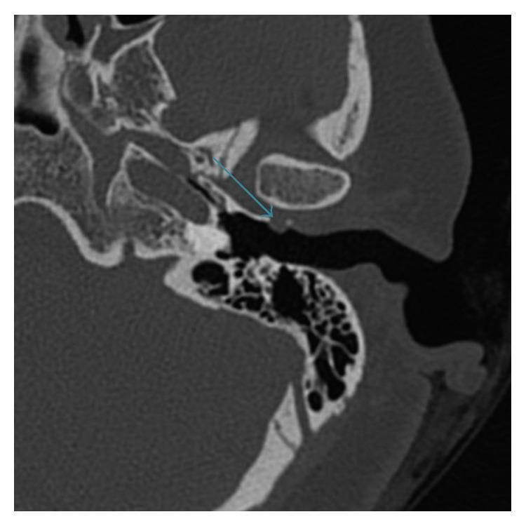
This diagnostic image is an axial non-contrast computed tomography (CT) scan of the left temporal bone, presented in bone window settings. It demonstrates the anatomical relationships between the temporomandibular joint (TMJ), external auditory canal (EAC), and middle ear structures. A blue arrow highlights a small, well-defined, rounded cystic lesion with fluid density arising from the anterior-inferior wall of the external auditory canal. The lesion is located in close proximity to the posterior aspect of the TMJ condyle, consistent with a TMJ ganglion cyst. The surrounding temporal bone anatomy, including the mastoid air cells, ossicular chain, and petrous portion of the temporal bone, appears normal. This image illustrates a rare cause of EAC masses, highlighting the importance of radiological imaging in differentiating TMJ-related pathologies from primary otologic conditions. It is a key educational example for radiology and otolaryngology trainees regarding the differential diagnosis of cystic lesions of the ear canal.
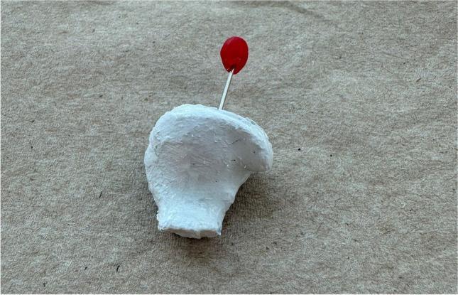
This clinical photograph shows a custom-molded therapeutic earplug designed for the treatment of temporomandibular joint (TMJ) disorders. The specimen is fabricated from a white, opaque, rigid material (Fotoplast) with a granular, unpolished surface texture that reflects the unique anatomy of an individual's external auditory canal. A key feature is the inclusion of a thin silver-colored metal handle protruding from the body of the plug, capped with a bright red spherical marker. This marker serves a dual purpose: facilitating easy insertion and removal by the patient and acting as a lateralization indicator (red for the right ear). The plug is designed to sit flush with the outer edge of the ear canal and features a longitudinal perforation (not directly visible from this angle) to prevent hearing impairment. This device illustrates a near-field therapeutic approach to managing myogenous TMD and bruxism by stabilizing the joint space through the ear canal.
TMJ Arthralgia and Dizziness - Causes and Mechanisms
What Is Costen Syndrome?
- Tinnitus
- Vertigo/dizziness
- Pain in front of the ear, radiating to the temple and face
- Blocked sensation in the ear
- Headache (frontal, parietal, occipital)
Why Does TMJ Disease Cause Dizziness? - Key Mechanisms
Why the Right Side Specifically?
- The right and left TMJs function as one unit, but unilateral TMJ arthralgia causes asymmetric muscle tension, bite deviation toward the affected side, and asymmetric sensory input to the brainstem
- Malocclusion or molar loss on the right side shifts condylar loading right, causing the mandible to deviate to the right on opening
- This asymmetric loading preferentially inflames the right joint and irritates the right auriculotemporal nerve, explaining why symptoms are lateralized
Other Conditions to Rule Out
| Condition | Key Distinguishing Features |
|---|---|
| Benign paroxysmal positional vertigo (BPPV) | Position-triggered, nystagmus on Dix-Hallpike |
| Meniere's disease | Episodic vertigo + unilateral hearing loss + tinnitus |
| Cervicogenic dizziness | Neck pain + restricted movement trigger dizziness |
| Referred otalgia from cervical spine | C2/C3 nerve irritation causes ear + jaw-area pain |
| Internal derangement of TMJ + labyrinthitis | Combined pathology |
| Rheumatoid arthritis / connective tissue disease | Systemic joint involvement, including TMJ |
Recent Evidence
Management Approach
- Dental/maxillofacial assessment - bite adjustment, occlusal splint
- NSAIDs/aspirin for joint inflammation
- Physical therapy - jaw and neck exercises, manual therapy
- Low-dose amitriptyline at bedtime for chronic pain (as noted in Adams & Victor)
- Vestibular rehabilitation if dizziness is prominent
- ENT assessment to rule out concurrent primary ear disease
- MRI of TMJ if effusion or disc displacement suspected
- KJ Lee's Essential Otolaryngology, p. 40 (Costen Syndrome)
- Scott-Brown's Otorhinolaryngology Head & Neck Surgery, Vol 2 (TMJ anatomy)
- Adams and Victor's Principles of Neurology 12th Ed., p. 2928
- Cummings Otolaryngology Head and Neck Surgery (Aural fullness chapter)
what could be the reason with vestibular dizziness ??
vestibular disorders vertigo causes diagnosis
Causes of Vestibular Dizziness (In the Context of Right-Sided Pain + TMJ Arthralgia)
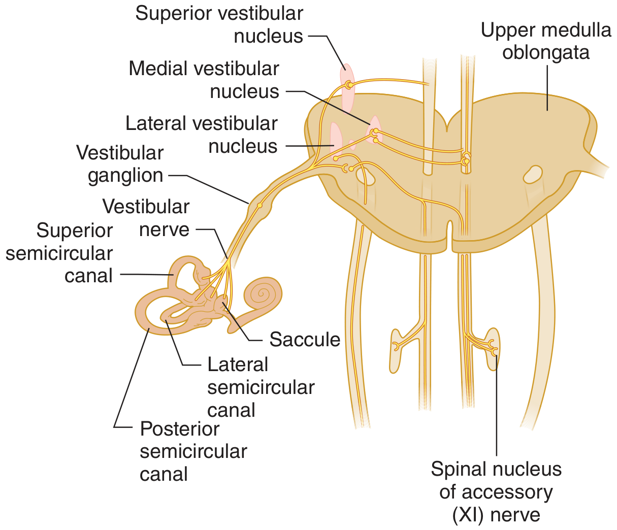
PERIPHERAL VESTIBULAR CAUSES
1. Benign Paroxysmal Positional Vertigo (BPPV) - Most Common
- Characterized by brief episodes (<1 min) of intense rotational vertigo triggered by head position changes (rolling over in bed, lying down, tilting the head back)
- Caused by dislodged otoconia (calcium carbonate crystals) in the semicircular canals - most commonly the posterior canal
- The right labyrinth is more commonly affected (Brandt et al.)
- Diagnosed at bedside with the Dix-Hallpike maneuver - reproduces transient mixed vertical-torsional nystagmus
- No associated hearing loss or tinnitus
- Link to TMJ: Head position changes that provoke BPPV also mechanically stress the TMJ, so both conditions can co-exist and reinforce each other
"Individual episodes last for less than a minute, but these may recur periodically for several days or for many months." - Adams and Victor's Principles of Neurology
2. Vestibular Neuritis - Second Most Common
- Likely viral (herpes simplex reactivation in the vestibular ganglion is the proposed mechanism)
- Prolonged continuous vertigo lasting days, with severe nausea/vomiting, that gradually improves over weeks to months
- No hearing loss, no tinnitus, no ear pain (this distinguishes it from labyrinthitis)
- Abnormal head impulse test toward the affected side (catch-up saccade when head rotated toward weak side)
3. Labyrinthitis
- Similar to vestibular neuritis but also affects the cochlea, causing concurrent:
- Ear pain
- Hearing loss
- Tinnitus
- Vertigo
- Often a complication of acute otitis media or viral infection
- Highly relevant here - the TMJ's proximity to the middle ear means TMJ inflammation can predispose to or co-exist with labyrinthitis
4. Meniere's Disease - Classic Triad
- Endolymphatic hydrops (excess endolymph in the inner ear) is the underlying pathology
- Classic triad:
- Episodic rotational vertigo (minutes to hours)
- Fluctuating unilateral sensorineural hearing loss (low-frequency)
- Tinnitus + aural fullness
- Attacks are abrupt, severe - patient "cannot stand or walk"
- Slow phase of nystagmus toward the affected ear during an attack
- Patients fall toward the affected side and prefer to lie with the bad ear uppermost
- Progressive deafness with recurrent attacks
- Treatment: diuretics, sodium restriction; intratympanic glucocorticoids or gentamicin if refractory
"The main pathologic changes consist of an increase in the volume of endolymph and distention of the endolymphatic system." - Adams and Victor's Principles of Neurology
5. Vestibular Schwannoma (Acoustic Neuroma)
- Slow-growing tumor of the Schwann cells of CN VIII at the cerebellopontine angle
- Causes gradually progressive unilateral sensorineural hearing loss + vestibular hypofunction
- Typically no vertigo - because the slow vestibular deficit is compensated centrally as it develops
- Diagnosis: MRI of the internal auditory canals
- Unilateral unexplained sensorineural hearing loss = MRI mandatory
CENTRAL VESTIBULAR CAUSES (Must Not Be Missed)
6. Cerebellar/Brainstem Stroke
- Most feared cause of vestibular dizziness
- Can mimic vestibular neuritis closely
- Red flags suggesting central cause:
- Nystagmus that changes direction with gaze (direction-changing)
- No corrective saccade on head impulse test (unlike peripheral)
- Vertical nystagmus (downbeat or upbeat) - always central
- Diplopia, dysarthria, dysphagia, limb ataxia
- Vertical skew deviation on cover test
- Use the HINTS exam (Head Impulse, Nystagmus, Test of Skew) to differentiate peripheral from central
7. Vestibular Migraine - Common but Under-diagnosed
- The most common cause of episodic vertigo in young women
- Vertigo can precede, accompany, or occur without headache
- Duration: minutes to hours; some patients have prolonged disequilibrium lasting days
- Associated features: photophobia, phonophobia, visual aura, motion sensitivity, sensitivity to visual motion (movies)
- Treated with migraine prophylactics
"Vestibular migraine is a common yet undiagnosed cause of episodic vertigo. Vertigo sometimes precedes a typical migraine headache but more often occurs without headache." - Harrison's Principles of Internal Medicine 22E
Key Distinguishing Features - Peripheral vs. Central
| Feature | Peripheral | Central |
|---|---|---|
| Nystagmus direction | Unidirectional, horizontal | Direction-changing with gaze |
| Visual fixation effect | Suppresses nystagmus | Does NOT suppress |
| Head impulse test | Abnormal (catch-up saccade) | Normal (no saccade) |
| Hearing loss | Can be present | Absent (unless CN VIII affected) |
| Severity of vertigo | Usually severe | Often milder but persistent |
| Other neuro signs | Absent | Diplopia, dysarthria, ataxia |
| Pure vertical nystagmus | Never | Always central |
How This Relates to the Right-Sided Pain + TMJ Arthralgia
- Right-sided TMJ arthralgia inflames structures adjacent to the right inner ear, potentially triggering or worsening right-sided labyrinthine dysfunction
- The resulting asymmetric vestibular input (right side disrupted) is interpreted by the brain as movement, producing dizziness
- BPPV of the right ear is statistically the most common first diagnosis to consider
- Labyrinthitis must be considered since ear pain + vertigo + hearing symptoms fit, especially with TMJ-related Eustachian tube dysfunction
- Vestibular migraine should be asked about if there is a personal/family history of migraine
- Cerebellar stroke must be actively excluded with the HINTS bedside exam before a benign peripheral diagnosis is accepted
Management Steps
- Bedside HINTS exam - rule out central cause first
- Dix-Hallpike test - diagnose BPPV and treat with Epley maneuver
- Audiogram - low-frequency hearing loss suggests Meniere's; high-frequency loss + tinnitus suggests acoustic neuroma
- MRI internal auditory canals if unilateral SNHL present
- Treat underlying TMJ disorder (reduces labyrinthine irritation)
- Vestibular rehabilitation exercises for residual imbalance
- Adams and Victor's Principles of Neurology 12th Ed. (BPPV, Meniere's)
- Harrison's Principles of Internal Medicine 22E (Vestibular migraine, Meniere's, schwannoma)
- Tintinalli's Emergency Medicine (Vestibular neuritis vs. labyrinthitis vs. stroke)