
Hyperactive bladder
"overactive bladder" AND "treatment"
overactive bladder detrusor overactivity
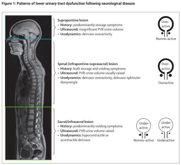
Summary : This figure illustrates the patterns of lower urinary tract dysfunction following neurological disease, categorised by lesion location (suprapontine, spinal, sacral/infrasacral) and their associated clinical, ultrasound, and urodynamic findings, as well as bladder and sphincter activity states.
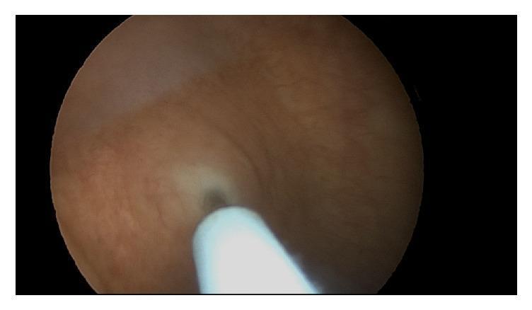
This cystoscopic procedural image shows an intravesical injection into the bladder wall. The visual perspective is from a 30-degree operative cystoscope, focusing on the posterior bladder wall's urothelium. The urothelial surface appears healthy, with a smooth, pinkish-tan texture and uniform coloration, showing no evidence of gross inflammation or lesions. A metallic, reflective injection needle (Laborie injeTAK) is visible in the foreground, angled as it penetrates the urothelium. A small, dark puncture site is evident at the point of needle entry. The image demonstrates the technique for delivering onabotulinumtoxinA injections, typically performed at a depth of approximately 3mm into the detrusor muscle or suburothelial space for the treatment of overactive bladder or neurogenic detrusor overactivity. This serves as an educational reference for urological procedural techniques and intraoperative bladder visualization.
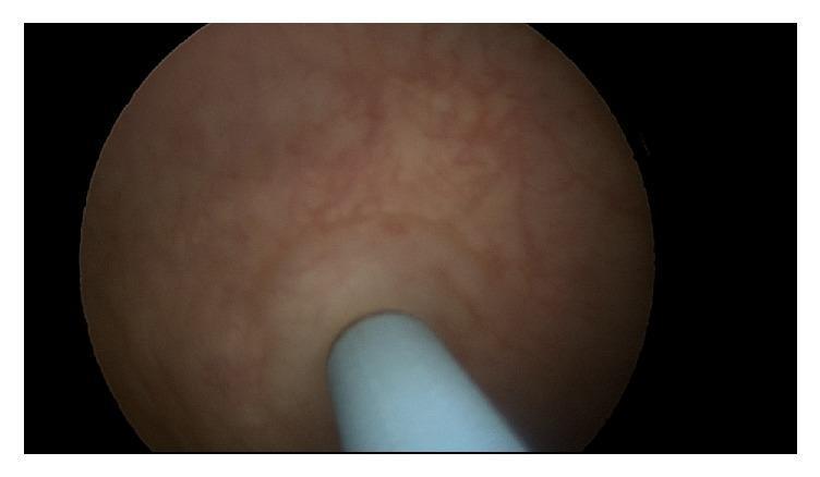
This clinical diagnostic image is a cystoscopic view within a human urinary bladder, capturing an intravesical injection procedure. The image depicts the healthy-appearing uroepithelium, which exhibits a smooth, glossy, pinkish-tan surface with a visible underlying submucosal vascular network. Centrally in the foreground, a rigid, cylindrical, light-blue endoscopic injection needle (such as an injeTAK needle) is shown making contact with the bladder wall. The visual context illustrates the clinical technique for administering intravesical onabotulinumtoxinA (Botox) for conditions such as overactive bladder or neurogenic detrusor overactivity. The procedure involves localized delivery of the toxin at a depth of approximately 3mm into the detrusor muscle or suburothelial space. This image serves as an educational reference for urological endoscopy, demonstrating normal bladder mucosa appearance and the standard positioning of an injection needle during minimally invasive urological therapy.
AUA/SUFU overactive bladder guideline 2024 first-line second-line third-line treatment
Overactive bladder (OAB)
Definition
Epidemiology
- About 40% of older adults have OAB, with or without incontinence. Symptoms are urgency, frequency (voiding every 2 hours or more often) and nocturia. - Harrison's Principles of Internal Medicine 22E, p. 3933
- Prevalence rises with age. In one European study of people over 75, 41.9% of men and 31.3% of women reported at least one OAB symptom. - Campbell Walsh Wein Urology
- Falls from rushing to the toilet are the most important complication. Others are skin irritation, social isolation and depression. - Harrison's, p. 3933
Pathophysiology
- Neurogenic: loss of central inhibition of the micturition reflex, as in stroke, Parkinson disease, MS and spinal cord disease.
- Myogenic: altered detrusor excitability and smooth muscle coupling.
- Urothelial/afferent: increased afferent signalling, with central sensitization contributing in some patients. - Campbell Walsh Wein Urology, Afferent Mechanisms in OAB
Evaluation
- History:
- Ask directly about incontinence, because many older people hide it. A useful question is "Do you ever wear padding to protect yourself?" Many answer "no" to the first two questions but "yes" to this one.
- Look for reversible contributors: UTI, atrophic vaginitis, diuretics, caffeine, constipation, delirium, restricted mobility, hyperglycemia and volume overload. - Harrison's
- Review the medication list, including anticholinergic burden.
- Examination: abdominal, pelvic and rectal examination, neurological screen, and a cough stress test with a comfortably full bladder to detect stress incontinence.
- Tests:
- Urinalysis.
- Post-void residual (PVR).
- Bladder diary.
- Urodynamics only if the diagnosis is unclear or first-line treatment fails.
- In men, consider PSA and assessment for BPH. - Goldman-Cecil Medicine
- Red flags that need further work-up: hematuria, recurrent UTI, pelvic pain, neurological signs, a high PVR, or a pelvic mass.
Management (stepwise)
- Fluid management, and less caffeine, alcohol and bladder irritants.
- Weight loss and treating constipation.
- Bladder training and timed voiding.
- Pelvic floor muscle exercises to suppress urgency.
- Can be combined with drugs.
- Antimuscarinics (oxybutynin, tolterodine, solifenacin, darifenacin, trospium, fesoterodine). They relax the detrusor, which raises bladder capacity and reduces urgency, frequency and incontinence. - Lippincott Illustrated Reviews: Pharmacology
- The most common adverse effects are dry mouth and constipation.
- Use caution in older patients because of the risk of cognitive effects. - Hinman's Atlas of Urologic Surgery
- Avoid them in narrow-angle glaucoma and in urinary retention or a high PVR.
- β3-agonists (mirabegron, vibegron). These are comparably effective with low rates of dry mouth, constipation and retention, so they are better tolerated. - Hinman's Atlas of Urologic Surgery
- Mirabegron can raise blood pressure and is a CYP2D6 inhibitor. Check blood pressure and interacting drugs.
- Vibegron avoids the CYP2D6 interaction.
- Intradetrusor onabotulinumtoxinA injection. The main risks are urinary retention and UTI, so patients must be willing to do clean intermittent catheterization if needed.
- Neuromodulation: sacral (SNM) or percutaneous tibial nerve stimulation (PTNS). - Hinman's Atlas of Urologic Surgery
- Rarely, augmentation cystoplasty or urinary diversion for severe, refractory cases.
Points for practice
- Exclude infection, hematuria, retention and neurological causes before labeling a patient as having idiopathic OAB.
- In men with BPH, treat the outlet obstruction first, usually with an α-blocker. Add an antimuscarinic or β3-agonist for persistent storage symptoms, and check PVR first. - Goldman-Cecil Medicine
- In frail older adults, prefer a β3-agonist or a low-anticholinergic option, because strong anticholinergics are linked to cognitive impairment.
- Persistence is low with oral drugs, so review at 4 to 8 weeks and switch agents if the response is inadequate or side effects limit use.
Recent evidence check
- A 2025 systematic review of mirabegron against placebo and other treatments (PMID 39343691).
- A 2025 meta-analysis of refractory OAB treatments including botulinum toxin and electrical stimulation (PMID 41150180).
- A meta-analysis of trospium (PMID 42687548).
Urinary retention
"urinary retention"[MeSH Terms] AND "catheterization"
urinary retention bladder distension ultrasound or catheterization
Urinary retention
Definition and types
- Acute: a painful inability to void, with pain relieved by catheterization.
- Chronic: painless, with a raised residual volume after voiding. - Bailey and Love's Short Practice of Surgery, 28th ed.
Epidemiology
- Acute retention is mostly seen in elderly men with BPH. About 1 in 10 men in their 70s and 1 in 3 men in their 80s have an episode.
- Recurrence within 6 months is about 20% in BPH-related retention.
- It is uncommon in women, who account for roughly 3 per 100,000 cases a year. The usual causes in women are bladder masses, gynecologic surgery and pelvic prolapse. - Tintinalli's Emergency Medicine, Ch. 92
Pathophysiology
Causes
| Domain | Examples |
|---|---|
| Obstructive | BPH, prostate or bladder cancer, urethral stricture, bladder stones, blood clots, prolapse, pelvic mass, fecal impaction |
| Neurogenic | Diabetic neuropathy, MS, Parkinson disease, spinal cord compression, cauda equina syndrome |
| Pharmacologic | Opiates, anticholinergics, α-agonists (decongestants), TCAs, antihistamines, sedatives |
| Infectious/inflammatory | Prostatitis, urethritis, genital herpes (sacral involvement) |
| Postoperative | Spinal or epidural anesthesia, pelvic, anorectal or hernia surgery, surgery longer than 2 hours, excess IV fluids |
| Other | Psychogenic, trauma |
- The incidence is 5 to 70%.
- Risk factors are age over 50, male sex, BPH, neurological disease, neuraxial anesthesia and anticholinergic, α/β-blocker or sedative drugs.
- Catheterize prophylactically if surgery is expected to last 3 hours or more, or if large fluid volumes are given. - Bailey and Love's Short Practice of Surgery
Clinical features
- Acute: inability to void, suprapubic pain and distension, urgency, and agitation in the elderly. Consider it in anyone with lower abdominal pain, even without urinary complaints.
- Chronic: often painless. It may present with overflow incontinence, recurrent UTI, a palpable suprapubic mass, or new hydronephrosis or renal impairment. - Campbell Walsh Wein Urology
Evaluation
- History: prostatism, prior urethral instrumentation or catheters, cancer, stones, medications, neurological symptoms (back pain, saddle numbness, bowel or sexual dysfunction), recent surgery or anesthesia.
- Examination: abdominal (palpable or percussible bladder), genitalia and meatus, digital rectal exam (prostate size, rectal tone, fecal loading), pelvic exam in women, and a neurological exam including perineal sensation.
- Bladder ultrasound confirms retention and quantifies volume. A post-void residual (PVR) above about 125 mL suggests outlet obstruction. - Tintinalli's Emergency Medicine
- Labs: urinalysis and culture, creatinine and electrolytes, and PSA in suspected BPH (interpret with caution, as it rises with catheterization and retention).
- Upper tract imaging (renal ultrasound) if renal impairment or chronic retention is suspected.
- Further tests: cystoscopy for hematuria, stricture or mass, and urodynamics for suspected neurogenic or detrusor failure. MRI of the spine urgently if cauda equina or cord compression is suspected.
Management
- Decompress with a urethral catheter as quickly as possible. Use a suprapubic catheter if urethral passage fails.
- Difficult catheterization:
- In BPH, use a coude catheter, keeping the curved tip at the 12 o'clock position. A common mistake is to use a smaller catheter. A larger 18 to 20F catheter is stiffer and is more likely to pass into the bladder than curl in the prostatic urethra.
- If resistance is met close to the meatus, suspect a urethral stricture. A smaller catheter (12 to 14F) may then help. - Schwartz's Principles of Surgery
- If these fail, call urology for a suprapubic catheter or cystoscopic or filiform-guided placement.
- Monitor for post-obstructive diuresis, a physiologic response to retained salt and water. It occurs in about 0.5 to 50% of cases, depending on duration and comorbidity. Watch for output above 200 mL/h.
- Use a fluid chart, daily weights, and serial creatinine and electrolytes.
- Give IV replacement only if the patient cannot keep up with losses. - Tintinalli's; Bailey and Love's
- Absence of diuresis in a patient with a significantly raised creatinine predicts poor renal recovery. - Tintinalli's
- Expect hematuria after decompression. It usually settles within a couple of days. The speed of bladder drainage has not been shown to affect it. - Bailey and Love's; Campbell Walsh Wein Urology
- Treat the cause:
- Stop or reduce offending drugs.
- Treat constipation.
- Manage infection.
- Treat the spinal cause if neurogenic.
- Start an α-blocker (tamsulosin, alfuzosin) and leave the catheter in for a trial without catheter (TWOC) after a few days. A 5α-reductase inhibitor may be added for larger prostates. These steps raise the chance of voiding successfully.
- Recurrence is common, so refer to urology. Definitive surgery such as TURP is considered if the TWOC fails.
- The best catheter duration is not well established. - Pfenninger and Fowler's Procedures for Primary Care
- Men with a low residual volume, no infection and good renal function may not need pre-operative catheterization before prostatectomy.
- Uremic patients need urgent catheterization so that renal function can recover. They are often dehydrated and anemic. - Bailey and Love's
- Long-term options are clean intermittent self-catheterization, which is generally preferred, or an indwelling urethral or suprapubic catheter, or surgery for the underlying obstruction.
- After orthotopic neobladder surgery, retention is best managed by intermittent self-catheterization. Drugs do not help. - Campbell Walsh Wein Urology
Red flags
- Retention with back pain, saddle anesthesia, lower limb weakness or bowel dysfunction needs emergency MRI for cauda equina or cord compression.
- Retention with renal failure or hydronephrosis needs urgent decompression and monitoring.
- Retention with fever or sepsis needs cultures and antibiotics, and care with instrumentation.
Recent evidence check
- A 2025 systematic review on bladder ultrasound to reduce unnecessary catheterization in suspected acute retention (PMID 40032047).