
process of spermatogenesis
diagram of spermatogenesis stages seminiferous tubule
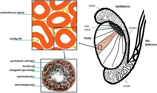
This medical illustration depicts the gross anatomy and histology of the human male reproductive system, specifically the testis and its associated ducts. The main diagram shows a sagittal-like section of the testis, highlighting internal lobules containing seminiferous tubules that converge at the rete testis. The epididymis is shown as a coiled ductal system attached to the posterior testis, divided into the caput (head), corpus (body), and cauda (tail), which then continues as the vas deferens. A magnified histological inset focuses on the interstitial space, showing Leydig cells clustered between cross-sections of seminiferous tubules. A second, high-magnification cross-section of a single seminiferous tubule illustrates the stages of spermatogenesis. Labeled structures include the peritubular cell layer (basal lamina), Sertoli cells for structural support, and germ cells in various stages of maturation: spermatogonia at the periphery, followed by spermatocytes I, and elongated spermatids positioned near the central lumen. This diagram serves as an educational tool for understanding the relationship between macroscopic anatomy and microscopic gametogenesis.

A multi-panel medical infographic illustrating the sequential stages of male reproductive function and the pharmacological impact of small molecule medications. The diagram is divided into four annotated sections linked by arrows: (A) Testicular weight, featuring a schematic of the male reproductive tract including the testes and vas deferens; (B) Testicular microenvironment, showing a cross-sectional view of a seminiferous tubule with various germ cell layers and a central lumen containing spermatozoa; (C) Spermatogenesis, a branching lineage diagram depicting the differentiation from spermatogonia through meiosis to mature spermatozoa; and (D) Sperm capacitation & Fertilization, illustrating sperm cell activation and interaction with an oocyte (egg). Each section includes an icon of a medication bottle and pills, symbolizing the points at which clinical drugs (e.g., SSRIs, antihypertensives, analgesics) can exert positive or negative effects. Panel D specifically includes a red upward arrow, signifying enhanced sperm motility or activity. This educational graphic serves as a summary of drug-induced changes in male fertility, from organ morphology to gamete function.
Spermatogenesis
Structural setting: Sertoli cells and the blood-testis barrier
- Basal compartment - contains diploid spermatogonia
- Luminal (adluminal) compartment - contains haploid spermatocytes and spermatids, immunologically shielded from the bloodstream
The three phases
- Each primary spermatocyte (46 chromosomes, diploid) undergoes meiosis I, a division lasting up to ~22 days, producing two secondary spermatocytes.
- Each secondary spermatocyte then quickly (within hours) undergoes meiosis II, producing spermatids with a haploid, reduced chromosome number (23 chromosomes, 1n, 1d).
- Guyton & Hall notes this is where the diploid chromosome complement is halved, with 23 chromosomes going to each spermatid - Guyton and Hall Textbook of Medical Physiology, p. 998-999.
- Condensation and elongation of the nucleus
- Formation of the acrosomal cap (a lysosome-like organelle containing enzymes for ovum penetration)
- Development of the flagellum (tail), with mitochondria helically wrapping its proximal segment
- Loss of most cytoplasm
Hormonal regulation
- Hypothalamic GnRH stimulates the anterior pituitary to release LH and FSH.
- LH acts on Leydig cells to produce testosterone.
- FSH and locally high testosterone act on Sertoli cells (which bear the relevant receptors), making Sertoli cells the primary regulators of the process.
- Intratesticular testosterone concentration can reach roughly 200 times circulating levels - necessary for spermatogenic cell proliferation and differentiation.
- Sertoli cells also secrete androgen-binding protein (ABP) to maintain high local androgen levels, and inhibin, which provides negative feedback on pituitary FSH secretion - Histology: A Text and Atlas, p. 2086-2087.
Clinical relevance
functions of testesterone
Functions of Testosterone
1. Fetal development
- From about the 7th week of gestation, the SRY gene triggers testicular differentiation and testosterone secretion.
- Drives development of the penis, scrotum, prostate, seminal vesicles, and male genital ducts, while suppressing female genital organ formation.
- Causes descent of the testes into the scrotum during the last 2-3 months of gestation.
2. Puberty - primary and secondary sexual characteristics
- Enlarges the penis, scrotum, and testes (roughly 8-fold by age 20).
- Body hair: growth over pubis, abdomen (linea alba), face, chest.
- Male pattern baldness: reduces scalp hair growth in genetically predisposed men.
- Voice: causes laryngeal hypertrophy and enlargement, producing the deeper adult male voice.
- Skin: thickens skin and increases sebaceous gland secretion (can contribute to acne).
- Larynx, pelvis: narrows and lengthens the pelvic outlet into a funnel shape and strengthens it for load-bearing (versus the broad female pelvis).
3. Protein and musculoskeletal effects
- Protein anabolism: increases muscle mass (~50% greater than in females) and protein deposition generally.
- Bone: increases bone matrix and calcium retention, thickens bones; also promotes epiphyseal closure, which ultimately limits final height despite an earlier growth spurt.
4. Metabolic effects
- Increases basal metabolic rate by roughly 5-10% (up to 15% with high doses), likely via its protein-anabolic action.
- Increases red blood cell count (~15-20%, accounting for men having ~700,000 more RBCs/mm³ than women on average) - this appears to be an indirect effect (not via direct erythropoietin stimulation), possibly related to increased metabolic rate.
- Mild effect on electrolyte and water balance: increases renal sodium reabsorption, contributing to a 5-10% increase in blood and extracellular fluid volume after puberty (much weaker effect than mineralocorticoids).
5. Reproductive/gametogenic function
- Maintains spermatogenesis via high intratesticular concentrations (up to ~200x circulating levels), acting through Sertoli cells.
- Sustains libido and other CNS-mediated sexual behaviors.
Mechanism of action
Regulation and clinical notes
- Secretion is controlled by the hypothalamic-pituitary-gonadal axis: GnRH -> LH -> Leydig cell testosterone production, with negative feedback from testosterone/inhibin.
- Levels decline modestly with age; only about 10-20% of men over 60 and 50% of men over 80 have testosterone below the "low" threshold (<300 ng/dL). Obesity and type 2 diabetes accelerate age-related decline.
- Testosterone replacement in elderly men has been associated with possible increased cardiovascular risk in some studies, so use requires caution.
oogenesis steps and hormones involved
oogenesis stages diagram oogonia primary oocyte secondary oocyte polar body
ovarian follicle development primordial follicle to corpus luteum diagram
Oogenesis: Steps and Hormonal Regulation
Prenatal phase (before birth)
- Migration and mitosis: Primordial germ cells migrate from the yolk sac to the developing ovary and proliferate by mitosis, becoming oogonia. By gestational weeks 20-24, there are roughly 6-7 million oogonia.
- Entry into meiosis I: Starting around gestational weeks 8-9 (continuing through mid-fetal life), oogonia enter meiotic prophase I and become primary oocytes. Each primary oocyte becomes surrounded by a single layer of granulosa cells, forming a primordial follicle.
- Meiotic arrest: Meiosis I arrests at the diplotene/dictyate stage of prophase I. This arrest persists for years to decades - not resuming until just before ovulation, potentially 13-50 years later.
- Attrition: Oocyte numbers fall dramatically through programmed atresia - about 2 million remain at birth, and only around 300,000-400,000 remain by puberty.
Postnatal/reproductive phase (puberty onward)
-
Follicular development (in three stages, paralleling the menstrual cycle):
- Stage 1 (years-long): primordial follicle -> primary follicle; granulosa cells proliferate, theca interna forms.
- Stage 2 (~70-85 days): antral fluid (containing steroids, FSH) accumulates; follicle becomes a graafian follicle (2-5 mm).
- Stage 3 (5-7 days, the final follicular phase of a cycle): one dominant follicle is selected, grows rapidly to ~20 mm, while others become atretic.
-
Completion of meiosis I: Just before ovulation (day 14 of a 28-day cycle), the primary oocyte completes its first meiotic division, producing a haploid secondary oocyte (23 duplicated chromosomes) and a small, non-functional first polar body.
-
Ovulation: The dominant (graafian) follicle ruptures, releasing the secondary oocyte into the fallopian tube.
-
Meiosis II - arrested again: The secondary oocyte begins meiosis II but arrests at metaphase II, and stays arrested unless fertilization occurs.
-
Fertilization completes meiosis: If a sperm fertilizes the oocyte, meiosis II completes, producing the mature haploid ovum (23 chromosomes) plus a second polar body, which degenerates. If fertilization does not occur, the oocyte degenerates unfertilized.
-
Corpus luteum formation: The ruptured follicle's remnants (granulosa + theca cells, capillaries, fibroblasts) form the corpus luteum, which secretes progesterone and estrogen to support a possible pregnancy; it regresses (~14 days) if fertilization doesn't occur, or is sustained by hCG if it does.
Hormones involved
| Hormone | Source | Role in oogenesis |
|---|---|---|
| GnRH | Hypothalamus | Pulsatile release drives pituitary FSH/LH secretion |
| FSH | Anterior pituitary | Stimulates granulosa cell proliferation, follicular growth, antral fluid/estrogen production; recruits the cohort of follicles each cycle |
| LH | Anterior pituitary | Mid-cycle LH surge triggers resumption of meiosis I, ovulation, and luteinization (corpus luteum formation); stimulates theca cells to produce androgen precursors for estrogen synthesis |
| Estrogen (estradiol) | Granulosa cells (from theca-derived androgens, via aromatase) | Promotes follicular growth, triggers the LH surge via positive feedback at high sustained levels, thickens endometrium |
| Progesterone | Corpus luteum (theca and granulosa-derived cells) | Maintains endometrium for implantation; provides negative feedback on GnRH/LH-FSH after ovulation |
| Inhibin | Granulosa cells | Selectively suppresses FSH release, helping ensure only one dominant follicle matures |
Follicular development steps and hormones involved
Follicular Development: Steps and Hormones
Steps of follicular development
- Converts granulosa/theca cells toward progesterone secretion (estrogen falls, progesterone rises just before ovulation)
- Triggers release of proteolytic enzymes that digest the follicular capsule
- Stimulates new blood vessel growth and local prostaglandin release, causing capsule rupture at the stigma
- Results in ovulation: release of the oocyte (with its surrounding corona radiata cells) into the peritoneal cavity/fallopian tube, on approximately day 14 of a 28-day cycle. Meiosis I completes just before this release.
Hormones involved
| Hormone | Source | Role |
|---|---|---|
| GnRH | Hypothalamus | Pulsatile stimulation of pituitary gonadotropes |
| FSH | Anterior pituitary | Recruits and drives growth of the follicle cohort; stimulates granulosa cell proliferation, antrum formation, and aromatase activity (estrogen synthesis) |
| LH | Anterior pituitary | Stimulates theca cell androgen production (substrate for estrogen); the LH surge triggers final follicular maturation, capsule rupture, ovulation, and luteinization |
| Estrogen (estradiol) | Granulosa cells (from theca-derived androgens) | Drives follicular growth; low levels give negative feedback on FSH (causing atresia of non-dominant follicles); high sustained levels from the dominant follicle switch to positive feedback, triggering the LH surge |
| Progesterone | Granulosa/theca cells (rising just before ovulation), then corpus luteum | Supports final follicular maturation and, after ovulation, maintains the endometrium; provides negative feedback on GnRH/LH-FSH during the luteal phase |
| Inhibin | Granulosa cells | Selectively suppresses FSH, reinforcing single-follicle dominance |
| Prostaglandins | Follicular tissue (LH-induced) | Local vasodilation and enzymatic weakening of the follicular wall, enabling rupture |
Create a comparison table: male vs female gametogenesis4th edition
Male vs Female Gametogenesis: Comparison Table
| Feature | Spermatogenesis (Male) | Oogenesis (Female) |
|---|---|---|
| Site | Seminiferous tubules of the testis | Ovarian follicles (cortex of the ovary) |
| Onset | Begins at puberty (~13 years) | Begins in fetal life (weeks 8-9 of gestation) |
| Continuity | Continuous from puberty until old age (slows with aging) | Discontinuous - arrested for years/decades; a fixed, non-renewable pool of oocytes is established before birth and only depletes |
| Stem cell pool | Spermatogonia are continuously replenished by mitosis throughout life | Oogonia proliferate only during fetal life; no new oogonia form after birth |
| Duration of full process | ~74 days per cycle | Variable - years to decades (from primordial follicle arrest to ovulation); final maturation ~85-90 days actively |
| Meiotic arrest | No prolonged arrest; meiosis proceeds relatively continuously once initiated | Two arrest points: (1) prophase I (dictyate stage) - from fetal life until just before ovulation; (2) metaphase II - from ovulation until fertilization |
| Number of gametes produced per parent cell | 4 functional spermatozoa per primary spermatocyte | 1 functional ovum per primary oocyte (plus 2-3 degenerating polar bodies) |
| Cell division symmetry | Equal cytoplasmic division at each meiotic step | Unequal division - oocyte retains most cytoplasm/nutrients; polar bodies get minimal cytoplasm and degenerate |
| Supporting/nurse cells | Sertoli cells (form blood-testis barrier, secrete ABP, inhibin, MIS) | Granulosa cells and theca cells (secrete estrogen, progesterone, inhibin) |
| Key intermediate stages | Spermatogonium -> primary spermatocyte -> secondary spermatocyte -> spermatid -> spermatozoon (spermiogenesis) | Oogonium -> primary oocyte -> secondary oocyte + 1st polar body -> ovum + 2nd polar body (only completes with fertilization) |
| Follicular/tubular development | No follicle-equivalent structure; germ cells mature within a stable tubular architecture | Follicle itself matures alongside the oocyte: primordial -> primary -> secondary (antral) -> graafian (dominant) -> ovulation -> corpus luteum |
| Release mechanism | Spermiation - continuous release into tubule lumen | Ovulation - cyclical, one dominant follicle ruptures per cycle (~monthly) |
| Endocrine control (HPG axis) | GnRH -> LH stimulates Leydig cells (testosterone); FSH acts on Sertoli cells | GnRH -> LH and FSH act cyclically on granulosa/theca cells |
| Principal steroid hormone | Testosterone (from Leydig cells) | Estrogen (granulosa cells, via aromatization of theca-derived androgens) and progesterone (corpus luteum) |
| Key feedback hormone | Inhibin (Sertoli cells) suppresses FSH | Inhibin (granulosa cells) suppresses FSH; estrogen gives negative feedback at low levels but triggers the LH surge (positive feedback) at high sustained levels |
| Trigger for final maturation/release | Sustained high intratesticular testosterone; no single hormonal "surge" event | Preovulatory LH surge triggers resumption of meiosis I and ovulation |
| Post-release remnant structure | None (tubule continues producing new cells) | Corpus luteum forms from ruptured follicle; secretes progesterone, regresses to corpus albicans if no fertilization |
| Chromosome/DNA content of final gamete | Haploid, 23 chromosomes (1n, 1d) | Haploid, 23 chromosomes (1n, 1d) |
| Effect of aging | Gradual decline in sperm quality/quantity; testosterone dips modestly | Sharp, quantifiable decline in oocyte number and quality, ending at menopause |
| Selected textbook sources | Guyton and Hall Textbook of Medical Physiology, p. 998-1008; Histology: A Text and Atlas, p. 2163 | Guyton and Hall Textbook of Medical Physiology, p. 1013-1017; Costanzo Physiology 7th ed., p. 475-476 |
Key conceptual differences to remember
- Continuous vs. cyclical: Spermatogenesis is an ongoing assembly line; oogenesis is cyclical and tied to the ~28-day ovarian/menstrual cycle.
- Quantity vs. quality/scarcity: Males produce millions of sperm daily from a renewable stem cell pool; females are born with a finite, depleting oocyte reserve and typically ovulate only one mature oocyte per cycle.
- Symmetric vs. asymmetric division: Meiosis in the male yields four equivalent gametes; meiosis in the female is deliberately asymmetric to concentrate cytoplasm/organelles into a single large, viable ovum.
- Timing of meiotic completion: Sperm complete meiosis entirely within the testis before release. Oocytes only complete meiosis II after fertilization - meaning the "final" female gamete (a true haploid ovum) technically only exists transiently at the moment of fertilization.
follicular development steps and hormones involved using 14th edition guyton and hall textbook
ISBN 9780443111013 Guyton and Hall Textbook of Medical Physiology edition
Follicular Development: Steps and Hormones (Guyton and Hall Textbook of Medical Physiology, 15th ed., Ch. 82, "Female Physiology Before Pregnancy and Female Hormones")
Steps
- A preovulatory LH surge (6- to 10-fold rise, peaking ~16 hours before ovulation) is essential; FSH also rises 2- to 3-fold.
- LH converts granulosa/theca cells to progesterone-secreting cells, so estrogen falls and progesterone rises just before ovulation.
- Proteolytic enzymes digest the follicular capsule; new blood vessels and prostaglandins promote swelling and rupture.
Hormones involved
| Hormone | Source | Role |
|---|---|---|
| GnRH | Hypothalamus | Stimulates pituitary FSH/LH release |
| FSH | Anterior pituitary | Drives granulosa cell proliferation and antral growth; recruits follicle cohort |
| LH | Anterior pituitary | Supports theca androgen production; LH surge triggers final maturation, ovulation, and luteinization |
| Estrogen | Granulosa cells | Drives follicular growth; suppresses FSH in non-dominant follicles (negative feedback), then triggers the LH surge at high sustained levels (positive feedback) |
| Progesterone | Theca/granulosa cells pre-ovulation, then corpus luteum | Rises just before ovulation; maintains endometrium post-ovulation |
| Prostaglandins | Follicular tissue | Local vasodilation aiding capsule rupture |