
Tumors of skin for mbbs exam point of view and easy to learn
basal cell carcinoma squamous cell carcinoma melanoma skin tumor histology
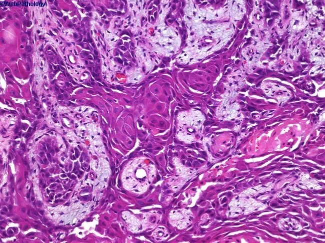
High-quality hematoxylin and eosin stained histology slide of cutaneous tissue shows invasive squamous cell carcinoma. The epidermis exhibits atypical squamous cells with pleomorphic, hyperchromatic nuclei and abundant eosinophilic cytoplasm. In several foci, nests and cords of neoplastic squamous cells invade the underlying dermis, producing a desmoplastic stromal response. Keratinization is evident, with formation of squamous keratin pearls and foci of intracellular keratinization within aggregates of tumor cells. Intercellular bridges are conspicuous at higher magnification. Mitoses are present, including atypical figures, supporting malignant proliferation. The surrounding stroma contains chronic inflammatory infiltrate. Some areas reveal perineural or lymphovascular invasion are suspected but not definitively demonstrable at this magnification. The lesion most consistent with cutaneous squamous cell carcinoma (epidermoid carcinoma) in sun-damaged skin; differential considerations include keratoacanthoma, desmoplastic melanoma, or basal cell carcinoma with squamous differentiation, but the conspicuous keratin production and invasive cords favor SCC. Clinically, this entity is associated with sun exposure, metastasis risk to regional lymph nodes, and requires surgical excision with adequate margins; prognosis depends on depth of invasion and perineural involvement. This image is relevant for dermatopathology, surgical oncology, and educational training in histologic grading of skin cancers.
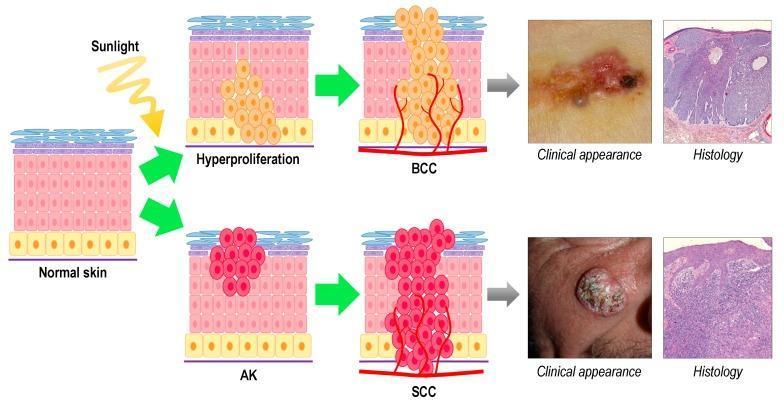
This medical infographic illustrates the progression, clinical presentation, and histology of Non-Melanoma Skin Cancer (NMSC), specifically Basal Cell Carcinoma (BCC) and Squamous Cell Carcinoma (SCC). The diagram starts with 'Normal skin' exposed to 'Sunlight' (UV radiation). The upper pathway shows BCC development originating from interfollicular basal cells, progressing through 'Hyperproliferation' to an invasive 'BCC' mass with prominent angiogenesis. The corresponding clinical photograph displays a pearly, erythematous lesion with ulceration and pigmented areas, while the H&E histology shows characteristic basaloid nests with peripheral palisading and retraction artifacts. The lower pathway depicts SCC development, starting from keratinocytes in the spinous layer, progressing through 'AK' (Actinic Keratosis) to invasive 'SCC'. The SCC clinical photograph shows a hyperkeratotic, crusty, and elevated nodule. The SCC histology reveals disordered epithelial growth, dermal invasion, and keratin pearls. This comparative chart serves as a diagnostic and educational tool for distinguishing these common cutaneous malignancies based on cellular origin, visual morphology, and tissue architecture.
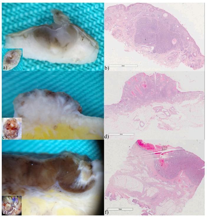
A comparison chart featuring ex vivo dermoscopy (a, c, e) and corresponding histopathology (b, d, f) of common skin malignancies. (a) Vertical ex vivo dermoscopy of an adenoid-cystic basal cell carcinoma (BCC) showing translucent tumor tissue with internal pigmentation invading the dermis. (b) Histology (H&E stain, x15) reveals classic reticulated pseudoglandular basaloid cell clusters and cystic formations. (c) Vertical dermoscopy of invasive squamous cell carcinoma (SCC) illustrating a dense, whitish, elevated mass with irregular borders and epidermal thickening. (d) Histology (H&E, x7) confirms a well-differentiated carcinoma penetrating the reticular dermis with disordered cellular architecture. (e) Vertical dermoscopy of superficial spreading melanoma (Breslow 6mm) depicting a linear, deeply pigmented, asymmetric brown-to-black mass extending into the hypodermis. (f) Histology (H&E, x7) shows asymmetric atypical melanocyte proliferation, ulceration, and focal vascular invasion. This panel demonstrates the correlation between vertical dermoscopic findings—such as tumor color, depth, and invasiveness—and gold-standard histopathological diagnosis in dermatopathology.
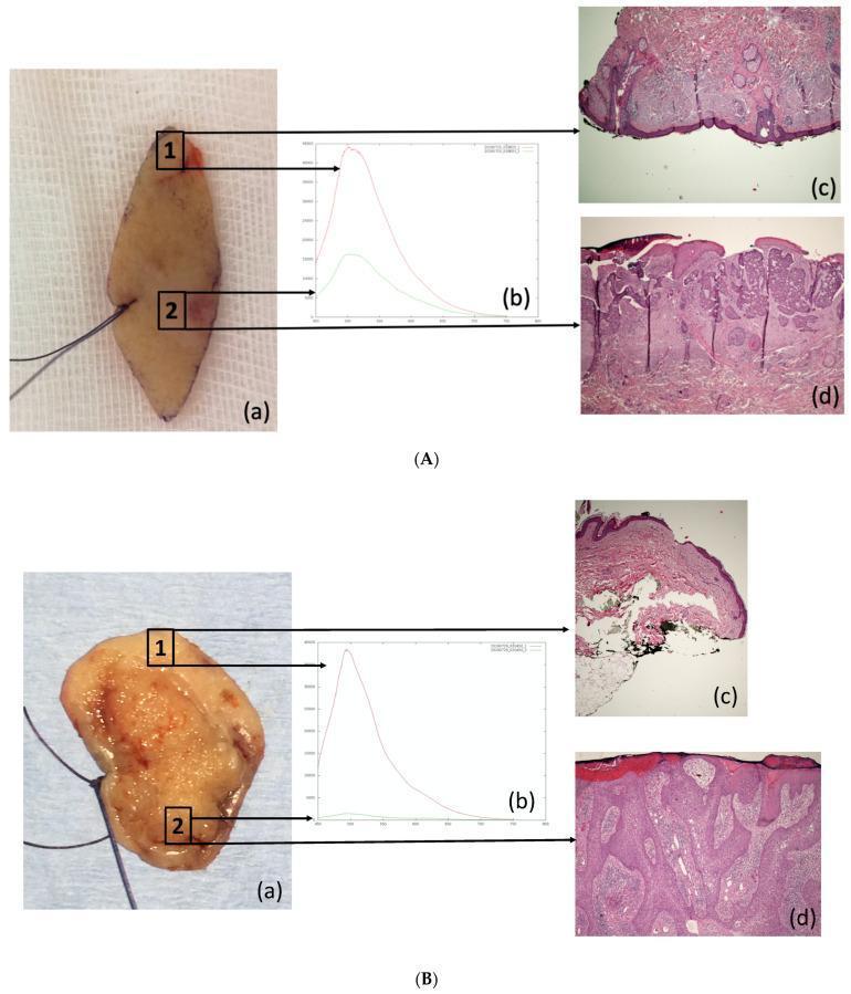
This composite educational image demonstrates the clinicopathological correlation of Autofluorescence (AF) alterations in Non-Melanoma Skin Cancer (NMSC). The image is divided into two primary panels: (A) Basal Cell Carcinoma (BCC) and (B) Squamous Cell Carcinoma (SCC). Both panels follow a consistent structure: (a) Clinical photographs of excised skin specimens with marked control points (1, healthy skin) and lesional points (2, tumor); (b) AF emission spectra showing significantly higher fluorescence intensity in healthy tissue (red line) compared to the tumor (green line), typically peaking at 500 nm; (c) Histopathological sections (H&E stain, 4x magnification) of the healthy skin at point 1, showing normal epidermal and dermal architecture; and (d) Histopathological sections of the malignant lesions at point 2. Panel A(d) illustrates infiltrating BCC with irregular nests of basaloid cells. Panel B(d) displays poorly differentiated SCC characterized by disorganized epithelial proliferation and cellular atypia. This visual resource illustrates how optical biopsy techniques like AF spectroscopy correlate with gold-standard histopathology for diagnosing skin malignancies.
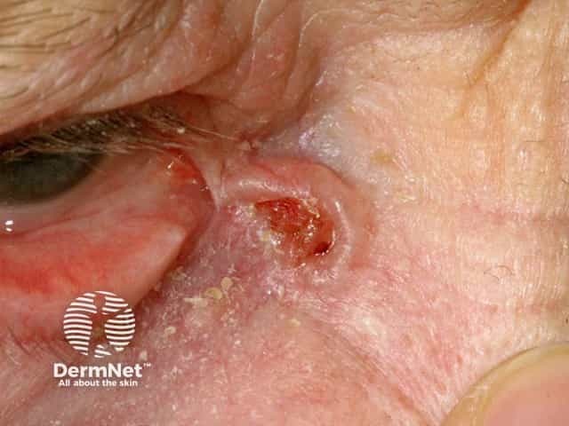
Clinical photography of an eyelid lesion consistent with basal cell carcinoma (BCC). Periocular skin of the eyelid margin shows a solitary, irregularly shaped, ulcerated papule with crusting and a translucent, pearly-like border. Telangiectatic vessels are visible within the raised lesion and surrounding erythema is present, indicating local invasion risk. The lesion is positioned at the anterior eyelid/lid margin with potential medial canthal involvement; the target tissue includes squamous-epidermal junction and periocular dermis. The image highlights classic BCC features, including indurated base, rolled borders, and a friable surface that bleeds on contact. Notably, eyelid BCC tends toward slow growth with a high cure rate when excised with adequate margins, while sparing adjacent ocular structures is essential to preserve function. Differential considerations include sebaceous carcinoma, squamous cell carcinoma, pigmented melanoma, and chalazion-like inflammatory lesions. Diagnostic significance lies in recognizing peril signs requiring biopsy for histopathology—basaloid tumor nests with peripheral palisading and stromal retraction on histology confirm diagnosis. This image is valuable for clinical education, dermatology and ophthalmology training, and referral triage, aiding early recognition, patient counseling, and surgical planning for margin-controlled excision or Mohs micrographic surgery. It also supports radiologic or histopathologic correlation when assessing periocular tumor extent. Clinical documentation and research.
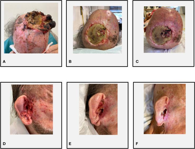
This set of clinical photographs illustrates the therapeutic progression of two distinct types of non-melanoma skin cancer (NMSC) in a single patient. Figures A-C depict a timeline of a bulky, recurrent cutaneous squamous cell carcinoma (cSCC) on the cranial vertex. Image A shows a large, exophytic, and friable mass with significant necrosis and ulceration. Figures B and C demonstrate a partial response and subsequent stable disease under combined cemiplimab and sonidegib therapy, characterized by marked flattening of the lesion, re-epithelialization of the margins, and reduction in hemorrhage. Figures D-F show an infiltrative basal cell carcinoma (BCC) extending into the right external auditory meatus. Image D displays an infiltrative, ulcerated lesion involving the auricular canal. Images E and F show the progression from a partial to a complete response, with Figure F demonstrating total resolution of the tumor and restoration of the normal anatomical contours of the ear canal. The series serves as an educational example of the efficacy of immune checkpoint inhibitors combined with Hedgehog pathway inhibitors in treating synchronous, advanced keratinocyte carcinomas.
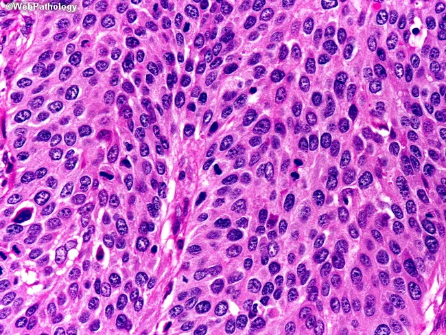
Comprehensive Description: This high-magnification light microscopic image depicts invasive cutaneous squamous cell carcinoma, moderately differentiated, derived from a skin biopsy stained with Hematoxylin and Eosin. Total magnification is approximately 400x (40x objective with 10x ocular). Tumor tissue forms irregular nests and interconnecting cords of polygonal keratinocytes infiltrating the dermal stroma, with invasive fronts delineated by jagged borders. The malignant cells exhibit pleomorphism, enlarged hyperchromatic nuclei, conspicuous nucleoli, and abundant eosinophilic cytoplasm. Increased mitotic activity is evident, and there is evidence of squamous differentiation with intercellular bridges; focal keratinization may yield keratin pearls in some areas. The tumor front is infiltrative, often accompanied by desmoplastic stromal reaction and a modest inflammatory infiltrate at the periphery. Epidermal involvement with extension toward adnexal structures can be observed, reflecting true invasion rather than in situ change. Overall, the histologic pattern supports a diagnosis of invasive cutaneous SCC with moderate differentiation, suggesting intermediate keratinization and nuclear atypia and a propensity for local invasion. Clinically, this finding informs surgical margins, staging, and prognosis, and is relevant for differential with basal cell carcinoma, keratoacanthoma, and melanoma in cutaneous lesions, as well as for educational and research contexts in tumor biology.
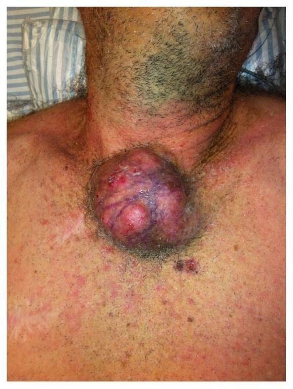
Clinical photograph of a large, exophytic non-melanoma skin cancer (likely basal cell or squamous cell carcinoma) located in the suprasternal and superior sternal region. The lesion presents as a massive, multinodular, and irregularly shaped growth with variegated coloring, including deep red, purple, and violaceous hues suggesting high vascularity or internal hemorrhage. The surface is uneven with visible areas of ulceration, crusting, and focal erosions. Clinically, the mass demonstrates local invasion, potentially involving the manubrium, clavicles, and underlying thoracic structures. The surrounding integument shows significant actinic damage, generalized erythema, and mottled hyperpigmentation. A prominent vertical surgical scar is visible on the left anterior chest wall, suggesting prior intervention. Small satellite lesions and numerous pigmented macules are scattered across the field, indicative of extensive field cancerization in a geriatric patient. This image serves as a clinical example of advanced skin malignancy and the necessity for aggressive surgical resection in cases of deep local invasion.
🧬 Tumors of the Skin - MBBS Exam Notes
QUICK CLASSIFICATION (Learn This First!)
SKIN TUMORS
│
├── BENIGN / PREMALIGNANT EPITHELIAL
│   ├── Seborrheic Keratosis
│   └── Actinic Keratosis (premalignant)
│
├── MALIGNANT EPITHELIAL (Keratinocyte Carcinomas)
│   ├── Basal Cell Carcinoma (BCC) — most common skin cancer
│   └── Squamous Cell Carcinoma (SCC)
│
├── MELANOCYTIC TUMORS
│   ├── Nevus (benign)
│   └── Melanoma (malignant)
│
└── OTHERS
    ├── Dermatofibroma
    ├── Xanthoma
    └── Kaposi Sarcoma
1. SEBORRHEIC KERATOSIS
| Feature | Detail |
|---|---|
| Type | Benign |
| Age | Middle-aged / elderly |
| Site | Trunk, face, extremities |
| Gross | "Stuck-on" appearance, tan-brown, velvety, coin-like plaques |
| Micro | Sheets of monotonous basaloid cells + horn cysts + pseudo-horn cysts (pathognomonic) |
| Mutation | FGFR3 (activating) → RAS/PI3K pathway |
| Key association | Sign of Leser-Trelat = sudden appearance of hundreds of seborrheic keratoses → paraneoplastic syndrome (GI carcinoma) |
| Variant | Dermatosis papulosa nigra (darker-skinned individuals, face/neck) |
| Malignant transformation | Extremely rare |
Exam tip: "Stuck-on" + horn cysts = Seborrheic keratosis. Leser-Trelat = always remember!
2. ACTINIC KERATOSIS (Solar Keratosis)
| Feature | Detail |
|---|---|
| Type | Premalignant |
| Cause | UV-induced DNA damage → TP53 mutations |
| Site | Sun-exposed skin: face, dorsum of hands, arms |
| Who | Fair-skinned, elderly |
| Gross | <1 cm, tan-brown or red, rough/sandpaper feel |
| Micro | Cytologic atypia in lower epidermis + solar elastosis (blue-gray elastic fibers in dermis) + parakeratosis |
| Progression | 0.1%-2.6%/year → SCC; most regress or remain stable |
| Full-thickness atypia | = Squamous cell carcinoma in situ |
| Treatment | Cryotherapy or topical agents |
Exam tip: Actinic = Sun damage → premalignant → can → SCC. TP53 mutation links both.
3. BASAL CELL CARCINOMA (BCC)
| Feature | Detail |
|---|---|
| Origin | Basal cells of epidermis / hair follicle stem cells |
| Risk factors | UV radiation (most important), fair skin, PTCH1 mutation (Hedgehog pathway) |
| Site | Face (nose, inner canthus - most common), rarely occurs on palms/soles |
| Gross | Pearly/translucent papule with rolled edges and telangiectasia, may ulcerate ("rodent ulcer") |
| Behavior | Locally invasive, rarely metastasizes (key exam point!) |
| Micro | Nests of basaloid cells with peripheral palisading + retraction artifact (cleft between tumor and stroma) |
| Syndrome | Gorlin syndrome (Basal Cell Nevus Syndrome) = PTCH1 germline mutation → hundreds of BCCs + jaw cysts + skeletal anomalies |
| Treatment | Excision / Mohs surgery; vismodegib (Hedgehog inhibitor) for advanced cases |
Exam tip: BCC = Locally invasive, NEVER metastasizes (classic exam trap!). Pearly papule + rolled border + telangiectasia = BCC.
4. SQUAMOUS CELL CARCINOMA (SCC)
| Feature | Detail |
|---|---|
| Origin | Keratinocytes (spinous layer) |
| Risk factors | UV light, fair skin, HPV (oncogenic strains), chemical carcinogens (arsenic), chronic inflammation/scarring (Marjolin's ulcer), immunosuppression, TP53 mutations |
| Precursor | Actinic keratosis |
| Site | Sun-exposed areas: lip (lower), ear, hands |
| Gross | Irregular ulcerated lesion or exophytic nodule with raised/indurated borders |
| Micro | Atypical squamous cells invading dermis + keratin (squamous) pearls + intercellular bridges |
| Behavior | Can metastasize (to regional LN) - key distinction from BCC |
| Marjolin's ulcer | SCC arising in chronic scars/burns/ulcers - aggressive, prone to metastasize |
| In situ form | Bowen's disease = SCC in situ (full-thickness epidermal atypia) |
Exam tip: SCC vs BCC - SCC can metastasize, BCC almost never does. Keratin pearls = SCC. Rolled pearly border = BCC.
5. MELANOMA
| Feature | Detail |
|---|---|
| Origin | Melanocytes |
| Risk factors | UV exposure, fair skin, dysplastic nevus syndrome, CDKN2A mutations, family history |
| Key mutations | BRAF V600E (most common, ~50%), NRAS, KIT, CDKN2A |
| Radial growth phase | Horizontal spread within epidermis - better prognosis |
| Vertical growth phase | Invasion into dermis - worse prognosis, metastatic potential |
Clinical Types (MBBS must-know):
| Type | Features |
|---|---|
| Superficial spreading | Most common (70%); pagetoid spread; ABCDE features |
| Nodular | Most aggressive; vertical growth from start; blue-black nodule |
| Lentigo maligna | Elderly; sun-exposed face; slowest growing; flat pigmented patch → lentigo maligna melanoma |
| Acral lentiginous | Most common in dark-skinned individuals; palms, soles, subungual |
ABCDE Rule (Clinical Diagnosis Aid):
- A - Asymmetry
- B - Border irregularity
- C - Color variation (brown, black, red, white, blue)
- D - Diameter >6 mm
- E - Evolution (changing over time)
Breslow Thickness (Prognosis):
| Thickness | Prognosis |
|---|---|
| <1 mm | Excellent |
| 1-2 mm | Good |
| 2-4 mm | Intermediate |
| >4 mm | Poor |
Treatment:
- Surgery (wide excision + sentinel lymph node biopsy)
- BRAF inhibitors (vemurafenib, dabrafenib) for BRAF V600E mutation
- Immune checkpoint inhibitors (pembrolizumab, nivolumab) - remarkable responses including complete remissions
Exam tip: Breslow thickness = single most important prognostic factor in melanoma. BRAF V600E = most common mutation. Nodular type = most aggressive.
6. BENIGN MELANOCYTIC NEVI (Moles)
| Feature | Detail |
|---|---|
| Type | Junctional nevus (at DEJ) → Compound nevus (DEJ + dermis) → Intradermal nevus (dermis only) |
| Key distinction | Absence of atypia, symmetry, uniform color |
| Dysplastic nevus | Atypical/Clark's nevus; precursor to melanoma; management with excision if high atypia |
7. OTHER TUMORS (Quick Points)
Dermatofibroma
- Benign spindle-cell proliferation in dermis
- Common in legs of women
- "Dimple sign" on pinching
- Micro: spindle cells + overlying epidermal hyperplasia
Xanthoma
- Lipid-laden macrophages (foam cells) in dermis
- Association: hyperlipidemia, biliary cirrhosis
- Types: xanthelasma (eyelid), tendon xanthoma (Achilles), tuberous xanthoma
Kaposi Sarcoma
- Origin: endothelial cells/vascular
- Associated with HHV-8 (Human Herpesvirus 8)
- Forms:
- Classic: elderly Mediterranean men, lower extremities
- AIDS-related (epidemic): most common; widespread, aggressive
- Gross: purple/violaceous plaques/nodules
- Micro: spindle cells + slit-like vascular spaces
HIGH-YIELD COMPARISON TABLE (BCC vs SCC vs Melanoma)
| Feature | BCC | SCC | Melanoma |
|---|---|---|---|
| Cell of origin | Basal cell | Keratinocyte | Melanocyte |
| Most common? | Most common skin cancer | 2nd most common | 3rd (but deadliest) |
| UV association | Yes | Yes | Yes |
| Metastasis | Rare (almost never) | Yes (regional LN) | Yes (widespread) |
| Key histology | Peripheral palisading + retraction cleft | Keratin pearls + intercellular bridges | Atypical melanocytes, pagetoid spread |
| Key mutation | PTCH1 (Hedgehog) | TP53 | BRAF V600E |
| Site | Face (nose) | Lip, ear, hands | Any site, back (M), leg (F) |
| Appearance | Pearly, rolled border | Ulcerated, indurated | Pigmented, irregular ABCDE |
| Prognosis | Excellent | Good if localized | Depends on Breslow thickness |
MNEMONICS TO REMEMBER
- Pearly nodule, Erodent ulcer, Almost never metastasizes, Rolled edges, Locally invasive, Young-ish sun exposure → Basal, Inner canthus most common, Low metastatic risk, Ledge (palisading)
- Superficial spreading (most common)
- Nodular (most aggressive)
- Lentigo maligna (slowest)
- Acral lentiginous (dark skin, palms/soles)
- <1 mm = best; 1-2 mm = moderate; 2-4 mm = worse; >4 mm = worst
EXAM TRAPS TO AVOID
- BCC is the most common skin cancer BUT almost never metastasizes - students confuse this.
- Melanoma Breslow thickness = most important prognostic factor (not Clark's level for MBBS).
- Sign of Leser-Trelat = seborrheic keratoses + internal malignancy (usually GI).
- Marjolin's ulcer = SCC in a chronic scar/burn - always aggressive.
- Acral lentiginous melanoma = most common type in dark-skinned individuals (NOT superficial spreading).
- Kaposi sarcoma = HHV-8, NOT HIV directly (HIV → immunosuppression → HHV-8 reactivation).
- Gorlin syndrome = PTCH1 mutation → multiple BCCs (not SCCs).
Only malignant tumors
🔴 Malignant Tumors of the Skin — MBBS Exam Notes
CLASSIFICATION AT A GLANCE
MALIGNANT SKIN TUMORS
│
├── KERATINOCYTE (Epithelial) CARCINOMAS
│   ├── Basal Cell Carcinoma (BCC)
│   └── Squamous Cell Carcinoma (SCC)
│       └── Special: Bowen's Disease (SCC in situ)
│           Marjolin's Ulcer
│
├── MELANOCYTIC
│   └── Malignant Melanoma
│       ├── Superficial Spreading
│       ├── Nodular
│       ├── Lentigo Maligna Melanoma
│       └── Acral Lentiginous
│
└── VASCULAR / OTHER
    └── Kaposi Sarcoma
1. BASAL CELL CARCINOMA (BCC)
"Most common cancer in humans. Locally evil, rarely travels."
| Feature | Detail |
|---|---|
| Cell of origin | Basal cells of epidermis / hair follicle stem cells |
| Most common site | Face (nose, inner canthus of eye) |
| Gross appearance | Pearly/translucent papule with rolled (everted) edges + surface telangiectasia |
| Ulceration | Central ulcer = "Rodent ulcer" |
| Metastasis | Extremely rare (almost NEVER) — #1 exam trap |
| Behavior | Locally invasive — destroys bone, cartilage, orbit |
Histology (Micro):
- Nests/islands of basaloid cells in dermis
- Peripheral palisading (cells arranged like a fence at the periphery)
- Retraction artifact = cleft/gap between tumor nests and stroma (artifact of fixation but diagnostically useful)
Key Genetics:
- PTCH1 mutation → loss of Hedgehog pathway inhibition → uncontrolled growth
- Gorlin Syndrome (Basal Cell Nevus Syndrome): PTCH1 germline mutation → hundreds of BCCs + jaw keratocysts + skeletal anomalies + calcified falx cerebri
Treatment:
- Excision / Mohs micrographic surgery
- Vismodegib (Hedgehog/SMO inhibitor) for advanced/metastatic
2. SQUAMOUS CELL CARCINOMA (SCC)
"Second most common skin cancer. Can metastasize — unlike BCC."
| Feature | Detail |
|---|---|
| Cell of origin | Keratinocytes (spinous/prickle cell layer) |
| Risk factors | UV light, TP53 mutation, HPV (6, 11, 16, 18), arsenic, chronic scars, immunosuppression |
| Precursor lesion | Actinic keratosis (→ SCC, low rate) |
| Common sites | Lower lip, ear, dorsum of hand, sun-exposed skin |
| Gross | Irregular ulcer with indurated/raised borders, or exophytic nodule |
| Metastasis | Yes — to regional lymph nodes |
Histology (Micro):
- Atypical keratinocytes invading dermis
- Keratin (squamous) pearls = concentric whorls of keratin (hallmark!)
- Intercellular bridges (desmosomes) visible
- Desmoplastic stromal reaction
Special Variants:
| Variant | Key Point |
|---|---|
| Bowen's Disease | SCC in situ — full-thickness epidermal atypia, no basement membrane breach |
| Marjolin's Ulcer | SCC arising in chronic scars, burns, venous ulcers — aggressive, high metastatic risk |
| Verrucous Carcinoma | Low-grade, well-differentiated SCC; warty appearance; low metastatic risk |
Exam tip: Bowen's = in situ (no invasion). Marjolin's = chronic wound → SCC = always aggressive.
BCC vs SCC — Quick Comparison (High Yield!)
| Feature | BCC | SCC |
|---|---|---|
| Origin | Basal cell | Keratinocyte |
| Most common site | Nose, inner canthus | Lower lip, ear |
| Appearance | Pearly, rolled border, telangiectasia | Ulcerated, indurated, crusted |
| Key histology | Peripheral palisading + retraction cleft | Keratin pearls + intercellular bridges |
| Metastasis | Almost NEVER | Yes (regional LN) |
| Key mutation | PTCH1 | TP53 |
| Precursor | None (de novo) | Actinic keratosis |
| Treatment | Excision, Mohs | Excision ± RT |
| Syndrome | Gorlin syndrome | None classic |
3. MALIGNANT MELANOMA
"Least common of the three, but the deadliest. All about Breslow."
| Feature | Detail |
|---|---|
| Cell of origin | Melanocytes |
| Risk factors | UV exposure, fair skin, dysplastic nevus syndrome, CDKN2A mutation, family history |
| Key mutation | BRAF V600E (~50%), NRAS, KIT, CDKN2A |
| Most common site in men | Back |
| Most common site in women | Leg |
Growth Phases:
| Phase | Description | Prognosis |
|---|---|---|
| Radial growth phase | Horizontal spread within epidermis & superficial dermis | Better — low metastatic risk |
| Vertical growth phase | Invasion deep into dermis | Worse — metastatic potential |
ABCDE Clinical Criteria:
| Letter | Meaning |
|---|---|
| A | Asymmetry |
| B | Border irregularity |
| C | Color variation (brown, black, red, white, blue — multiple colors) |
| D | Diameter >6 mm |
| E | Evolution (changing lesion) |
4 Clinical Types (Mnemonic: "Super Nodular Lentigo Acral"):
| Type | % | Key Features |
|---|---|---|
| Superficial Spreading | 70% | Most common; pagetoid spread (single cells in epidermis); ABCDE pattern |
| Nodular | 15-20% | Most aggressive; vertical growth from start; blue-black dome-shaped nodule; worst prognosis |
| Lentigo Maligna | 5-10% | Elderly; sun-damaged face; slowest growing; flat, tan-brown patch → LM melanoma |
| Acral Lentiginous | 2-8% | Most common in dark-skinned individuals; palms, soles, subungual (nail bed); not UV-related |
Exam tip: Acral lentiginous = dark skin people. NOT superficial spreading. Remember this!
Histology:
- Atypical melanocytes at DEJ (dermal-epidermal junction) and dermis
- Pagetoid spread = single melanocytes scattered through epidermis like buckshot
- Large cells with prominent nucleoli ("owl eye" nucleoli in some)
Breslow Thickness — Most Important Prognostic Factor:
| Thickness | 5-year Survival | Exam Memory |
|---|---|---|
| <1 mm | ~95% | Thin = excellent |
| 1–2 mm | ~80% | |
| 2–4 mm | ~60% | |
| >4 mm | ~50% | Thick = poor |
Breslow thickness = single most important prognostic factor — not Clark's level (outdated for MBBS).
Metastasis Pattern:
- Regional LN (first) → Liver, Lung, Brain, Bone (in situ)
- Brain metastasis = common in melanoma (one of the top 3 causes of brain mets)
Treatment:
- Surgery: wide local excision + sentinel lymph node biopsy
- BRAF inhibitors: vemurafenib, dabrafenib (for BRAF V600E mutant tumors)
- Immune checkpoint inhibitors: pembrolizumab, nivolumab (anti-PD1) — dramatic responses, even complete remissions
4. KAPOSI SARCOMA
"Purple nodules = HHV-8 = Think AIDS"
| Feature | Detail |
|---|---|
| Origin | Endothelial cells (vascular tumor) |
| Cause | HHV-8 (Human Herpesvirus 8) — necessary but not sufficient |
| Gross | Purple/violaceous plaques and nodules |
| Micro | Spindle cells + slit-like vascular spaces + extravasated RBCs + hemosiderin |
4 Clinical Forms:
| Form | Who | Features |
|---|---|---|
| Classic | Elderly Mediterranean/Eastern European men | Lower extremities; indolent |
| Endemic (African) | Sub-Saharan Africa, young adults/children | Lymph node involvement, aggressive |
| Iatrogenic | Transplant recipients (immunosuppressed) | Regresses with stopping immunosuppression |
| Epidemic (AIDS-related) | HIV+ patients (CD4 <200) | Most common form; widespread; skin, mucosa, viscera |
Exam tip: AIDS-related KS = most common form today. HHV-8 is the direct oncogenic virus — HIV causes immunosuppression allowing HHV-8 to reactivate.
MASTER COMPARISON TABLE
| Feature | BCC | SCC | Melanoma | Kaposi Sarcoma |
|---|---|---|---|---|
| Cell | Basal | Keratinocyte | Melanocyte | Endothelial |
| Frequency | Most common | 2nd | 3rd | Rare |
| Deadliest | No | No | Yes | In AIDS |
| Metastasis | Rare | Yes (LN) | Yes (widespread) | Yes (AIDS form) |
| Key histology | Palisading + retraction | Keratin pearls | Pagetoid spread | Spindle + slit vessels |
| Key mutation | PTCH1 | TP53 | BRAF V600E | HHV-8 |
| UV cause | Yes | Yes | Yes | No |
| Prognosis tool | Size/invasion | Depth/LN | Breslow thickness | CD4 count |
POWER MNEMONICS
TOP EXAM TRAPS ⚠️
- BCC almost never metastasizes — students wrongly say it can spread widely
- Marjolin's ulcer = SCC in chronic scar, NOT BCC
- Acral lentiginous melanoma = most common in dark-skinned people, not superficial spreading
- Nodular melanoma = most aggressive (vertical growth from day 1), despite not being most common
- Breslow thickness = most important prognostic factor, not Clark's level
- Kaposi sarcoma is caused by HHV-8, not directly by HIV
- Bowen's disease = SCC in situ (no basement membrane breach = no metastasis)
- Gorlin syndrome = multiple BCCs, NOT SCCs (PTCH1 mutation)
Ewing tumors mbbs
Ewing sarcoma bone tumor histology onion skin periosteal reaction xray
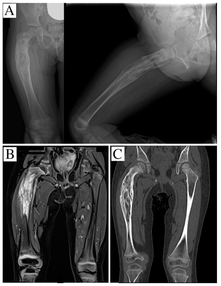
This composite of diagnostic images demonstrates a primary malignant bone tumor, specifically Ewing's sarcoma, in the right femur of a pediatric patient. Panel A displays plain radiographs (anteroposterior and lateral views) showing an aggressive osteolytic lesion in the proximal and middle third of the femoral shaft. Key findings include a pathological bowing deformity and a classic 'onion-skin' periosteal reaction, indicating rapid, multilayered new bone formation. Panel B is a STIR-weighted coronal MRI reconstruction showing high signal intensity, which evidences significant intramedullary marrow infiltration and extensive peritumoral soft tissue edema. Panel C provides a coronal CT scan, highlighting geographic osteolysis and significant cortical thinning/disruption. These images collectively illustrate the radiographic hallmarks of aggressive musculoskeletal neoplasms: permeative bone destruction, exuberant periosteal response, and soft tissue involvement. The preservation of the growth plates is visible, though the lesion extends near the proximal physis. This material is suitable for advanced medical education regarding orthopedic oncology and pediatric radiology.
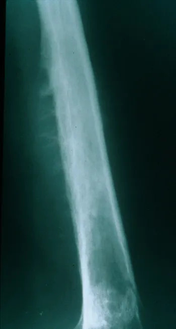
Plain Radiography of a long bone diaphysis demonstrates an aggressive, ill-defined osteolytic lesion with cortical destruction and periosteal reaction. The radiograph shows a moth-eaten destruction pattern with diffuse bone loss extending along a substantial portion of the shaft. Periosteal elevation produces Codman triangle in the swept cortex, while layered deposition of reactive new bone beneath the periosteum can create an onion-skin appearance; in some cases perpendicular radiating spicules or sunburst-like projections may be seen. The lesion often breaches the cortex or expands to form a soft tissue mass that extends beyond the bone. Although ultrasound or CT can delineate the soft tissue component, CT and MRI best characterize the extent, relation to surrounding structures, and marrow involvement. The radiographic pattern is highly suggestive of an aggressive small round blue cell tumor, most notably Ewing sarcoma, particularly in children and adolescents. Differential considerations include osteosarcoma, osteomyelitis, lymphoma, metastasis, or eosinophilic granuloma, but the combination of an diaphyseal, permeative lesion with onion-skin periosteal reaction strongly favors Ewing. Clinical correlation with pain, fever, raised inflammatory markers, and prompt biopsy is essential to confirm diagnosis and guide treatment, which typically combines chemotherapy, limb-sparing surgery, and possibly radiotherapy.
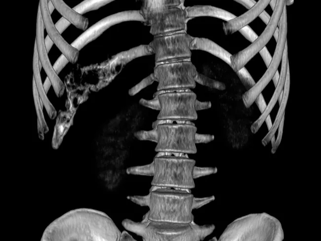
Imaging modality: Computed Tomography (CT) with three-dimensional (3D) volume rendering of the chest wall. The study localizes a lytic, destructive lesion of the right 11th rib with cortical disruption and destruction extending into the adjacent chest wall soft tissues. The appearance is aggressive with permeative margins, focal cortical breakthrough, possible small soft tissue mass, and associated chest wall remodeling. No definitive matrix mineralization is noted on the given reconstruction, though an onion-skin periosteal reaction may be present in some Ewing sarcoma cases; differential considerations include Ewing sarcoma/of the chest wall, osteogenic sarcoma, osteomyelitis, metastatic neuroectodermal tumor, Langerhans cell histiocytosis, or rhabdomyosarcoma. The findings are highly suggestive of a malignant small round blue cell tumor of bone; radiologic-pathologic correlation is recommended. Imaging helps assess extent for biopsy planning, surgical resection margins, and staging workup; chest wall involvement raises prognosis considerations and chemotherapy responsiveness typical of Ewing sarcoma family tumors. This lesion is consistent with chest wall Ewing sarcoma (Askin tumor) in pediatric patients; correlation with histopathology confirms the diagnosis. The 3D CT technique provides spatial relationships to nearby pleura, lung, diaphragm, and mediastinal structures aiding thoracic surgical planning and multidisciplinary treatment.
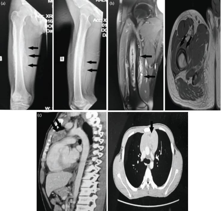
This composite of diagnostic images illustrates multi-modal findings of synchronous pathologies in a pediatric patient. Panel (a) contains anterior-posterior and lateral radiographs of the right femur, demonstrating a prominent lamellated (onion-skin) periosteal reaction along the femoral diaphysis, accompanied by significant soft tissue swelling—features classic for aggressive bone tumors like Ewing sarcoma or parosteal osteosarcoma. Panel (b) shows MRI sequences (coronal T2-weighted fat-saturated and axial T1-weighted) revealing a large intramedullary mass with extensive extraosseous soft tissue extension and cortical involvement. Panel (c) presents contrast-enhanced CT images of the thorax in sagittal and axial views. These images show a large, lobulated anterior mediastinal mass (indicated by arrows) with internal hypodense areas, suggestive of necrosis or cystic components. The collection serves as an educational example of oncological staging, highlighting how secondary findings in the thorax may represent distinct primary entities (such as a germ cell tumor) rather than metastatic disease.
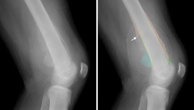
Imaging modality and technique: Plain radiography of the left distal femur and knee is performed with lateral views, showing aggressive bony lesion. Anatomy: Distal femur near the knee joint; left lower limb. Visual features: The lesion demonstrates irregular, partly destructive cortical bone with permeative medullary involvement, aggressive periosteal reaction, Codman triangle formation, an indistinct zone of transition, perpendicular spiculations producing a sunburst appearance, and a large soft tissue mass extending into adjacent compartments. Radiographic interpretation: These radiographic hallmarks strongly suggest a malignant primary bone tumor in a teenager, with Ewing sarcoma in this histologically proven case; differential includes osteosarcoma and occult infection. Clinical significance: The combination of cortical destruction, aggressive periosteal reaction, and soft-tissue extension correlates with higher tumor grade, local invasion risk, and the need for systemic staging and multidisciplinary therapy. Potential clinical uses: initial suspicion prompting MRI for local staging, CT or MRI to assess skip lesions, CT chest to evaluate metastasis, biopsy planning, treatment monitoring, and education. Context: Ewing sarcoma typically affects long bones of adolescents; this case confirms distal femur involvement with radiographic characteristics of a malignant diaphyseal/metaphyseal lesion. Additional keywords: pediatric bone tumor, onion-skin periosteal reaction (notes), EWSR1-FLI1 translocation, CD99 positivity and biopsy correlation recommended.
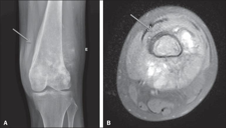
This composite diagnostic image demonstrates an aggressive periosteal reaction associated with osteosarcoma of the distal femur. Image A is a conventional radiograph (CR) of the knee in an anteroposterior view, showing a classic laminated, or 'onion skin,' periosteal reaction along the lateral aspect of the femoral diaphysis and metaphysis (arrow). This reaction is characterized by thin, concentric, radiopaque layers of new bone formation. Image B is an axial MRI scan of the same region, providing a cross-sectional view of the pathology. The MRI highlights the circumferential signal abnormalities at the periphery of the femoral cortex (arrow), corresponding to the laminated periosteal reaction and its extension into the adjacent soft tissues. The images illustrate key radiographic markers of aggressive bone lesions, emphasizing the high-velocity nature of the underlying disease process. These findings are clinically significant for the diagnosis of primary bone malignancies such as osteosarcoma or Ewing sarcoma, where rapid periosteal elevation occurs.
🦴 Ewing Sarcoma — MBBS Exam Notes
QUICK IDENTITY CARD
| Feature | Detail |
|---|---|
| Full name | Ewing Sarcoma / Peripheral Primitive Neuroectodermal Tumor (pPNET) |
| Type | Primary malignant bone tumor |
| Rank | 2nd most common primary bone malignancy in children (after osteosarcoma) |
| Age | 5-25 years (80% under 20) — peak in 2nd decade |
| Sex | Slight male predominance |
| Race | Extremely rare in individuals of African descent (mostly Caucasians) |
| Cell of origin | Mesenchymal stem cells / primitive neuroectodermal cells |
Exam tip: Ewing sarcoma = most common bone tumor in children under 10. Osteosarcoma = most common overall in young patients. Know this distinction!
PATHOGENESIS — THE TRANSLOCATION (MOST HIGH-YIELD!)
t(11;22)(q24;q12)
     ↓
EWSR1 gene (chr 22) + FLI1 gene (chr 11)
     ↓
EWS-FLI1 chimeric fusion protein
     ↓
Binds chromatin → dysregulates transcription
     ↓
Uncontrolled proliferation + abnormal differentiation
| Key Fact | Detail |
|---|---|
| Translocation | t(11;22) — present in >90% of cases |
| Fusion gene | EWSR1-FLI1 |
| Chromosome | EWSR1 on chr 22, FLI1 on chr 11 |
| Mechanism | Chimeric protein dysregulates transcription |
Mnemonic: "11 and 22 — Ewing's clue" — t(11;22) = Ewing's hallmark translocation
SITES OF INVOLVEMENT
Most common:
  → DIAPHYSIS of long bones (femur, tibia, humerus, fibula)
  → Metaphysis of long bones (with extension into diaphysis)
  → Flat bones: pelvis, scapula, ribs (shoulder & pelvic girdle)

Less common:
  → Vertebral column (sacrum most common; 50% sacral involvement)
  → Small bones of feet/hands (rare)

20% are EXTRASKELETAL (soft tissue Ewing's)
Exam tip: Ewing's = diaphysis of long bones. Osteosarcoma = metaphysis. Giant cell tumor = epiphysis. Know the locations!
Location Comparison (Must-Know!):
| Tumor | Location | Age |
|---|---|---|
| Osteosarcoma | Metaphysis | 10-20 yrs |
| Ewing Sarcoma | Diaphysis | 5-25 yrs |
| Giant Cell Tumor | Epiphysis | 20-40 yrs |
| Chondrosarcoma | Metaphysis/diaphysis | >40 yrs |
CLINICAL FEATURES
| Feature | Detail |
|---|---|
| Pain | Almost universal; onset insidious; initially mild/intermittent |
| Swelling | Enlarging, painful soft tissue mass |
| Local signs | Affected site is tender, warm, swollen — mimics osteomyelitis/infection! |
| Systemic signs | Fever, raised ESR/CRP, leukocytosis — further mimics infection |
| Diagnostic delay | Average 34 weeks from symptoms to diagnosis |
| Pathologic fracture | Can occur |
Exam trap: Ewing's mimics osteomyelitis clinically! Fever + warmth + bone pain in a child → always biopsy to distinguish!
GROSS PATHOLOGY
- Arises in medullary cavity
- Invades cortex → periosteum → soft tissue
- Soft, tan-white tumor
- Frequently shows areas of hemorrhage and necrosis
- Periosteal reaction → "Onion skin" appearance on X-ray
HISTOLOGY (MICRO) — KEY!
| Feature | Detail |
|---|---|
| Cells | Sheets of uniform small round cells |
| Size | Slightly larger than lymphocytes |
| Cytoplasm | Scant, clear cytoplasm (rich in glycogen — PAS positive!) |
| Nuclear features | Monotonous, minimal atypia |
| Special structures | Homer-Wright rosettes (circular groupings with central fibrillary core) — indicates neuroectodermal differentiation |
| Matrix | No bone or cartilage production (distinguishes from osteosarcoma) |
| IHC marker | CD99 (MIC2) strongly positive — most important marker! |
Mnemonic for SRBCT: "Wretched Little Rascal Eating" = Wilms, Lymphoma, Rhabdomyosarcoma, Ewing's
RADIOLOGY — IMAGING FEATURES
Plain X-Ray:
| Feature | Description |
|---|---|
| "Onion skin" periosteal reaction | Layers of reactive new bone — PATHOGNOMONIC |
| Permeative/moth-eaten pattern | Ill-defined bone destruction |
| Cortical destruction | Breakthrough into soft tissue |
| Soft tissue mass | Extends beyond bone |
| Codman's triangle | May be present (periosteal elevation) |
MRI (Investigation of Choice for Staging):
- Best for local staging and soft tissue extent
- Isointense on T1, hyperintense on T2
- Gadolinium enhancement (due to hypercellularity)
- Shows intramedullary extent, skip lesions
Bone Scan:
- Used for detecting bone metastases
FDG-PET/CT:
- New standard for initial staging and detection of recurrence/metastasis
INVESTIGATIONS
| Investigation | Purpose |
|---|---|
| X-ray bone | First line; onion skin periosteal reaction |
| MRI | Local staging (investigation of choice) |
| CT chest | Lung metastases |
| Bone scan | Skeletal metastases |
| FDG-PET/CT | Full staging, recurrence detection |
| Biopsy | Definitive diagnosis |
| CBC, ESR | Leukocytosis, elevated ESR (mimics infection) |
| LDH | Elevated = poor prognosis marker |
EWING SARCOMA FAMILY OF TUMORS (ESFT)
| Tumor | Location | Notes |
|---|---|---|
| Classic Ewing Sarcoma | Bone (diaphysis) | Most common |
| Extraskeletal Ewing | Soft tissue | 20% of cases |
| pPNET | Bone/soft tissue | More neural differentiation, Homer-Wright rosettes prominent |
| Askin Tumor | Chest wall | pPNET of chest wall; thoracopulmonary |
Exam tip: Askin tumor = Ewing's family tumor of chest wall (thoracopulmonary pPNET). High-yield fact!
METASTASIS
| Site | Notes |
|---|---|
| Lung | Most common site of metastasis |
| Bone | Second most common |
| Regional LN | Less common |
- 25% have metastases at diagnosis — key poor prognostic sign
- Metastatic disease = dramatically worse prognosis
PROGNOSIS
| Factor | Good Prognosis | Poor Prognosis |
|---|---|---|
| Location | Distal extremities | Pelvis/axial skeleton |
| Tumor size | <8 cm | >8 cm |
| Metastasis | None at diagnosis | Present at diagnosis |
| Histologic response to chemo | Good response (>90% necrosis) | Poor response |
| LDH | Normal | Elevated |
| Age | Younger | Older |
- 5-year survival (localized): ~70-80%
- 5-year survival (metastatic): ~20-30%
TREATMENT
MULTIMODAL APPROACH
│
├── CHEMOTHERAPY (always first)
│   └── Neoadjuvant chemo → surgery → adjuvant chemo
│   └── Regimen: VDC/IE
│       (Vincristine + Doxorubicin + Cyclophosphamide
│        alternating with Ifosfamide + Etoposide)
│
├── SURGERY
│   └── Wide resection / limb-salvage surgery preferred
│   └── Amputation if cannot achieve adequate margins
│
└── RADIOTHERAPY
    └── For unresectable tumors (e.g., pelvis, spine)
    └── Ewing's is radiosensitive!
    └── Avoid in growing children if possible (growth disturbance)
Key point: Ewing sarcoma is radiosensitive (unlike osteosarcoma). This is a classic exam distinction!
EWING vs OSTEOSARCOMA — HIGH-YIELD COMPARISON
| Feature | Ewing Sarcoma | Osteosarcoma |
|---|---|---|
| Age | 5-25 yrs (peak 10-15) | 10-20 yrs (peak 15-19) |
| Most common in <10 yr | Yes | No |
| Location | Diaphysis | Metaphysis |
| Bones affected | Femur, pelvis, fibula | Distal femur, proximal tibia |
| Flat bones | Yes (pelvis, scapula) | Less common |
| Radiology | Onion skin periosteal reaction | Sunburst pattern + Codman's triangle |
| Histology | Small round blue cells, NO matrix | Malignant cells + osteoid/bone matrix |
| IHC | CD99+ | Vimentin+ |
| Translocation | t(11;22) EWSR1-FLI1 | No consistent translocation |
| Radiosensitive | YES | No |
| Race | Rare in Black patients | All races |
| Chemo regimen | VDC/IE | MAP (Methotrexate, Adriamycin, Cisplatin) |
MNEMONICS
- Small round blue cells
- Onion skin periosteal reaction
- Age 5-25 (peak 2nd decade)
- Pelvis + Diaphysis of long bones
- Diaphysis (NOT metaphysis)
- Radiosensitive
EXAM TRAPS ⚠️
- Ewing's mimics osteomyelitis — fever, warmth, elevated ESR. Always biopsy!
- Onion skin = Ewing (not osteosarcoma — that's sunburst)
- Diaphysis = Ewing (Metaphysis = Osteosarcoma, Epiphysis = GCT)
- Radiosensitive = Ewing (osteosarcoma is NOT radiosensitive)
- t(11;22) = always Ewing — CD99 positive on IHC
- No bone/cartilage matrix in Ewing's histology — distinguishes from osteosarcoma
- Askin tumor = Ewing's family, chest wall location
- Rare in Black patients — important epidemiology fact
- LDH elevated = poor prognostic marker
- Ewing's is the most common bone tumor in children under 10 (osteosarcoma peaks at 10-20)
Only i slide comprises all information
mkdir -p /tmp/workspace/ewing-slide && ls /tmp/workspace/
~/ewing-slide/create_slide.py
from pptx import Presentation
from pptx.util import Inches, Pt, Emu
from pptx.dml.color import RGBColor
from pptx.enum.text import PP_ALIGN, MSO_ANCHOR
from pptx.oxml.ns import qn
from lxml import etree
import copy

# ── Colour Palette ──────────────────────────────────────────────────────────
DARK_BG    = RGBColor(0x0D, 0x1B, 0x2A)   # deep navy background
ACCENT1    = RGBColor(0xE8, 0x3A, 0x3A)   # red accent (title bar, headings)
ACCENT2    = RGBColor(0xF5, 0xA6, 0x23)   # amber (highlight boxes)
ACCENT3    = RGBColor(0x2E, 0xCC, 0xB0)   # teal (section headings)
WHITE      = RGBColor(0xFF, 0xFF, 0xFF)
LIGHT_GRAY = RGBColor(0xCC, 0xD6, 0xE0)
DARK_CARD  = RGBColor(0x16, 0x2B, 0x40)   # card background
WARN_RED   = RGBColor(0xFF, 0x4C, 0x4C)

def add_rect(slide, x, y, w, h, fill_rgb, alpha=None):
    shape = slide.shapes.add_shape(1, Inches(x), Inches(y), Inches(w), Inches(h))
    shape.line.fill.background()
    shape.fill.solid()
    shape.fill.fore_color.rgb = fill_rgb
    return shape

def add_textbox(slide, x, y, w, h, text, font_size, bold=False,
                color=WHITE, align=PP_ALIGN.LEFT, word_wrap=True, italic=False):
    tb = slide.shapes.add_textbox(Inches(x), Inches(y), Inches(w), Inches(h))
    tf = tb.text_frame
    tf.word_wrap = word_wrap
    tf.margin_left = Inches(0.05)
    tf.margin_right = Inches(0.05)
    tf.margin_top = Inches(0.02)
    tf.margin_bottom = Inches(0.02)
    p = tf.paragraphs[0]
    p.alignment = align
    run = p.add_run()
    run.text = text
    run.font.size = Pt(font_size)
    run.font.bold = bold
    run.font.italic = italic
    run.font.color.rgb = color
    run.font.name = "Calibri"
    return tf

def add_multiline_textbox(slide, x, y, w, h, lines, font_size,
                           bold=False, color=WHITE, align=PP_ALIGN.LEFT,
                           line_spacing=1.0):
    tb = slide.shapes.add_textbox(Inches(x), Inches(y), Inches(w), Inches(h))
    tf = tb.text_frame
    tf.word_wrap = True
    tf.margin_left = Inches(0.06)
    tf.margin_right = Inches(0.04)
    tf.margin_top = Inches(0.03)
    tf.margin_bottom = Inches(0.02)
    first = True
    for line in lines:
        if first:
            p = tf.paragraphs[0]
            first = False
        else:
            p = tf.add_paragraph()
        p.alignment = align
        if isinstance(line, tuple):
            # (text, bold, color, size)
            txt, lb, lc, ls = line
        else:
            txt, lb, lc, ls = line, bold, color, font_size
        run = p.add_run()
        run.text = txt
        run.font.size = Pt(ls)
        run.font.bold = lb
        run.font.color.rgb = lc
        run.font.name = "Calibri"
    return tf

# ── Presentation Setup ──────────────────────────────────────────────────────
prs = Presentation()
prs.slide_width  = Inches(20)    # very wide A3-like landscape
prs.slide_height = Inches(11.25)

blank = prs.slide_layouts[6]
slide = prs.slides.add_slide(blank)

W = 20.0
H = 11.25

# ── Background ──────────────────────────────────────────────────────────────
add_rect(slide, 0, 0, W, H, DARK_BG)

# ═══════════════════════════════════════════════════════════════════════════
# TITLE BAR
# ═══════════════════════════════════════════════════════════════════════════
add_rect(slide, 0, 0, W, 0.85, ACCENT1)
add_textbox(slide, 0.15, 0.04, 12, 0.75,
            "EWING SARCOMA  —  MBBS COMPLETE REVISION SLIDE",
            26, bold=True, color=WHITE, align=PP_ALIGN.LEFT)
add_textbox(slide, 12.2, 0.04, 7.6, 0.75,
            "Robbins & Kumar Basic Pathology  |  Campbell's Orthopaedics",
            11, bold=False, color=RGBColor(0xFF,0xCC,0xAA), align=PP_ALIGN.RIGHT, italic=True)

# ═══════════════════════════════════════════════════════════════════════════
# COLUMN LAYOUT  (5 columns)
# ═══════════════════════════════════════════════════════════════════════════
COL_W = 3.78
GAP   = 0.06
TOP   = 0.92
BOT   = 10.30
COL_H = BOT - TOP

cols_x = [0.06 + i*(COL_W + GAP) for i in range(5)]

def card(slide, cx, cy, cw, ch, heading, lines, font_size=8.5):
    # card background
    add_rect(slide, cx, cy, cw, ch, DARK_CARD)
    # heading bar
    add_rect(slide, cx, cy, cw, 0.30, ACCENT3)
    add_textbox(slide, cx+0.05, cy+0.01, cw-0.1, 0.28,
                heading, 9.5, bold=True, color=DARK_BG, align=PP_ALIGN.LEFT)
    # content
    add_multiline_textbox(slide, cx+0.05, cy+0.33, cw-0.1, ch-0.38,
                          lines, font_size, color=LIGHT_GRAY)

# ═══════════════════════════════════════════════════════════════════════════
# COL 1 — Identity + Epidemiology
# ═══════════════════════════════════════════════════════════════════════════
cx = cols_x[0]
card(slide, cx, TOP, COL_W, 2.80, "IDENTITY & EPIDEMIOLOGY", [
    ("▸ 2nd most common primary bone malignancy in children", False, WHITE, 8.5),
    ("  (after osteosarcoma)", False, LIGHT_GRAY, 7.5),
    ("▸ Most common bone tumor in children < 10 yrs", False, WHITE, 8.5),
    ("▸ Age: 5–25 yrs  (80% < 20 yrs)  |  Peak: 2nd decade", False, LIGHT_GRAY, 8),
    ("▸ Slight male predominance", False, LIGHT_GRAY, 8),
    ("▸ RARE in individuals of African descent", False, ACCENT2, 8.5),
    ("▸ No known predisposing factors", False, LIGHT_GRAY, 7.5),
    ("▸ ~200 new cases/year (USA)", False, LIGHT_GRAY, 7.5),
])

card(slide, cx, TOP+2.87, COL_W, 3.25, "PATHOGENESIS (KEY!)", [
    ("t(11;22)(q24;q12)", True, ACCENT2, 11),
    ("EWSR1 (chr 22)  +  FLI1 (chr 11)", True, WHITE, 9),
    ("       ↓", False, ACCENT3, 9),
    ("EWS-FLI1 chimeric fusion protein", True, ACCENT3, 9),
    ("       ↓", False, ACCENT3, 9),
    ("Dysregulates transcription", False, LIGHT_GRAY, 8.5),
    ("       ↓", False, ACCENT3, 8.5),
    ("Uncontrolled proliferation", False, LIGHT_GRAY, 8.5),
    ("", False, LIGHT_GRAY, 5),
    ("▸ >90% of Ewing's carry this translocation", False, WHITE, 8),
    ("▸ Cell of origin: mesenchymal stem cells /", False, LIGHT_GRAY, 8),
    ("  primitive neuroectodermal cells", False, LIGHT_GRAY, 8),
])

card(slide, cx, TOP+6.20, COL_W, 4.10-0.08, "EWING SARCOMA FAMILY (ESFT)", [
    ("All share EWSR1 translocation:", False, ACCENT3, 8.5),
    ("", False, LIGHT_GRAY, 4),
    ("▸ Classic Ewing Sarcoma  → bone (diaphysis)", False, WHITE, 8.5),
    ("▸ Extraskeletal Ewing   → soft tissue (20%)", False, WHITE, 8.5),
    ("▸ pPNET  → bone/soft tissue, neural diff.", False, WHITE, 8.5),
    ("▸ Askin Tumor  → chest wall (thoracopulmonary)", True, ACCENT2, 9),
    ("  ★ Askin = pPNET of chest wall — HIGH YIELD", True, ACCENT2, 8.5),
])

# ═══════════════════════════════════════════════════════════════════════════
# COL 2 — Sites + Clinical
# ═══════════════════════════════════════════════════════════════════════════
cx = cols_x[1]
card(slide, cx, TOP, COL_W, 3.10, "SITES OF INVOLVEMENT", [
    ("PRIMARY SITES:", True, ACCENT3, 9),
    ("▸ Diaphysis of long bones (most common)", True, WHITE, 9),
    ("  – Femur, tibia, fibula, humerus", False, LIGHT_GRAY, 8),
    ("▸ Flat bones: PELVIS, scapula, ribs", False, WHITE, 8.5),
    ("▸ Vertebral column (sacrum in 50%)", False, LIGHT_GRAY, 8),
    ("▸ 20% extraskeletal (soft tissue)", False, LIGHT_GRAY, 8),
    ("", False, LIGHT_GRAY, 4),
    ("LOCATION COMPARISON:", True, ACCENT2, 8.5),
    ("Osteosarcoma → METAPHYSIS  (10–20 yrs)", False, LIGHT_GRAY, 8),
    ("Ewing's      → DIAPHYSIS   (5–25 yrs)", True, ACCENT2, 8.5),
    ("Giant Cell   → EPIPHYSIS   (20–40 yrs)", False, LIGHT_GRAY, 8),
])

card(slide, cx, TOP+3.17, COL_W, 3.50, "CLINICAL FEATURES", [
    ("▸ PAIN — almost universal; insidious onset", True, WHITE, 9),
    ("▸ Swelling — enlarging painful soft tissue mass", False, WHITE, 8.5),
    ("▸ Tender, WARM, swollen site", True, ACCENT2, 9),
    ("▸ Fever + raised ESR + leukocytosis", True, ACCENT2, 9),
    ("  ⚠ MIMICS OSTEOMYELITIS!", True, WARN_RED, 9.5),
    ("▸ Average diagnostic delay: 34 weeks", False, LIGHT_GRAY, 8),
    ("▸ Pathologic fracture may occur", False, LIGHT_GRAY, 8),
    ("▸ 25% have metastases at diagnosis", True, WARN_RED, 9),
    ("▸ Metastases: LUNG (MC) → bone → LN", False, LIGHT_GRAY, 8),
])

card(slide, cx, TOP+6.74, COL_W, 3.56, "INVESTIGATIONS", [
    ("▸ X-ray bone   — first line (onion skin!)", True, WHITE, 9),
    ("▸ MRI          — investigation of CHOICE", True, ACCENT2, 9),
    ("  for local staging; soft tissue extent", False, LIGHT_GRAY, 8),
    ("▸ CT chest     — lung metastases", False, LIGHT_GRAY, 8),
    ("▸ Bone scan    — skeletal metastases", False, LIGHT_GRAY, 8),
    ("▸ FDG-PET/CT   — new standard for staging", False, LIGHT_GRAY, 8),
    ("▸ Biopsy       — DEFINITIVE diagnosis", True, WHITE, 9),
    ("▸ LDH elevated — poor prognosis marker", False, ACCENT2, 8.5),
])

# ═══════════════════════════════════════════════════════════════════════════
# COL 3 — Histology + Radiology
# ═══════════════════════════════════════════════════════════════════════════
cx = cols_x[2]
card(slide, cx, TOP, COL_W, 4.10, "HISTOLOGY (MICRO)", [
    ("★ SMALL ROUND BLUE CELL TUMOR (SRBCT)", True, ACCENT2, 10),
    ("  Mnemonic: Wretched Little Rascal Eating", False, ACCENT3, 8),
    ("  = Wilms, Lymphoma, Rhabdo, Ewing's", False, LIGHT_GRAY, 8),
    ("", False, LIGHT_GRAY, 4),
    ("▸ Sheets of UNIFORM SMALL ROUND CELLS", True, WHITE, 9),
    ("  (slightly larger than lymphocytes)", False, LIGHT_GRAY, 8),
    ("▸ SCANT CLEAR CYTOPLASM (glycogen-rich)", True, WHITE, 9),
    ("  → PAS POSITIVE", True, ACCENT2, 8.5),
    ("▸ Homer-Wright rosettes", True, WHITE, 9),
    ("  (cells around central fibrillary core)", False, LIGHT_GRAY, 8),
    ("  → indicates neuroectodermal differentiation", False, LIGHT_GRAY, 8),
    ("▸ NO bone or cartilage production", True, WARN_RED, 9),
    ("  (key difference from osteosarcoma!)", False, LIGHT_GRAY, 8),
    ("▸ IHC: CD99 (MIC2) STRONGLY POSITIVE ★", True, ACCENT2, 9),
    ("▸ Gross: soft, tan-white + hemorrhage/necrosis", False, LIGHT_GRAY, 8),
])

card(slide, cx, TOP+4.17, COL_W, 3.50, "RADIOLOGY", [
    ("PLAIN X-RAY:", True, ACCENT3, 9),
    ("▸ 'ONION SKIN' periosteal reaction ★★", True, ACCENT2, 10),
    ("  (layered reactive bone — PATHOGNOMONIC)", False, LIGHT_GRAY, 8),
    ("▸ Permeative / moth-eaten bone destruction", False, WHITE, 8.5),
    ("▸ Codman's triangle may be present", False, LIGHT_GRAY, 8),
    ("▸ Soft tissue mass beyond cortex", False, LIGHT_GRAY, 8),
    ("", False, LIGHT_GRAY, 4),
    ("MRI:", True, ACCENT3, 9),
    ("▸ T1: isointense  |  T2: hyperintense", False, WHITE, 8.5),
    ("▸ Gadolinium enhancement (hypercellular)", False, LIGHT_GRAY, 8),
    ("▸ Best for marrow extent + skip lesions", False, LIGHT_GRAY, 8),
])

card(slide, cx, TOP+7.74, COL_W, 2.56, "GROSS PATHOLOGY", [
    ("▸ Arises in MEDULLARY CAVITY", False, WHITE, 8.5),
    ("▸ Invades cortex → periosteum → soft tissue", False, WHITE, 8.5),
    ("▸ Soft, tan-white tumor", False, LIGHT_GRAY, 8),
    ("▸ Frequent hemorrhage and necrosis", False, LIGHT_GRAY, 8),
    ("▸ No osteoid/chondroid matrix", True, WARN_RED, 8.5),
])

# ═══════════════════════════════════════════════════════════════════════════
# COL 4 — Prognosis + Treatment + Ewing vs OS
# ═══════════════════════════════════════════════════════════════════════════
cx = cols_x[3]
card(slide, cx, TOP, COL_W, 3.40, "PROGNOSIS", [
    ("5-yr survival (localized):  ~70–80% ✓", True, ACCENT3, 9),
    ("5-yr survival (metastatic): ~20–30% ✗", True, WARN_RED, 9),
    ("", False, LIGHT_GRAY, 4),
    ("GOOD PROGNOSIS:            POOR PROGNOSIS:", True, ACCENT2, 8.5),
    ("▸ Distal extremity    ←→  Pelvis / axial", False, LIGHT_GRAY, 8),
    ("▸ Tumor <8 cm         ←→  Tumor >8 cm", False, LIGHT_GRAY, 8),
    ("▸ No metastasis       ←→  Metastasis at Dx", False, LIGHT_GRAY, 8),
    ("▸ Good chemo response ←→  Poor response", False, LIGHT_GRAY, 8),
    ("  (>90% necrosis)     ←→  (<90% necrosis)", False, LIGHT_GRAY, 7.5),
    ("▸ Normal LDH          ←→  Elevated LDH", False, LIGHT_GRAY, 8),
])

card(slide, cx, TOP+3.47, COL_W, 3.80, "TREATMENT", [
    ("MULTIMODAL — ALWAYS chemotherapy first!", True, ACCENT2, 9.5),
    ("", False, LIGHT_GRAY, 4),
    ("1. NEOADJUVANT CHEMOTHERAPY", True, ACCENT3, 9),
    ("   Regimen:  VDC / IE", True, WHITE, 9),
    ("   Vincristine + Doxorubicin + Cyclophosphamide", False, LIGHT_GRAY, 8),
    ("   alternating with Ifosfamide + Etoposide", False, LIGHT_GRAY, 8),
    ("", False, LIGHT_GRAY, 4),
    ("2. SURGERY", True, ACCENT3, 9),
    ("   Wide resection / limb-salvage (preferred)", False, WHITE, 8.5),
    ("   Amputation if margins inadequate", False, LIGHT_GRAY, 8),
    ("", False, LIGHT_GRAY, 4),
    ("3. RADIOTHERAPY", True, ACCENT3, 9),
    ("   ★ Ewing's is RADIOSENSITIVE!", True, ACCENT2, 9.5),
    ("   For unresectable tumors (pelvis, spine)", False, WHITE, 8.5),
    ("   Avoid in growing children (growth disturbance)", False, LIGHT_GRAY, 8),
])

card(slide, cx, TOP+7.34, COL_W, 2.96, "METASTASIS", [
    ("▸ 25% have mets AT diagnosis (poor prognosis)", True, WARN_RED, 9),
    ("▸ Most common site: LUNG ★", True, WHITE, 9),
    ("▸ 2nd most common: BONE", False, WHITE, 8.5),
    ("▸ Less common: regional lymph nodes", False, LIGHT_GRAY, 8),
    ("▸ Skip lesions in same bone possible", False, LIGHT_GRAY, 8),
])

# ═══════════════════════════════════════════════════════════════════════════
# COL 5 — VS Osteosarcoma + Mnemonics + Exam Traps
# ═══════════════════════════════════════════════════════════════════════════
cx = cols_x[4]
card(slide, cx, TOP, COL_W, 4.20, "EWING vs OSTEOSARCOMA", [
    ("FEATURE          EWING        OSTEO-SARC", True, ACCENT3, 8.5),
    ("Age               5–25 yrs     10–20 yrs", False, WHITE, 8),
    ("<10 yrs MC        YES ★        No", False, ACCENT2, 8),
    ("Location          DIAPHYSIS    METAPHYSIS", True, ACCENT2, 9),
    ("Flat bones        YES          Less common", False, LIGHT_GRAY, 8),
    ("X-ray sign        ONION SKIN   SUNBURST", True, ACCENT2, 9),
    ("Histology         Sm round     Osteoid", False, WHITE, 8.5),
    ("                  blue cells   matrix", False, LIGHT_GRAY, 7.5),
    ("Bone/cartilage    ABSENT ★     PRESENT", True, WARN_RED, 8.5),
    ("IHC               CD99+        Vimentin+", False, WHITE, 8),
    ("Translocation     t(11;22)     None classic", True, ACCENT2, 8.5),
    ("Radiosensitive    YES ★        NO", True, ACCENT2, 9),
    ("Race              Rare Black   All races", False, LIGHT_GRAY, 8),
    ("Chemo             VDC/IE       MAP protocol", False, LIGHT_GRAY, 8),
])

card(slide, cx, TOP+4.27, COL_W, 2.40, "MNEMONICS", [
    ('"11 and 22 — Ewing\'s Clue"', True, ACCENT2, 9),
    ("  → t(11;22) = Ewing's translocation", False, WHITE, 8.5),
    ("", False, LIGHT_GRAY, 3),
    ('"SOAP-DR"', True, ACCENT3, 9.5),
    ("S=Small round blue cells  O=Onion skin", False, WHITE, 8),
    ("A=Age 5–25  P=Pelvis+Diaphysis", False, WHITE, 8),
    ("D=Diaphysis (not metaphysis)", False, WHITE, 8),
    ("R=Radiosensitive", False, WHITE, 8),
])

card(slide, cx, TOP+6.74, COL_W, 3.56, "⚠  EXAM TRAPS", [
    ("1. Onion skin = EWING  (NOT osteosarcoma)", True, WARN_RED, 8.5),
    ("   Sunburst = osteosarcoma", False, LIGHT_GRAY, 8),
    ("2. Diaphysis = Ewing  |  Metaphysis = Osteo", True, WARN_RED, 8.5),
    ("3. RADIOSENSITIVE = Ewing  (Osteo is NOT)", True, WARN_RED, 8.5),
    ("4. t(11;22) = ALWAYS Ewing", True, ACCENT2, 8.5),
    ("5. NO bone/cartilage matrix in Ewing's", True, WARN_RED, 8.5),
    ("6. Mimics osteomyelitis clinically — BIOPSY!", True, ACCENT2, 8.5),
    ("7. Askin tumor = Ewing family, chest wall", False, WHITE, 8),
    ("8. CD99 (MIC2) = most important IHC marker", False, WHITE, 8),
    ("9. Rare in Black patients — epidemiology fact", False, WHITE, 8),
    ("10. LDH elevated = poor prognosis", False, LIGHT_GRAY, 8),
])

# ═══════════════════════════════════════════════════════════════════════════
# BOTTOM BANNER
# ═══════════════════════════════════════════════════════════════════════════
add_rect(slide, 0, 10.88, W, 0.37, RGBColor(0x1A, 0x35, 0x52))
add_textbox(slide, 0.15, 10.90, W-0.3, 0.30,
            "MNEMONIC: t(11;22) → EWS-FLI1  |  Small Round Blue Cells  |  Onion Skin X-ray  |  Diaphysis  |  Radiosensitive  |  CD99+  |  VDC/IE Chemo  |  Askin = Chest Wall",
            8.5, bold=False, color=ACCENT2, align=PP_ALIGN.CENTER)

# ═══════════════════════════════════════════════════════════════════════════
# SAVE
# ═══════════════════════════════════════════════════════════════════════════
out = "/tmp/workspace/ewing-slide/Ewing_Sarcoma_MBBS.pptx"
prs.save(out)
print(f"Saved → {out}")
python /tmp/workspace/ewing-slide/create_slide.py
Ewing Sarcoma MBBS
Presentation · PPTX
| Column | Content |
|---|---|
| 1 | Identity & Epidemiology + Pathogenesis (t(11;22) + EWS-FLI1) + ESFT variants (Askin tumor) |
| 2 | Sites of involvement + Location comparison table + Clinical features + Investigations |
| 3 | Histology (SRBCT, Homer-Wright rosettes, CD99, PAS+) + Radiology (onion skin) + Gross pathology |
| 4 | Prognosis (good vs poor factors) + Treatment (VDC/IE, surgery, radiotherapy) + Metastasis |
| 5 | Ewing vs Osteosarcoma comparison + Mnemonics (SOAP-DR) + 10 Exam Traps ⚠ |
Giant cells tumors
giant cell tumor bone epiphysis histology osteoclast radiograph soap bubble
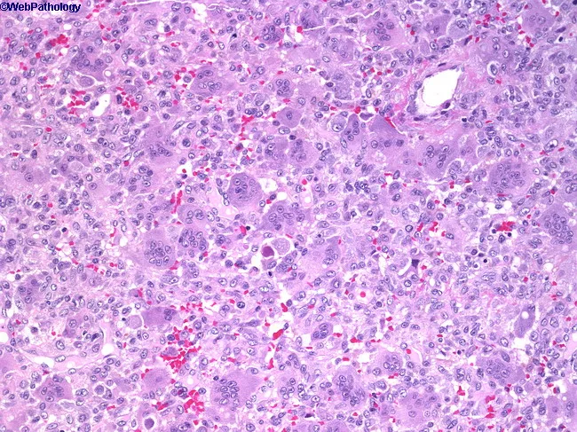
Giant cell tumor of bone (osteoclastoma) histopathology. This hematoxylin and eosin stained section shows numerous multinucleated osteoclast-like giant cells dispersed among a background of uniform, oval to spindle-shaped mononuclear stromal cells. The giant cells contain multiple nuclei with scant cytoplasm, while the mononuclear cells display vesicular nuclei, fine chromatin, and limited mitotic activity. The lesion often shows a vascular, hemorrhagic background and scattered hemosiderin deposits. There is typically minimal cytologic atypia; occasional atypical mitoses may be present in more aggressive variants. Architecturally, the tissue demonstrates a proliferative stromal framework with evenly distributed giant cells and occasional mitotic figures but not overt anaplasia. This histology correlates with radiologic eccentric, radiolucent, soap-bubble–appearing lesions that arise in the epiphysis of long bones. Clinically, patients present with pain and swelling; there is a female predominance in the first two decades, with peak incidence in the third decade. The diagnosis is important to distinguish from other giant cell–rich lesions, including aneurysmal bone cyst, brown tumor of hyperparathyroidism, fibrous dysplasia, chondroblastoma, and osteosarcoma with giant cells. Immunohistochemistry may help in uncertain cases, with stromal cells often expressing RANKL and osteoclast-like giant cells being TRAP-positive. Management relies on curettage with adjuvants or en bloc resection, with recurrence risk dependent on procedure.
This diagnostic radiograph illustrates a classic presentation of a Giant Cell Tumor (Osteoclastoma) involving the epiphysis of a long bone. The image displays a large, expansile, lytic lesion characterized by a 'soap-bubble' appearance, created by internal bony trabeculations and uneven bone absorption. The lesion is eccentrically located and extends to the articular surface, though the subarticular bone remains remarkably intact—a hallmark feature of this pathology. The tumor is well-demarcated from the normal bone by a regular, convex transition zone with a graded density, rather than a sharp sclerotic rim. There is significant cortical thinning and bulging (ballooning) due to the expansive growth of the lesion, yet it lacks an aggressive periosteal reaction. This image serves as a key educational tool for differentiating non-infiltrative benign but locally aggressive bone tumors from sarcomas based on their characteristic dissolution patterns and logical progression toward the joint surface.
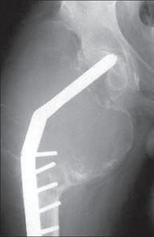
Anteroposterior (AP) radiograph of the right proximal femur demonstrating a massive, recurrent Giant Cell Tumor (GCT). The image shows a large, expansile lytic lesion involving the epiphysis and metaphysis. Notable features include significant cortical thinning and marked expansion of the bone contours, indicating an aggressive local progression. The lesion exhibits a 'soap-bubble' or trabeculated appearance characteristic of GCT, extending to the subchondral bone of the femoral head. An orthopedic fixation device, consisting of a dynamic hip screw (DHS) and side plate with multiple cortical screws, is visible. The implant's structural integrity is compromised due to the lack of adequate bony support from the extensive osteolysis, with the caption suggesting impending hardware failure. This case illustrates the challenge of managing recurrent GCT in the proximal femur, highlighting the relationship between tumor-induced bone destruction and the mechanical failure of orthopedic stabilization implants.
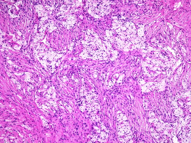
This histopathology image depicts a giant cell tumor of bone observed under light microscopy following Hematoxylin and Eosin staining. The cellular stroma consists of uniform spindle-shaped mononuclear cells interspersed with numerous osteoclast-like multinucleated giant cells. Notably, foam cells—lipid-laden macrophages—may populate the interstitium, contributing foamy cytoplasm in parts of the lesion. Giant cells contain multiple nuclei with even distribution and lack pronounced cytologic atypia; mitotic activity, if present, is typically limited. The architectural pattern can be variable, and in some cases the classic giant cell-rich areas are inconspicuous, complicating diagnosis without radiographic context. Clinically, GCTs arise in the epiphyseal/metaphyseal region of long bones in young to middle-aged adults and demonstrate local aggressiveness comparable to other bone tumors. Radiographs often reveal a soap-bubble lytic lesion with cortical thinning and expansion, providing a critical diagnostic adjunct. Differential considerations include fibrous histiocytoma and other giant cell-containing lesions. Definitive diagnosis relies on correlating histology with imaging and tumor location; immunohistochemical markers such as RANKL expression in stromal cells and tartrate-resistant acid phosphatase in giant cells can support confirmation.
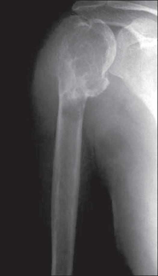
This anteroposterior radiograph of the shoulder demonstrates a large, locally aggressive lytic lesion in the proximal humerus, characteristic of a Giant Cell Tumor (GCT). The lesion is centered in the epiphysis but extends distally into the metaphysis and reaches the subchondral bone plate. Key radiographic features include a multi-loculated or 'soap-bubble' appearance created by prominent bony ridges and internal trabeculations. There is significant expansion of the bone contour with extensive cortical thinning and destruction, particularly along the lateral aspect. The margins of the lesion are relatively well-defined (geographic bone destruction) without a sclerotic rim, indicating its osteolytic nature. The absence of visible matrix mineralization and its eccentric placement in the subarticular region are classic diagnostic hallmarks of GCT in a skeletally mature patient. This image serves as an educational example of primary bone tumor morphology and the assessment of cortical integrity in musculoskeletal oncology.
~/ewing-slide/create_gct_slide.py
from pptx import Presentation
from pptx.util import Inches, Pt
from pptx.dml.color import RGBColor
from pptx.enum.text import PP_ALIGN

# ── Colour Palette ──────────────────────────────────────────────────────────
DARK_BG    = RGBColor(0x0A, 0x1A, 0x2E)   # deep navy
ACCENT1    = RGBColor(0x8B, 0x1A, 0x1A)   # deep red (title bar)
ACCENT2    = RGBColor(0xF5, 0xA6, 0x23)   # amber (highlights)
ACCENT3    = RGBColor(0x2E, 0xCC, 0xB0)   # teal (section heads)
WHITE      = RGBColor(0xFF, 0xFF, 0xFF)
LIGHT_GRAY = RGBColor(0xCC, 0xD6, 0xE0)
DARK_CARD  = RGBColor(0x14, 0x28, 0x3C)
WARN_RED   = RGBColor(0xFF, 0x55, 0x55)
GOLD       = RGBColor(0xFF, 0xD7, 0x00)

def add_rect(slide, x, y, w, h, fill_rgb):
    s = slide.shapes.add_shape(1, Inches(x), Inches(y), Inches(w), Inches(h))
    s.line.fill.background()
    s.fill.solid()
    s.fill.fore_color.rgb = fill_rgb
    return s

def tb(slide, x, y, w, h, text, sz, bold=False, color=WHITE,
       align=PP_ALIGN.LEFT, italic=False, wrap=True):
    t = slide.shapes.add_textbox(Inches(x), Inches(y), Inches(w), Inches(h))
    tf = t.text_frame
    tf.word_wrap = wrap
    tf.margin_left = Inches(0.05)
    tf.margin_right = Inches(0.04)
    tf.margin_top  = Inches(0.02)
    tf.margin_bottom = Inches(0.02)
    p = tf.paragraphs[0]
    p.alignment = align
    r = p.add_run()
    r.text = text
    r.font.size  = Pt(sz)
    r.font.bold  = bold
    r.font.italic = italic
    r.font.color.rgb = color
    r.font.name  = "Calibri"
    return tf

def multi(slide, x, y, w, h, lines, default_sz=8.5, default_color=LIGHT_GRAY):
    t = slide.shapes.add_textbox(Inches(x), Inches(y), Inches(w), Inches(h))
    tf = t.text_frame
    tf.word_wrap = True
    tf.margin_left   = Inches(0.06)
    tf.margin_right  = Inches(0.04)
    tf.margin_top    = Inches(0.03)
    tf.margin_bottom = Inches(0.02)
    first = True
    for line in lines:
        if isinstance(line, tuple):
            txt, bold, color, sz = line
        else:
            txt, bold, color, sz = line, False, default_color, default_sz
        if first:
            p = tf.paragraphs[0]; first = False
        else:
            p = tf.add_paragraph()
        p.alignment = PP_ALIGN.LEFT
        r = p.add_run()
        r.text = txt
        r.font.size  = Pt(sz)
        r.font.bold  = bold
        r.font.color.rgb = color
        r.font.name  = "Calibri"
    return tf

def card(slide, cx, cy, cw, ch, heading, lines, fsz=8.5, hcolor=DARK_BG):
    add_rect(slide, cx, cy, cw, ch, DARK_CARD)
    add_rect(slide, cx, cy, cw, 0.30, ACCENT3)
    tb(slide, cx+0.05, cy+0.01, cw-0.1, 0.28, heading,
       9.5, bold=True, color=hcolor)
    multi(slide, cx+0.05, cy+0.32, cw-0.1, ch-0.37, lines, fsz)

# ── Presentation ────────────────────────────────────────────────────────────
prs = Presentation()
prs.slide_width  = Inches(20)
prs.slide_height = Inches(11.25)
slide = prs.slides.add_slide(prs.slide_layouts[6])

W, H = 20.0, 11.25

# Background
add_rect(slide, 0, 0, W, H, DARK_BG)

# Title bar
add_rect(slide, 0, 0, W, 0.85, ACCENT1)
tb(slide, 0.15, 0.05, 14, 0.75,
   "GIANT CELL TUMOR (GCT) OF BONE  —  MBBS COMPLETE REVISION SLIDE",
   27, bold=True, color=WHITE)
tb(slide, 14.2, 0.05, 5.6, 0.75,
   "Osteoclastoma  |  Robbins & Kumar  |  Campbell's Orthopaedics",
   10, bold=False, color=RGBColor(0xFF,0xCC,0xAA),
   align=PP_ALIGN.RIGHT, italic=True)

# ── 5-column layout ─────────────────────────────────────────────────────────
COL_W = 3.78
GAP   = 0.06
TOP   = 0.92
cols_x = [0.06 + i*(COL_W + GAP) for i in range(5)]

# ═══════════════════════════════════════════════
# COL 1 — Identity + Epidemiology + Pathogenesis
# ═══════════════════════════════════════════════
cx = cols_x[0]
card(slide, cx, TOP, COL_W, 2.70, "IDENTITY & EPIDEMIOLOGY", [
    ("Also called: OSTEOCLASTOMA", True, ACCENT2, 10),
    ("", False, LIGHT_GRAY, 4),
    ("▸ Benign BUT locally aggressive tumor", True, WARN_RED, 9),
    ("▸ Age: 3rd–5th decade (20–45 yrs)", False, WHITE, 8.5),
    ("  → skeletally MATURE individuals ★", True, ACCENT2, 8.5),
    ("▸ Slight female predominance", False, LIGHT_GRAY, 8),
    ("▸ ~5% of all primary bone tumors", False, LIGHT_GRAY, 8),
    ("▸ Higher incidence in Asian populations", False, LIGHT_GRAY, 8),
    ("▸ No known predisposing factors", False, LIGHT_GRAY, 8),
])

card(slide, cx, TOP+2.77, COL_W, 3.30, "PATHOGENESIS", [
    ("TWO CELL POPULATIONS:", True, ACCENT3, 9),
    ("", False, LIGHT_GRAY, 3),
    ("1. NEOPLASTIC cells (mononuclear stromal)", True, WHITE, 9),
    ("   = primitive osteoblast precursors", False, LIGHT_GRAY, 8),
    ("   → express HIGH levels of RANKL", True, ACCENT2, 9),
    ("", False, LIGHT_GRAY, 3),
    ("2. NON-NEOPLASTIC giant cells (osteoclasts)", True, WHITE, 9),
    ("   → express RANK receptor", False, LIGHT_GRAY, 8),
    ("   → recruited by RANKL → bone resorption", False, LIGHT_GRAY, 8),
    ("", False, LIGHT_GRAY, 3),
    ("RANKL → RANK → osteoclast activation", True, ACCENT2, 9),
    ("    ↓", False, ACCENT3, 8),
    ("Loss of feedback → uncontrolled bone lysis", False, LIGHT_GRAY, 8),
    ("", False, LIGHT_GRAY, 3),
    ("Key mutation: H3.3 (H3F3A) gene mutation", False, WHITE, 8),
])

card(slide, cx, TOP+6.14, COL_W, 4.16, "GRADES (JAFFE's Classification)", [
    ("GRADE I — Benign", True, ACCENT3, 9),
    ("  ▸ Well-differentiated, minimal atypia", False, LIGHT_GRAY, 8),
    ("  ▸ Abundant giant cells", False, LIGHT_GRAY, 8),
    ("", False, LIGHT_GRAY, 3),
    ("GRADE II — Intermediate", True, ACCENT2, 9),
    ("  ▸ Moderate cellularity, mild atypia", False, LIGHT_GRAY, 8),
    ("  ▸ Most common grade", False, LIGHT_GRAY, 8),
    ("", False, LIGHT_GRAY, 3),
    ("GRADE III — Malignant GCT", True, WARN_RED, 9),
    ("  ▸ Prominent atypia + numerous mitoses", False, LIGHT_GRAY, 8),
    ("  ▸ Sarcomatous transformation", False, LIGHT_GRAY, 8),
    ("  ▸ Rare; can follow radiation therapy", False, LIGHT_GRAY, 8),
])

# ═══════════════════════════════════════════════
# COL 2 — Sites + Clinical
# ═══════════════════════════════════════════════
cx = cols_x[1]
card(slide, cx, TOP, COL_W, 3.60, "SITES OF INVOLVEMENT", [
    ("KEY: EPIPHYSIS of long bones ★★★", True, ACCENT2, 10.5),
    ("(extends to subchondral bone plate)", False, LIGHT_GRAY, 8),
    ("", False, LIGHT_GRAY, 3),
    ("MOST COMMON SITES (50% around knee):", True, ACCENT3, 9),
    ("  1. Distal FEMUR (most common overall)", True, WHITE, 9),
    ("  2. Proximal TIBIA", True, WHITE, 9),
    ("  3. Distal RADIUS", False, WHITE, 8.5),
    ("  4. Proximal HUMERUS", False, WHITE, 8.5),
    ("  5. Pelvis / Sacrum", False, WHITE, 8.5),
    ("", False, LIGHT_GRAY, 3),
    ("LOCATION COMPARISON:", True, ACCENT2, 8.5),
    ("Ewing's  → DIAPHYSIS  (5–25 yrs)", False, LIGHT_GRAY, 8),
    ("Osteosar → METAPHYSIS (10–20 yrs)", False, LIGHT_GRAY, 8),
    ("GCT      → EPIPHYSIS  (20–45 yrs) ★", True, ACCENT2, 8.5),
])

card(slide, cx, TOP+3.67, COL_W, 3.20, "CLINICAL FEATURES", [
    ("▸ PAIN — most common symptom", True, WHITE, 9),
    ("  dull, aching; worse on movement", False, LIGHT_GRAY, 8),
    ("▸ Swelling / local tenderness", False, WHITE, 8.5),
    ("▸ Joint symptoms (arthritis-like)", False, WHITE, 8.5),
    ("  (due to proximity to joint)", False, LIGHT_GRAY, 8),
    ("▸ Pathologic fracture — may occur", True, WARN_RED, 9),
    ("▸ Limited ROM of adjacent joint", False, LIGHT_GRAY, 8),
    ("▸ Rarely: lung metastases (4%)", True, ACCENT2, 8.5),
    ("  → behave benignly — excision curative", False, LIGHT_GRAY, 8),
])

card(slide, cx, TOP+6.94, COL_W, 3.36, "INVESTIGATIONS", [
    ("▸ X-ray          — first line", True, WHITE, 9),
    ("▸ MRI             — best for soft tissue", True, ACCENT2, 9),
    ("  extent + intraosseous spread", False, LIGHT_GRAY, 8),
    ("▸ CT scan         — cortical destruction", False, WHITE, 8.5),
    ("▸ Bone scan       — multifocal disease", False, LIGHT_GRAY, 8),
    ("▸ Biopsy          — definitive Dx", True, WHITE, 9),
    ("▸ Serum Ca²⁺      — normal (vs Brown tumor!)", True, ACCENT2, 9),
    ("▸ Serum PTH       — normal", False, LIGHT_GRAY, 8),
])

# ═══════════════════════════════════════════════
# COL 3 — Histology + Radiology
# ═══════════════════════════════════════════════
cx = cols_x[2]
card(slide, cx, TOP, COL_W, 4.50, "HISTOLOGY (MICRO) ★", [
    ("TWO CELL TYPES:", True, ACCENT3, 9.5),
    ("", False, LIGHT_GRAY, 3),
    ("1. MULTINUCLEATED GIANT CELLS", True, WHITE, 10),
    ("   → osteoclast-type, NON-neoplastic", False, LIGHT_GRAY, 8),
    ("   → 100+ nuclei per cell", True, ACCENT2, 8.5),
    ("   → TRAP positive (tartrate-resistant", False, LIGHT_GRAY, 8),
    ("     acid phosphatase)", False, LIGHT_GRAY, 8),
    ("", False, LIGHT_GRAY, 3),
    ("2. MONONUCLEAR STROMAL CELLS", True, WHITE, 10),
    ("   → uniform, oval — THE NEOPLASTIC cells", True, ACCENT2, 9),
    ("   → RANKL expressing", False, LIGHT_GRAY, 8),
    ("", False, LIGHT_GRAY, 3),
    ("★ KEY: Nuclei of giant cells LOOK IDENTICAL", True, GOLD, 9.5),
    ("  to nuclei of stromal cells", True, GOLD, 9),
    ("  (all nuclei appear similar — pathognomonic)", False, LIGHT_GRAY, 8),
    ("", False, LIGHT_GRAY, 3),
    ("Gross: Red-brown mass + cystic degeneration", False, WHITE, 8.5),
    ("Hemorrhagic background + hemosiderin", False, LIGHT_GRAY, 8),
])

card(slide, cx, TOP+4.57, COL_W, 2.80, "RADIOLOGY ★★", [
    ("PLAIN X-RAY:", True, ACCENT3, 9.5),
    ("▸ SOAP BUBBLE appearance ★★★", True, ACCENT2, 10.5),
    ("  (internal trabeculations)", False, LIGHT_GRAY, 8),
    ("▸ ECCENTRIC lytic lesion", True, WHITE, 9),
    ("▸ Extends to subchondral bone", True, WHITE, 9),
    ("▸ NO sclerotic rim (aggressive)", True, WARN_RED, 9),
    ("▸ Cortical thinning + ballooning", False, WHITE, 8.5),
    ("▸ NO periosteal reaction (unlike Ewing's)", True, WARN_RED, 9),
    ("▸ NO matrix calcification", False, LIGHT_GRAY, 8),
])

card(slide, cx, TOP+7.44, COL_W, 2.86, "GROSS PATHOLOGY", [
    ("▸ Typically destroys overlying cortex", False, WHITE, 8.5),
    ("▸ Bulging soft tissue mass bounded by", False, WHITE, 8.5),
    ("  thin shell of reactive bone", False, LIGHT_GRAY, 8),
    ("▸ Grossly: RED-BROWN mass ★", True, ACCENT2, 9),
    ("▸ Frequently undergoes cystic degeneration", False, LIGHT_GRAY, 8),
    ("▸ Hemorrhagic areas common", False, LIGHT_GRAY, 8),
])

# ═══════════════════════════════════════════════
# COL 4 — Treatment + Recurrence + Prognosis
# ═══════════════════════════════════════════════
cx = cols_x[3]
card(slide, cx, TOP, COL_W, 4.40, "TREATMENT", [
    ("PRIMARY: CURETTAGE + ADJUVANTS", True, ACCENT3, 10),
    ("", False, LIGHT_GRAY, 3),
    ("▸ Extended curettage with high-speed burr", False, WHITE, 8.5),
    ("  + large cortical window", False, LIGHT_GRAY, 8),
    ("", False, LIGHT_GRAY, 3),
    ("ADJUVANTS (reduce recurrence):", True, ACCENT2, 9),
    ("  ▸ Phenol (chemical cautery)", False, WHITE, 8.5),
    ("  ▸ Cryosurgery (liquid nitrogen)", False, WHITE, 8.5),
    ("  ▸ PMMA bone cement ★", True, ACCENT2, 8.5),
    ("    (polymethylmethacrylate — thermal effect)", False, LIGHT_GRAY, 8),
    ("  ▸ Argon beam coagulation", False, LIGHT_GRAY, 8),
    ("", False, LIGHT_GRAY, 3),
    ("DENOSUMAB (RANKL inhibitor) ★★", True, GOLD, 10),
    ("  ▸ Monoclonal Ab against RANKL", False, WHITE, 8.5),
    ("  ▸ For unresectable / recurrent cases", False, LIGHT_GRAY, 8),
    ("  ▸ When resection would be deforming", False, LIGHT_GRAY, 8),
    ("", False, LIGHT_GRAY, 3),
    ("Wide resection — for aggressive/recurrent", False, WHITE, 8.5),
    ("Amputation — rarely, if unresectable", False, LIGHT_GRAY, 8),
    ("After pathologic fracture — wide excision", False, WHITE, 8),
    ("  ± reconstruction / amputation", False, LIGHT_GRAY, 8),
])

card(slide, cx, TOP+4.47, COL_W, 2.50, "RECURRENCE", [
    ("Local recurrence rate: 40–60% ⚠", True, WARN_RED, 10),
    ("(simple curettage alone)", False, LIGHT_GRAY, 8),
    ("", False, LIGHT_GRAY, 3),
    ("▸ Adjuvants reduce recurrence to ~10–15%", False, WHITE, 8.5),
    ("▸ Most recurrences within 2–3 years", False, LIGHT_GRAY, 8),
    ("▸ Serial follow-up mandatory", False, LIGHT_GRAY, 8),
])

card(slide, cx, TOP+7.04, COL_W, 3.26, "PROGNOSIS & BEHAVIOR", [
    ("▸ Generally BENIGN — curable with surgery", True, ACCENT3, 9),
    ("▸ HIGH local recurrence (40–60%)", True, WARN_RED, 9),
    ("▸ Lung metastases in ~4% cases", True, ACCENT2, 9),
    ("  → BENIGN behavior even in lung", False, LIGHT_GRAY, 8),
    ("  → most cured by excision of mets", False, LIGHT_GRAY, 8),
    ("▸ Malignant transformation: RARE (~1–2%)", True, WARN_RED, 9),
    ("  (especially post-radiation — sarcoma)", False, LIGHT_GRAY, 8),
    ("▸ Multicentric GCT: extremely rare", False, LIGHT_GRAY, 8),
])

# ═══════════════════════════════════════════════
# COL 5 — Differentials + Comparison + Exam Traps
# ═══════════════════════════════════════════════
cx = cols_x[4]
card(slide, cx, TOP, COL_W, 3.60, "DIFFERENTIAL DIAGNOSIS", [
    ("▸ BROWN TUMOR of hyperparathyroidism", True, WARN_RED, 9.5),
    ("  (most important DDx!)", False, LIGHT_GRAY, 8),
    ("  → multiple lesions, ↑Ca²⁺, ↑PTH", True, ACCENT2, 8.5),
    ("  → GCT: single lesion, normal Ca²⁺", True, ACCENT3, 8.5),
    ("", False, LIGHT_GRAY, 3),
    ("▸ Aneurysmal Bone Cyst (ABC)", False, WHITE, 8.5),
    ("  → younger age, blood-filled cysts", False, LIGHT_GRAY, 8),
    ("▸ Chondroblastoma", False, WHITE, 8.5),
    ("  → also epiphyseal, <20 yrs, calcifications", False, LIGHT_GRAY, 8),
    ("▸ Osteosarcoma with giant cells", False, WHITE, 8.5),
    ("▸ Non-ossifying fibroma", False, LIGHT_GRAY, 8),
    ("▸ Fibrous histiocytoma", False, LIGHT_GRAY, 8),
])

card(slide, cx, TOP+3.67, COL_W, 2.80, "MNEMONICS", [
    ('"GREAT KNEES GET GCT"', True, ACCENT2, 9.5),
    ("→ Distal femur + Proximal tibia = MC sites", False, WHITE, 8.5),
    ("", False, LIGHT_GRAY, 3),
    ('"SOAP BUBBLE + RED-BROWN + EPIPHYSIS"', True, GOLD, 9.5),
    ("= GCT until proven otherwise", False, WHITE, 8.5),
    ("", False, LIGHT_GRAY, 3),
    ('"RANK + RANKL = GCT mechanism"', True, ACCENT3, 9),
    ("→ Denosumab blocks RANKL = treatment", False, WHITE, 8.5),
    ("", False, LIGHT_GRAY, 3),
    ("Age 20–45 + SKELETALLY MATURE ★", True, ACCENT2, 8.5),
])

card(slide, cx, TOP+6.54, COL_W, 3.76, "⚠  EXAM TRAPS", [
    ("1. GCT = EPIPHYSIS (NOT metaphysis/diaphysis)", True, WARN_RED, 9),
    ("2. Skeletally MATURE — NOT children", True, WARN_RED, 9),
    ("3. SOAP BUBBLE X-ray = GCT ★", True, WARN_RED, 9),
    ("4. Nuclei of giant cells = SAME as stromal", True, GOLD, 9),
    ("   cells (identical nuclei — KEY micro feature)", False, LIGHT_GRAY, 8),
    ("5. Giant cells = NON-neoplastic (osteoclasts)", True, WARN_RED, 9),
    ("   Stromal cells = THE neoplastic cells", True, WARN_RED, 9),
    ("6. GCT mets to LUNG but behave BENIGNLY", True, ACCENT2, 8.5),
    ("7. Brown tumor: ↑Ca²⁺ / ↑PTH; GCT = normal", True, ACCENT2, 8.5),
    ("8. Local recurrence = 40–60% (very high!)", True, WARN_RED, 8.5),
    ("9. Denosumab = RANKL inhibitor for GCT", True, ACCENT2, 8.5),
    ("10. Malignant transformation rare but↑ with RT", False, LIGHT_GRAY, 8),
])

# Bottom banner
add_rect(slide, 0, 10.88, W, 0.37, RGBColor(0x12, 0x2A, 0x42))
tb(slide, 0.15, 10.90, W-0.3, 0.30,
   "KEY FACTS: Epiphysis  |  Age 20–45 yrs  |  Soap Bubble X-ray  |  Red-Brown Mass  |  Giant Cells = NON-neoplastic  |  Stromal cells = Neoplastic (RANKL+)  |  Recurrence 40–60%  |  Denosumab (RANKL inhibitor)",
   8.5, bold=False, color=ACCENT2, align=PP_ALIGN.CENTER)

out = "/tmp/workspace/ewing-slide/Giant_Cell_Tumor_MBBS.pptx"
prs.save(out)
print(f"Saved → {out}")
python /tmp/workspace/ewing-slide/create_gct_slide.py