
Hi
Who are you
Explain deeply mitral valve replacement
mitral valve replacement prosthetic valve surgery
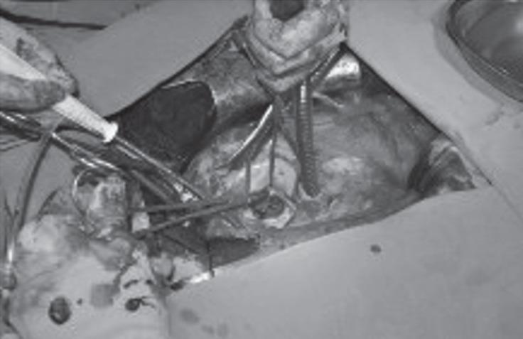
This grayscale clinical photograph provides an intraoperative view during a redo cardiac surgery, specifically a mitral valve replacement (MVR). The surgical field is exposed via a resternotomy and a left atriotomy, revealing the mitral position. Central to the image is a mechanical prosthetic valve that is visibly obstructed by a dense, adhesive thrombotic mass, characteristic of prosthetic valve thrombosis (PVT). This pathological finding results in a 'stuck' valve with immobile or restricted leaflets. Various surgical instruments, including retractors and forceps, are actively manipulating the tissue to provide exposure. Additionally, cardiopulmonary bypass (CPB) components, such as suction cannulae and perfusion tubing, are visible within the operative field. This image serves as an educational example of mechanical valve complications and the surgical approach required for emergency thrombectomy or redo valve replacement in the context of acute valve failure.
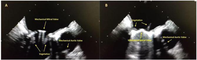
This diagnostic image consists of two side-by-side transesophageal echocardiography (TEE) frames (labeled A and B) captured at 120 degrees, focusing on the mitral and aortic valve regions. The ultrasound demonstrates a mechanical prosthetic bileaflet mitral valve characterized by bright, linear echogenic signals with acoustic shadowing. Pathological findings include multiple, large, irregular, and heterogeneous masses consistent with vegetations attached to both the atrial and ventricular surfaces of the mechanical mitral valve leaflets. In image A, vegetations are seen protruding downward into the left ventricle, while image B shows superiorly projecting masses. Adjacent to the mitral apparatus, a second echogenic mechanical prosthetic aortic valve is visible. These findings are clinically indicative of prosthetic valve endocarditis (PVE). This image serves as an educational tool for identifying cardiac vegetations, recognizing the appearance of mechanical valves on ultrasound, and diagnosing infectious complications following valve replacement surgery.
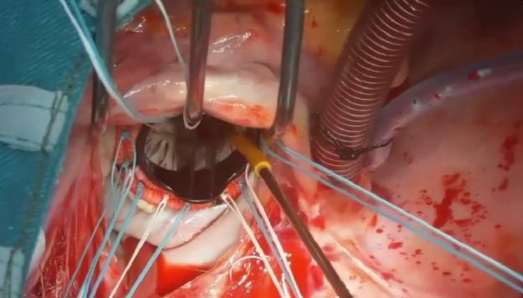
This clinical photograph captures an intraoperative view of a mitral valve replacement (MVR) using an eversion surgical technique. Centrally positioned within the left atriotomy is a St. Jude Masters bileaflet mechanical prosthetic valve. The valve features two dark carbon leaflets within a low-profile circular housing, surrounded by a sewing cuff. Multiple blue and white 2-0 polyester mattress sutures are visible, anchored through the mitral annulus and the prosthetic sewing ring to secure the valve in place. The surgical field includes specialized cardiac instruments, such as a metal retractor with three curved prongs providing exposure of the atrial chamber. In the upper right quadrant, surgical tubing and a ribbed cannula related to cardiopulmonary bypass or suction are visible. The surrounding cardiac tissue is partially covered by remnants of the native mitral valve leaflets, which have been everted to facilitate the implantation of a larger prosthetic valve. This image demonstrates key steps in advanced valvular cardiac surgery, emphasizing prosthetic seating and suture management.
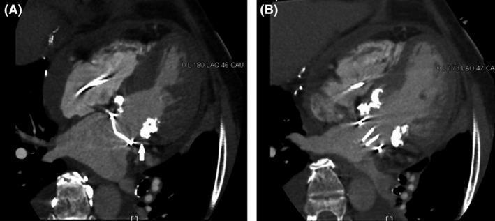
Postoperative multidetector computed tomography (MDCT) scans of the heart in a patient after mitral valve replacement (MVR) for severe mitral stenosis and extensive calcification. Image (A) displays the systolic phase, showing the St. Jude Medical (SJM) bileaflet mechanical valve in a closed position. A white arrow highlights the anatomical relationship where the prosthetic valve is displaced from the posterior mitral annulus due to significant, hyperdense calcification extending toward the left atrium. Image (B) displays the diastolic phase, demonstrating the adequate opening of the mechanical valve leaflets. The MDCT highlights that the prosthetic structure is unobstructed by the subannular apparatus, confirming functional integrity despite severe regional calcification. These cardiac images illustrate complex valve positioning in the presence of extensive annular calcification, relevant for cardiothoracic surgery and diagnostic radiology educational levels.
"mitral valve replacement" outcomes
Mitral Valve Replacement (MVR) - A Deep Explanation
1. Anatomy and Pathophysiology Basis
- Mitral stenosis (MS): obstruction to LV inflow, causing LA enlargement, pulmonary hypertension, and reduced cardiac output
- Mitral regurgitation (MR): backflow into the LA during systole, causing volume overload and progressive LV dilation/dysfunction
2. Indications for MVR (When Repair Is Not Enough)
- The valve is severely deformed (e.g., rheumatic disease with heavy calcification, dense fibrosis)
- Repair is technically not feasible or likely to fail
- Previous repair has failed
- Severe ischemic MR in select patients - the 2020 ACC/AHA guidelines state it is reasonable (Class IIb) to choose chordal-sparing MVR over downsized annuloplasty repair in symptomatic patients with chronic severe ischemic MR, given high rates of repair failure (~59% MR recurrence at 2 years vs. 3.8% after replacement)
- Severe MS not amenable to balloon mitral valvuloplasty (BMV) - calcified valve, dense subvalvular disease, or prior failed BMV
3. Surgical Technique
- Systemic anticoagulation with heparin; CPB initiated
- Aortic cross-clamp; cardioplegic cardiac arrest
- Left atriotomy (incision behind the interatrial groove)
- Diseased valve exposed and assessed
- Excision of leaflets and subvalvular apparatus (in non-chordal-sparing) OR preservation of chordae/papillary attachments
- Sizing the mitral annulus
- Prosthetic valve seated with interrupted mattress sutures through the annulus
- De-airing of the left heart (critical - to prevent air embolism)
- Atriotomy closure; CPB weaning
- Intraoperative transesophageal echocardiography (TOE/TEE) used to confirm adequate valve function
Chordal Sparing vs. Non-Chordal Sparing
| Feature | Chordal Sparing | Without Chordal Sparing |
|---|---|---|
| LV function | Better preserved | Risk of sudden LV dysfunction |
| When used | Most modern MVR | Severe rheumatic deformity |
| Hospital mortality | ~4-5% | Higher |
| Concept introduced | Lillehei, 1964 | Earlier era |
4. Prosthetic Valve Choice: Mechanical vs. Bioprosthetic
| Feature | Mechanical Valve | Bioprosthetic (Tissue) Valve |
|---|---|---|
| Durability | Lifelong (>25-30 yrs) | 10-15 years (faster failure in young patients) |
| Anticoagulation | Lifelong warfarin required | Usually short-term (3-6 months) post-op |
| Thromboembolism risk | Higher (need INR 2.5-3.5) | Lower |
| Bleeding risk | Higher (from anticoagulation) | Lower |
| Reoperation risk | Low (valve lasts) | Higher (structural deterioration) |
| Sound | Audible mechanical click | Resembles native valve sounds |
- Mechanical valve is preferred when: the patient already has another indication for anticoagulation (e.g., AF), is younger with a longer life expectancy and wants to minimize reoperation
- Bioprosthetic valve is preferred when: life expectancy <15 years, patient unable/unwilling to maintain warfarin, high bleeding risk, or patient preference
- Long-term survival with mitral bioprosthetic vs. mechanical valves is similar (unlike aortic position, where mechanical valves offer a survival advantage). - Goldman-Cecil Medicine
- Bioprosthetic valves fail faster in younger patients (<50 years), for poorly understood reasons
5. Postoperative Management
- All mechanical MVR patients require lifelong warfarin (VKA). Target INR: 3.0 for mitral mechanical valves (higher than aortic due to greater thromboembolic risk), plus low-dose aspirin (75-100 mg/day)
- Mitral mechanical valves carry twice the embolic risk of aortic mechanical valves
- Bioprosthetic MVR: VKA for 3-6 months post-op, then consider aspirin alone
- Baseline echocardiogram post-op (reference for future comparison)
- No routine echo follow-up unless symptoms or clinical findings change
- For any fever >100°F in a prosthetic valve patient: blood cultures mandatory, endocarditis must be excluded
- Endocarditis prophylaxis before high-risk dental/surgical procedures
| Complication | Rate / Notes |
|---|---|
| Operative mortality (elective) | ~4-5% (up to 5%) depending on LV function and comorbidities |
| Stroke | <3% in-hospital |
| Renal failure | ~3% |
| Prosthetic valve thrombosis | Emergency - presents as stuck valve, hemodynamic collapse |
| Prosthetic valve endocarditis | Life-threatening; usually requires redo surgery |
| Paravalvular leak | Incomplete seating of prosthesis; may require reoperation |
| LV dysfunction | From loss of mitral apparatus (non-chordal-sparing) |
| Complete heart block | Especially with concomitant tricuspid + mitral valve surgery (up to 28%) |
6. Transcatheter Mitral Valve Replacement (TMVR)
- Valve-in-Ring (ViR): Replacing a failed surgical annuloplasty ring with a transcatheter valve
- Valve-in-Valve (ViV): Replacing a failed bioprosthetic mitral valve (approved, good outcomes data)
- Native annulus TMVR: Used especially in severe mitral annular calcification (MAC) - the calcification provides rigid anchoring for the device. Technical success is promising but the procedure remains experimental for native valve disease
7. Repair vs. Replacement - The Central Decision
- 30-day mortality: 1.6% (repair) vs. 4% (replacement) - not significantly different
- 2-year survival: 81% vs. 76.8% (P = 0.39, not significant)
- MR recurrence: 59% after repair vs. only 3.8% after replacement (P <0.001)
- Cardiovascular rehospitalization: higher after repair (48.3 vs. 32.2 per 100 patient-years)
8. Prognosis
- Degree of preoperative LV dysfunction (EF and end-systolic dimension)
- Presence of pulmonary hypertension
- Comorbidities (coronary artery disease, AF, renal function)
- Whether concomitant CABG or tricuspid repair was needed
- Choice and sizing of prosthesis
Wow good explanation