
Sites to check pallor
- Conjunctiva (lower palpebral) - most reliable site
- Nail beds
- Palmar creases
- Tongue / oral mucosa
- Lips
- Ear lobes
- Skin (face, forehead)
pallor examination conjunctiva nail bed clinical signs
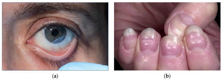
This composite of clinical photographs illustrates physical manifestations of severe anemia. Image (a) shows a close-up of a patient's eye with the lower eyelid retracted to reveal marked conjunctival pallor. The typically pink or reddish palpebral conjunctiva appears significantly pale and porcelain-white, a sign classically associated with a hemoglobin concentration below 90 g/L. Image (b) presents the fingernails of a patient demonstrating pronounced nail bed pallor. The distal nail beds lack the normal healthy pink hue. Additionally, horizontal white bands, known as Muehrcke's lines, are visible across the nail plates; these non-palpable transverse lines are associated with systemic conditions such as hypoalbuminemia and severe anemia. Together, these images demonstrate key external clinical signs used in the bedside assessment of hematological disorders and circulatory efficiency. The content is suitable for medical education regarding the physical examination findings of iron deficiency and other severe anemias.
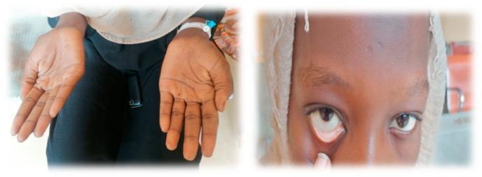
This composite clinical photograph illustrates common physical examination findings associated with severe anemia. The left panel shows the bilateral palms of a patient, demonstrating marked palmar pallor. The skin on the palms and creases appears significantly lighter and lacks the typical pinkish hue of healthy vascularized tissue, suggesting reduced hemoglobin concentration. The right panel is a close-up of the patient's face with the lower eyelid retracted to reveal the palpebral conjunctiva. Instead of the normal vibrant red or pink color, the conjunctiva exhibits conjunctival pallor, appearing whitish or very pale pink. These visual signs are critical diagnostic markers in hematology and general medicine for identifying systemic conditions such as iron-deficiency anemia or other forms of chronic blood loss. The image serves as a teaching tool for medical students and clinicians to recognize mucosal and cutaneous indicators of decreased red blood cell mass or oxygen-carrying capacity.
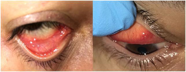
Clinical photograph showing an ophthalmological examination of a right eye. The image consists of two side-by-side panels demonstrating the eversion of the lower and upper eyelids to inspect the palpebral conjunctiva. Key visible clinical signs include pronounced conjunctival injection (redness) and mild chemosis (edema of the conjunctiva). A follicular reaction is evident, characterized by multiple small, raised, translucent bumps within the upper and lower conjunctival fornices. Additionally, a clear serous secretion is visible, indicating a non-purulent inflammatory process. The cornea appears clear in the visible portions. These findings are typical of acute follicular conjunctivitis, which in this clinical context was identified as a manifestation of viral infection, specifically SARS-CoV-2. The image serves as an educational reference for identifying follicular patterns and inflammatory changes in viral conjunctivitis.
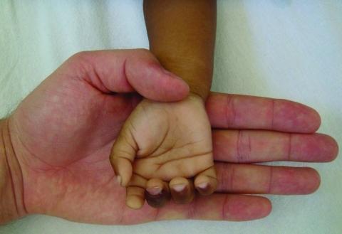
This clinical photograph provides a side-by-side comparison between the palmar surface of a 2-year-old child's hand and that of a healthy adult. The image serves as a classic visual demonstration of severe clinical pallor. The child's hand, associated with a diagnostic hemoglobin level of 3.6 g/dL (severe anemia), exhibits a marked loss of normal pinkish hue, appearing significantly lighter and almost white compared to the adult hand. This contrast is most evident in the palmar creases and the nail beds, which lack the typical capillary refill color. The adult hand displays normal skin perfusion and saturation, providing a reference point for healthy oxygenation. This visual aid is primarily used in hematology and pediatrics to teach the physical signs of extreme anemia, often secondary to conditions such as Plasmodium vivax malaria infection as indicated by the clinical context. It illustrates the importance of physical examination in identifying life-threatening reductions in red blood cell mass or hemoglobin concentration.
Sites to Check Pallor
Primary (Most Reliable) Sites
| Site | How to Examine | Clinical Significance |
|---|---|---|
| Lower palpebral conjunctiva | Pull down lower eyelid and inspect the inner surface | Most specific; pallor here generally indicates Hb < 9 g/dL |
| Palmar creases | Extend the fingers by pushing the palm flat | If creases fail to become pink when extended, Hb is usually ≤ 7-8 g/dL |
| Nail beds | Press the nail, release, observe capillary refill and color | Pink hue is normally present; pallor with poor capillary fill suggests anemia |
Secondary Sites
| Site | Notes |
|---|---|
| Oral mucosa / tongue | Pink normally; pale in anemia |
| Lips | Easily visible; useful in all skin types |
| Ear lobes | Thin skin, useful in pigmented patients |
| Face / forehead skin | Less specific; affected by ambient temperature and vasoconstriction |
| Fundus (retina) | Retinal pallor or hemorrhages in severe anemia (Hb very low) |
Why Conjunctiva and Palmar Creases Are Preferred
- These sites are free from melanin pigmentation
- The mucosa/vasculature is superficial and directly visible
- They are not affected by peripheral vasoconstriction as much as the skin
Clinical Images
Key Points to Remember
- Conjunctiva > Palms > Nail beds in terms of specificity for anemia
- Pallor of conjunctiva: Hb usually < 9 g/dL
- Pallor of palmar creases (flat on extension): Hb usually ≤ 7-8 g/dL
- Skin pallor alone is not specific - vasoconstriction, fear, cold, or shock can all cause it without anemia
- Always examine multiple sites before concluding presence or absence of pallor