
Dermatomyositis
"dermatomyositis"[MeSH Terms]
dermatomyositis heliotrope rash Gottron papules
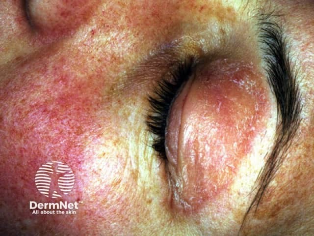
Clinical photography of the left periorbital region demonstrating the hallmark heliotrope rash associated with dermatomyositis. The image shows erythematous to violaceous papery skin surrounding the eyelids with mild edema and rough, flaky texture. The involved lid margin is reddened and slightly swollen; the ambient facial skin displays diffuse erythema and subtle scaling consistent with inflammatory dermatosis. In dermatomyositis, heliotrope rash is a pathognomonic cutaneous manifestation; it may precede or accompany proximal muscle weakness and elevated muscle enzymes. The appearance is typically bilateral and symmetric around the orbit, with sparing of mucous membranes. Additional signs to assess include Gottron papules over extensor surfaces and shawl sign; however these are not visible in this focal image. This photograph serves as documentation of cutaneous involvement used in clinical assessment, disease monitoring, and educational reference for dermatology, rheumatology, and neurology. The image is suitable for keyword-rich indexing: periorbital rash, heliotrope eruption, dermatomyositis cutaneous signs, eyelid edema, violaceous erythema, facial dermatitis, inflammatory dermatosis, autoimmune myopathy, DM, proximal muscle weakness correlation, differential diagnoses include eczema, lupus pernio, contact dermatitis, photosensitive dermatitis. Clinical relevance includes screening for underlying malignancy in adults and guiding multidisciplinary evaluation for myositis.
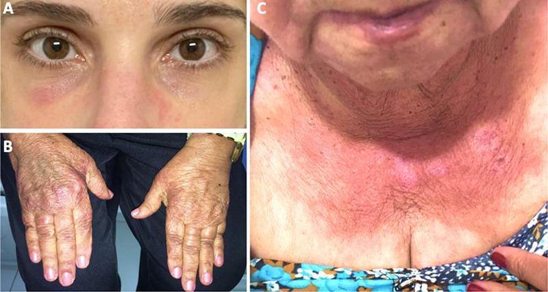
A composite of three clinical photographs demonstrating the classic cutaneous manifestations of dermatomyositis, an idiopathic inflammatory myopathy. Panel A: A heliotrope rash characterized by a reddish-purple (violaceous) discoloration in the periorbital region, involving the eyelids and malar area. Panel B: Gottron papules/sign, presenting as erythematous to violaceous, slightly scaly patches overlying the dorsal aspect of the metacarpophalangeal and interphalangeal joints. Panel C: The 'V-sign,' a confluent erythematous rash in a photosensitive distribution across the anterior lower neck and upper chest. These findings are pathognomonic for dermatomyositis and are critical for clinical diagnosis, often preceding or accompanying proximal muscle weakness. The images highlight key dermatologic markers used to differentiate dermatomyositis from other connective tissue diseases and inflammatory myopathies like polymyositis.
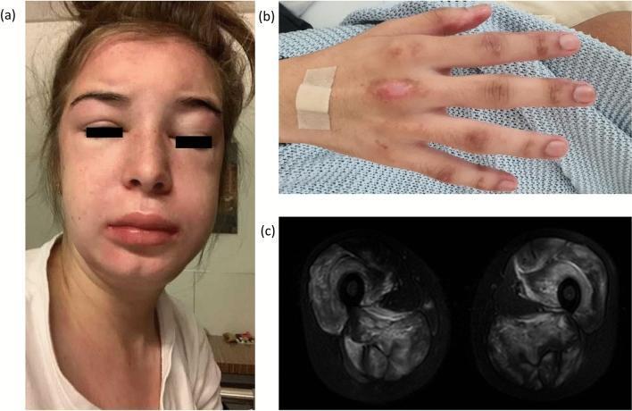
A multi-panel clinical figure documenting classic cutaneous and musculoskeletal manifestations of dermatomyositis. Panel (a) is a clinical photograph of a patient's face demonstrating diffuse mid-facial edema and heliotrope rash, characterized by violaceous erythema concentrated in the periorbital and malar regions. Panel (b) shows the dorsal aspect of a hand with Gottron papules—erythematous, slightly scaly, lichenoid papules overlying the metacarpophalangeal and interphalangeal joints. Panel (c) provides an axial T2-weighted magnetic resonance imaging (MRI) scan of the thighs, showing bilateral, symmetric hyperintensities within the adductor and extensor muscle compartments, indicative of active inflammatory myositis with intramuscular edema. This collection of images illustrates the diagnostic triad of dermatomyositis: pathognomonic skin lesions and objective evidence of proximal muscle inflammation.
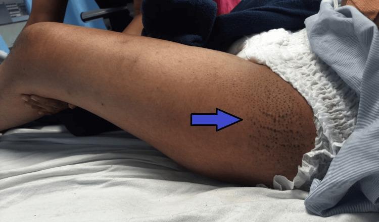
A clinical photograph displaying the lateral aspect of a patient's right thigh. A blue arrow highlights a localized area of reddish-purple to hyperpigmented discoloration with a mottled, reticulated appearance. This dermatological finding is known as the 'holster sign,' characterized by an erythematous rash on the lateral thigh, distal to the greater trochanter. The rash lacks sharply defined margins and fades into the surrounding skin. This finding is a classic cutaneous manifestation of dermatomyositis, an idiopathic inflammatory myopathy. The image serves as an educational tool for identifying systemic autoimmune disease markers and understanding the diverse dermatologic presentations of myositis beyond the more common heliotrope rash and Gottron papules.
Quick answer
Dermatomyositis
Epidemiology
- It has a bimodal distribution: juvenile (about 5 to 15 years) and adult (about 40 to 60 years).
- It is more common in women.
- In adults, it is associated with malignancy in roughly 15 to 25% of cases. The risk is highest within 3 years of diagnosis, and ovarian cancer is a notable association. Other associations are lung, breast, colorectal, pancreatic, gastric, and nasopharyngeal cancers (nasopharyngeal especially in Asian patients).
Clinical features
- Heliotrope rash: violaceous erythema of the upper eyelids, often with periorbital edema.
- Gottron papules: violaceous, flat-topped papules over the MCP, PIP, and DIP joints.
- Gottron sign: erythema over the elbows, knees, and malleoli.
- Photodistributed poikiloderma: shawl sign (shoulders and upper back), V-sign (neck and chest), and holster sign (lateral thighs).
- Nailfold changes: periungual telangiectasia and ragged cuticles (the "Samitz sign").
- Mechanic's hands: more typical of antisynthetase syndrome.
- Scalp involvement: psoriasiform, with itching.
- Calcinosis cutis: more common in juvenile DM.
- Symmetric proximal weakness: difficulty rising from a chair, climbing stairs, and raising the arms.
- Neck flexor weakness.
- Myalgia can occur.
- Interstitial lung disease.
- Dysphagia from pharyngeal and upper esophageal weakness.
- Cardiac involvement: arrhythmias and cardiomyopathy.
- Arthritis.
Myositis-specific antibodies
| Antibody | Associations |
|---|---|
| Anti-Mi-2 | Classic DM with a good prognosis; prominent cuticular overgrowth |
| Anti-TIF1-gamma (p155/140) | Cancer-associated DM in adults; severe skin disease |
| Anti-NXP2 | Calcinosis; cancer in adults; juvenile DM |
| Anti-MDA5 | Amyopathic DM; rapidly progressive ILD; skin ulcers |
| Anti-SAE | Initial skin disease, later myopathy; dysphagia |
| Anti-Jo-1 and other antisynthetase antibodies | Antisynthetase syndrome with ILD, mechanic's hands, and arthritis |
Diagnosis
- Muscle enzymes: CK (may be normal in DM or amyopathic disease), aldolase, AST, ALT, and LDH.
- Antibodies: myositis-specific and myositis-associated panels, and ANA.
- EMG: myopathic pattern with irritability.
- MRI: T2/STIR hyperintensity showing muscle edema, which can also guide biopsy site.
- Muscle biopsy:
- Perifascicular atrophy.
- Perivascular and perimysial inflammation (B cells, CD4+ T cells, and plasmacytoid dendritic cells).
- Capillary loss with membrane attack complex deposition.
- MxA staining.
- Skin biopsy: interface dermatitis with mucin deposition. This resembles lupus and cannot reliably distinguish it.
- Staging and screening:
- High-resolution CT chest and pulmonary function tests.
- Age-appropriate cancer screening plus targeted imaging, such as CT chest/abdomen/pelvis.
- Swallow evaluation if there is dysphagia.
- ECG.
- Polymyositis
- Inclusion body myositis
- Immune-mediated necrotizing myopathy (statin-associated)
- Lupus
- Systemic sclerosis and mixed connective tissue disease
- Hypothyroidism
- Drug myopathy
- Muscular dystrophies
- Metabolic myopathies
Management
- First line:
- Sun protection with sunscreen.
- Systemic glucocorticoids, such as prednisone 1 mg/kg/day, tapered over months.
- A steroid-sparing agent started early.
- Steroid-sparing agents: methotrexate, azathioprine, or mycophenolate. Mycophenolate is favored when there is ILD.
- Skin disease:
- Topical steroids or calcineurin inhibitors.
- Hydroxychloroquine, though it is less effective than in lupus and has a higher rash risk.
- Antimalarials.
- Refractory or severe disease:
- IVIG, which has randomized trial evidence (ProDERM) for DM and is useful for dysphagia and skin disease.
- Rituximab.
- Tacrolimus or cyclosporine for ILD.
- Cyclophosphamide.
- JAK inhibitors, such as tofacitinib or baricitinib, used especially in anti-MDA5 disease and refractory skin disease.
- Rapidly progressive ILD (anti-MDA5): early aggressive triple therapy with high-dose steroids, a calcineurin inhibitor, and cyclophosphamide or other agents.
- Supportive care:
- Physical therapy, with exercise once the disease is controlled.
- Bone protection.
- Monitoring for infections (PJP prophylaxis if on high-dose immunosuppression).
- Treatment of the underlying cancer when present.
Prognosis
Recent evidence flag
- A 2026 systematic review of clinically amyopathic DM (PMID 42301567).
- A 2026 systematic review of long-term juvenile DM outcomes (PMID 42167619).
- A 2024 JAMA Dermatology systematic review of drug-induced DM (PMID 38198130). Culprits include immune checkpoint inhibitors, hydroxyurea, and statins.
Images