
/generatehandwrittenimages The 2024 Nobel prize in physiology or medicine was awarded to victor ambros and Gary ruvkun for their discovery of micro rnas and their role in post transcriptional gene regulation. A) discuss the physiological significance of micro rnas in cellular differentiation and development (5 marks) B) explain how discovery of micro rnas has expanded our understanding of gene regulation beyond transcriptional control . - for md physiology 10 marks theory question. You can make many images and not just one for completing the answer
microRNA physiological significance cellular differentiation development post-transcriptional gene regulation Nobel 2024
microRNA biogenesis pathway RISC gene silencing diagram
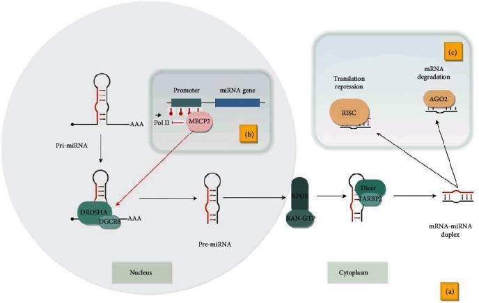
A multi-part pathophysiology diagram illustrating microRNA (miRNA) biogenesis and gene silencing mechanisms. Panel (a) shows the canonical pathway starting in the nucleus where primary miRNA (pri-miRNA) is cleaved by the Microprocessor complex (DROSHA and DGCR8) into precursor miRNA (pre-miRNA). This pre-miRNA is exported to the cytoplasm via XPO5 and RAN-GTP, where it is further processed by Dicer and TARBP2 into a mature mRNA-miRNA duplex. Panel (b) illustrates epigenetic regulation, highlighting how DNA methylation at the miRNA promoter attracts MECP2; this binding inhibits RNA Polymerase II (Pol II) elongation but facilitates co-transcriptional pri-miRNA processing. Panel (c) details the functional outcomes of mature miRNAs in the cytoplasm, specifically translation repression mediated by the RISC complex and target mRNA degradation facilitated by the AGO2 protein. This diagram serves as an educational resource for molecular biology and genetics, focusing on the regulation of gene expression and the lifecycle of non-coding RNAs.
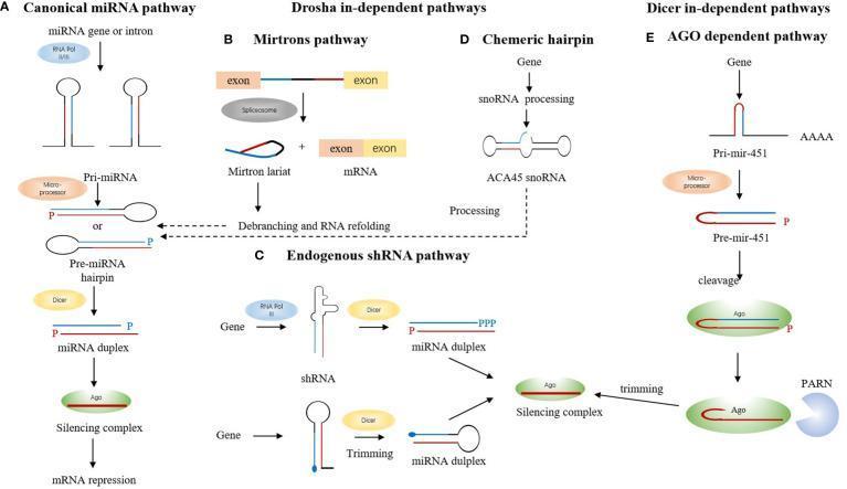
This pathophysiology diagram illustrates five distinct biogenesis pathways of microRNA (miRNA). (A) The Canonical pathway shows transcription of miRNA genes into pri-miRNA, followed by Microprocessor cleavage into pre-miRNA hairpins, Dicer-mediated processing into miRNA duplexes, and loading onto Argonaute (Ago) for mRNA repression. (B-D) Drosha-independent pathways are depicted: (B) The Mirtrons pathway involves spliceosome-mediated excision of introns into mirtron lariats, which bypass Drosha and undergo debranching to enter the canonical export path. (C) The Endogenous shRNA pathway shows direct processing of short hairpins by Dicer. (D) The Chemeric hairpin pathway shows snoRNA processing (e.g., ACA45) leading to silencing complexes. (E) The Dicer-independent pathway (AGO-dependent) focuses on miR-451, which is processed by the Microprocessor and loaded directly onto Ago for cleavage and subsequent trimming by the exonuclease PARN. The diagram highlights the diversity of RNA precursors and the selective involvement of Microprocessor (Drosha/DGCR8), Dicer, and Argonaute complexes in gene silencing mechanisms.
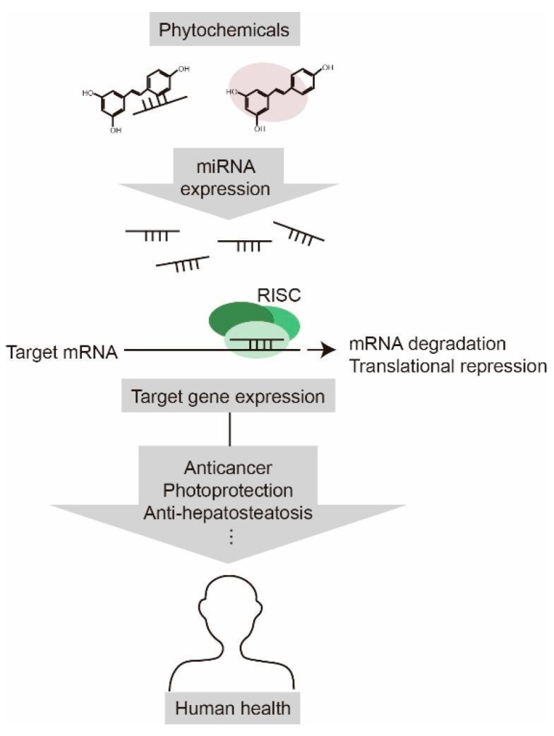
This pathophysiology diagram illustrates the signaling pathway through which phytochemicals modulate human health via microRNA (miRNA) regulation. The vertical flow begins with phytochemical molecules, represented by chemical structures, influencing 'miRNA expression'. The resulting miRNAs are shown interacting with the RNA-induced silencing complex (RISC), depicted as a green protein structure bound to 'Target mRNA'. This interaction leads to two primary molecular outcomes: 'mRNA degradation' and 'translational repression'. These inhibitory processes subsequently modify 'Target gene expression'. The diagram highlights the clinical significance of this pathway, specifically identifying 'anticancer', 'photoprotection', and 'anti-hepatosteatosis' effects as the therapeutic outcomes. The final stage of the algorithm links these molecular events to the broad promotion of 'Human health', represented by a silhouette icon. This educational visual is designed to explain the molecular mechanisms of nutriceuticals in human pathophysiology and gene silencing.
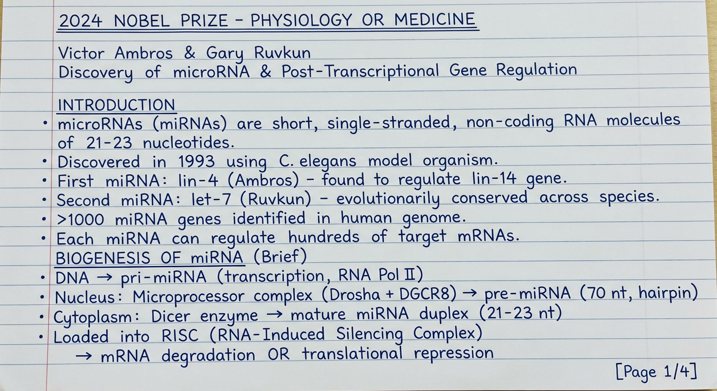
A handwritten medical physiology exam answer on white ruled notebook paper, written in blue ink in a neat student handwriting style. The content reads: "2024 NOBEL PRIZE - PHYSIOLOGY OR MEDICINE Victor Ambros & Gary Ruvkun Discovery of microRNA & Post-Transcriptional Gene Regulation INTRODUCTION microRNAs (miRNAs) are short, single-stranded, non-coding RNA molecules of 21-23 nucleotides. Discovered in 1993 using C. elegans model organism. First miRNA: lin-4 (Ambros) - found to regulate lin-14 gene. Second miRNA: let-7 (Ruvkun) - evolutionarily conserved across species. >1000 miRNA genes identified in human genome. Each miRNA can regulate hundreds of target mRNAs. BIOGENESIS OF miRNA (Brief) DNA → pri-miRNA (transcription, RNA Pol II) Nucleus: Microprocessor complex (Drosha + DGCR8) → pre-miRNA (70 nt, hairpin) Cytoplasm: Dicer enzyme → mature miRNA duplex (21-23 nt) Loaded into RISC (RNA-Induced Silencing Complex) → mRNA degradation OR translational repression [Page 1/4]" The handwriting is clear, uses underlining for headings, has slight natural variation, written on blue-lined notebook paper.
A handwritten medical physiology exam answer on white ruled notebook paper, written in blue ink in neat student handwriting. The content reads: "PART A: PHYSIOLOGICAL SIGNIFICANCE OF miRNAs IN CELLULAR DIFFERENTIATION AND DEVELOPMENT (5 Marks) 1. TEMPORAL CONTROL OF DEVELOPMENT - lin-4 miRNA represses lin-14 → controls timing of larval stage transitions (L1→L4) in C. elegans - let-7 miRNA represses lin-41 → ensures correct adult stage differentiation - miRNAs act as 'developmental timers' switching cell identity at right time 2. STEM CELL MAINTENANCE & DIFFERENTIATION - miR-302/367 cluster maintains embryonic stem cell (ESC) pluripotency - miR-34 promotes differentiation; miR-145 represses OCT4/SOX2/KLF4 (pluripotency factors) - Differentiation requires sequential activation of specific miRNAs - Without miRNAs (Dicer knockout): embryo fails to develop → embryonic lethal 3. NEURAL DIFFERENTIATION - miR-124: most abundant brain miRNA; promotes neuronal differentiation → Represses REST (neural gene suppressor) → allows neuron-specific genes to be expressed - miR-9: regulates neural progenitor proliferation vs. differentiation balance - miR-132: synaptic plasticity and dendritic growth [Page 2/4]" Written in neat blue ink on lined notebook paper with slight natural handwriting variation, headings underlined.
A handwritten medical physiology exam answer on white ruled notebook paper, written in blue ink in neat student handwriting. The content reads: "PART A (continued): Physiological Significance of miRNAs 4. CARDIAC & MUSCLE DEVELOPMENT - miR-1 & miR-133: cardiac and skeletal muscle differentiation → miR-1: promotes muscle gene expression, suppresses non-muscle genes → miR-133: regulates cardiomyocyte proliferation - Absence of miR-1-2 in mice → ventricular septal defects - miR-208: cardiac hypertrophy regulation (encoded within MHC gene) 5. HEMATOPOIETIC DIFFERENTIATION (Blood Cell Development) - miR-181: promotes B-lymphocyte differentiation - miR-155: T-cell immunity and B-cell function - miR-150: regulates megakaryocyte-to-platelet transition - miR-223: granulocyte differentiation; myeloid lineage commitment 6. ORGAN DEVELOPMENT & PATTERNING - miR-196: Hox gene regulation → limb patterning - miR-430 (zebrafish): clears maternal mRNA at embryo activation - miR-17~92 cluster: lung branching morphogenesis - DICER1 mutations (enzyme that processes pre-miRNA) → DICER1 syndrome: Multinodular goiter, pleuropulmonary blastoma, Wilms tumor SUMMARY BOX - Part A: miRNAs act as FINE-TUNERS of gene expression during development. They ensure: Right gene, Right cell, Right time ~60% of human protein-coding genes are regulated by miRNAs [Page 3/4]" Written in neat blue ink on lined notebook paper, headings underlined with a ruler, small box drawn around the summary section.
A handwritten medical physiology exam answer on white ruled notebook paper, written in blue ink in neat student handwriting. The content reads: "PART B: HOW miRNA DISCOVERY EXPANDED OUR UNDERSTANDING OF GENE REGULATION BEYOND TRANSCRIPTIONAL CONTROL (5 Marks) CLASSICAL VIEW OF GENE REGULATION (Pre-miRNA Era): DNA → (Transcription factors) → mRNA → Protein Gene regulation was thought to occur ONLY at transcriptional level (promoters, enhancers, transcription factors - 'one gene, one protein' concept) THE PARADIGM SHIFT - Post-Transcriptional Control: miRNA discovery revealed a NEW layer of regulation AFTER transcription: 1. POST-TRANSCRIPTIONAL SILENCING MECHANISM - miRNA binds 3'-UTR of target mRNA with imperfect complementarity - Outcome 1: Translational repression (ribosome stalling) - Outcome 2: mRNA deadenylation → decapping → degradation - One miRNA can regulate MULTIPLE mRNAs (gene networks, not just single genes) - One mRNA can be regulated by MULTIPLE miRNAs (combinatorial control) 2. EXPANSION BEYOND 'CENTRAL DOGMA' - Central dogma: DNA → RNA → Protein - miRNA adds: DNA → RNA → [miRNA regulation] → ↓Protein - Non-coding RNA is NOT 'junk' - it has regulatory function - ~98% of transcribed human genome is non-coding; miRNAs are a major functional class [Page 4/4 - continued overleaf]" Written in neat blue ink on lined notebook paper, headings underlined, arrows drawn with pen.
A handwritten medical physiology exam answer on white ruled notebook paper, written in blue ink in neat student handwriting. The content reads: "PART B (continued): Expanded Understanding of Gene Regulation 3. EPIGENETIC & MULTI-LEVEL REGULATORY INTEGRATION - miRNAs regulate transcription factors → indirect transcriptional effects - miRNAs interact with epigenetic machinery (DNA methylation, histone modification) - e.g. miR-29 targets DNMT3a/3b → controls DNA methylation patterns - Creates feedback loops: TF activates miRNA → miRNA suppresses TF's own targets 4. CONCEPT OF 'GENE REGULATORY NETWORKS' - Before miRNA: gene regulation was linear (one TF → one gene) - After miRNA: gene regulation is a NETWORK (many-to-many relationships) - miRNAs buffer gene expression noise → canalization of development - miRNAs fine-tune protein output without changing mRNA transcription rate 5. CLINICAL & DISEASE IMPLICATIONS (expansion of understanding) Disease | miRNA involved Cancer | miR-21 (oncomiR), miR-34 (tumor suppressor) Heart disease | miR-208, miR-1, miR-133 Congenital deafness | mutations in miRNA genes Eye disorders | miR-204, miR-211 Viral immunity | miR-122 (Hep C), miR-155 (influenza) 6. THERAPEUTIC POTENTIAL - NEW FRONTIERS - miRNA mimics: replace lost tumor-suppressor miRNAs - Antagomirs (anti-miRNA): block overactive oncomiRs - Miravirsen: anti-miR-122 drug for Hepatitis C (first clinical trial) - miRNAs as circulating biomarkers in blood (liquid biopsy for cancer/MI) CONCLUSION: miRNA discovery revealed that gene expression is regulated at multiple levels - transcriptional, post-transcriptional, translational, and epigenetic. It transformed our view from a simple 'on-off switch' to a complex, tunable, multi-layered regulatory system. [Nobel Prize 2024 - Ambros & Ruvkun] [Page 5/5 - END]" Written in neat blue ink on lined notebook paper, table drawn with pen/ruler, headings underlined, conclusion in a box.
Handwritten medical exam answer on lined notebook paper in blue ink, neat student handwriting. Title: "PART B continued - Expanded Gene Regulation". Content covers: epigenetic integration of miRNAs with DNA methylation (miR-29 targets DNMT3a), gene regulatory networks showing many-to-many miRNA-mRNA relationships, a small table listing diseases and their miRNAs (cancer: miR-21, heart: miR-208, hearing loss: miRNA mutations), therapeutic applications (antagomirs, miRNA mimics, Miravirsen for Hepatitis C), and a boxed conclusion stating that miRNA discovery revealed multi-layered gene regulation beyond transcription, transforming simple on-off switch concept to a complex tunable system. Nobel 2024 Ambros and Ruvkun. Page 5 of 5. Headings underlined.
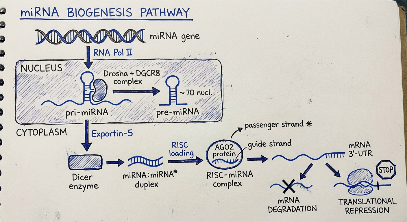
A hand-drawn diagram on white paper in blue and black pen, resembling a student's notebook sketch for a medical physiology exam. The diagram shows the miRNA biogenesis pathway with the following labeled steps drawn as simple sketches: At the top: DNA double helix labeled "miRNA gene" with arrow labeled "RNA Pol II" pointing down to "pri-miRNA" (long wavy line with hairpin loop). In a shaded box labeled "NUCLEUS": Drosha + DGCR8 complex (shown as oval shape) cutting pri-miRNA into "pre-miRNA" (stem-loop hairpin structure, ~70 nt). Arrow labeled "Exportin-5" pointing out of nucleus box downward. In area labeled "CYTOPLASM": Dicer enzyme (rectangle) processing pre-miRNA into "miRNA:miRNA* duplex" (two parallel lines). Arrow to "RISC loading" (AGO2 protein shown as circle). One strand discarded (labeled "passenger strand *"), other strand labeled "guide strand". Final RISC-miRNA complex binds to mRNA 3'-UTR. Two outcome arrows: left arrow to "mRNA DEGRADATION" (wavy line with X), right arrow to "TRANSLATIONAL REPRESSION" (ribosome blocked with stop sign). Title at top: "miRNA BIOGENESIS PATHWAY" underlined. Simple clean educational diagram style.
Generate the image
📄 Page 1 — Introduction & Biogenesis
📄 Page 2 — Part A: Cellular Differentiation (Points 1-3)
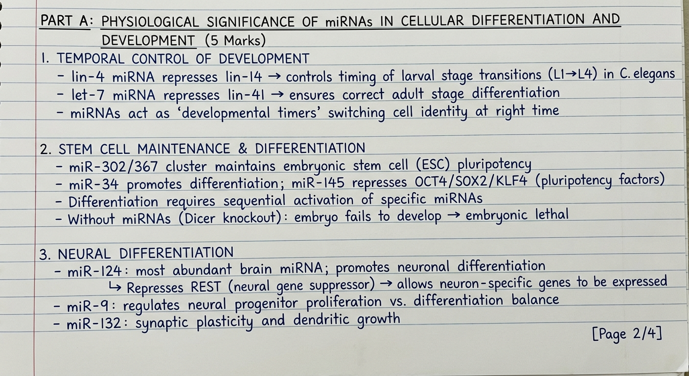
📄 Page 3 — Part A: Cellular Differentiation (Points 4-6 + Summary)
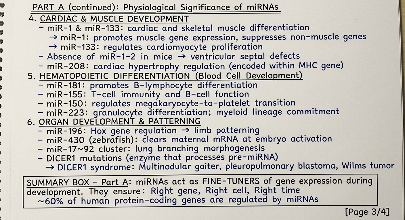
📄 Page 4 — Part B: Beyond Transcriptional Control (Points 1-2)
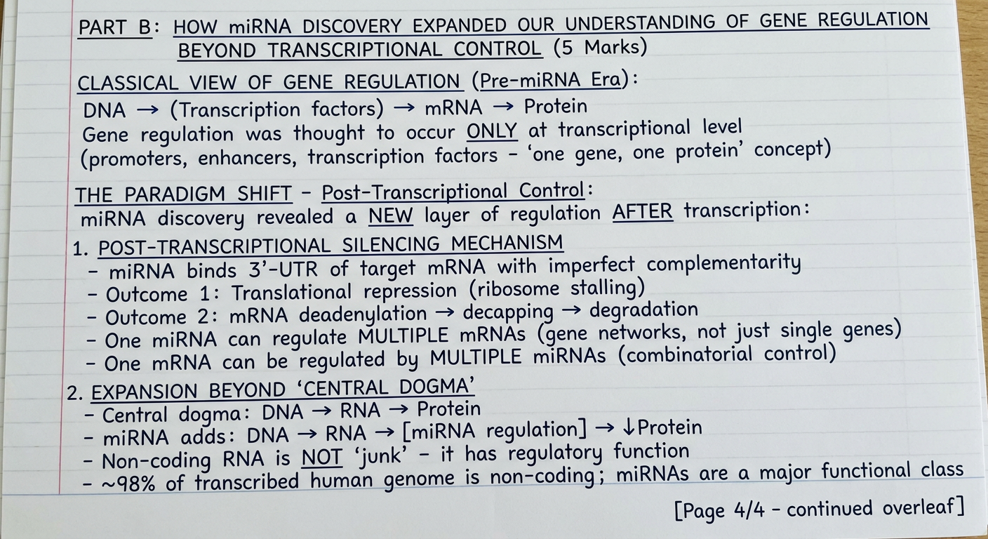
📄 Page 5 — Part B: Clinical Implications, Therapeutics & Conclusion
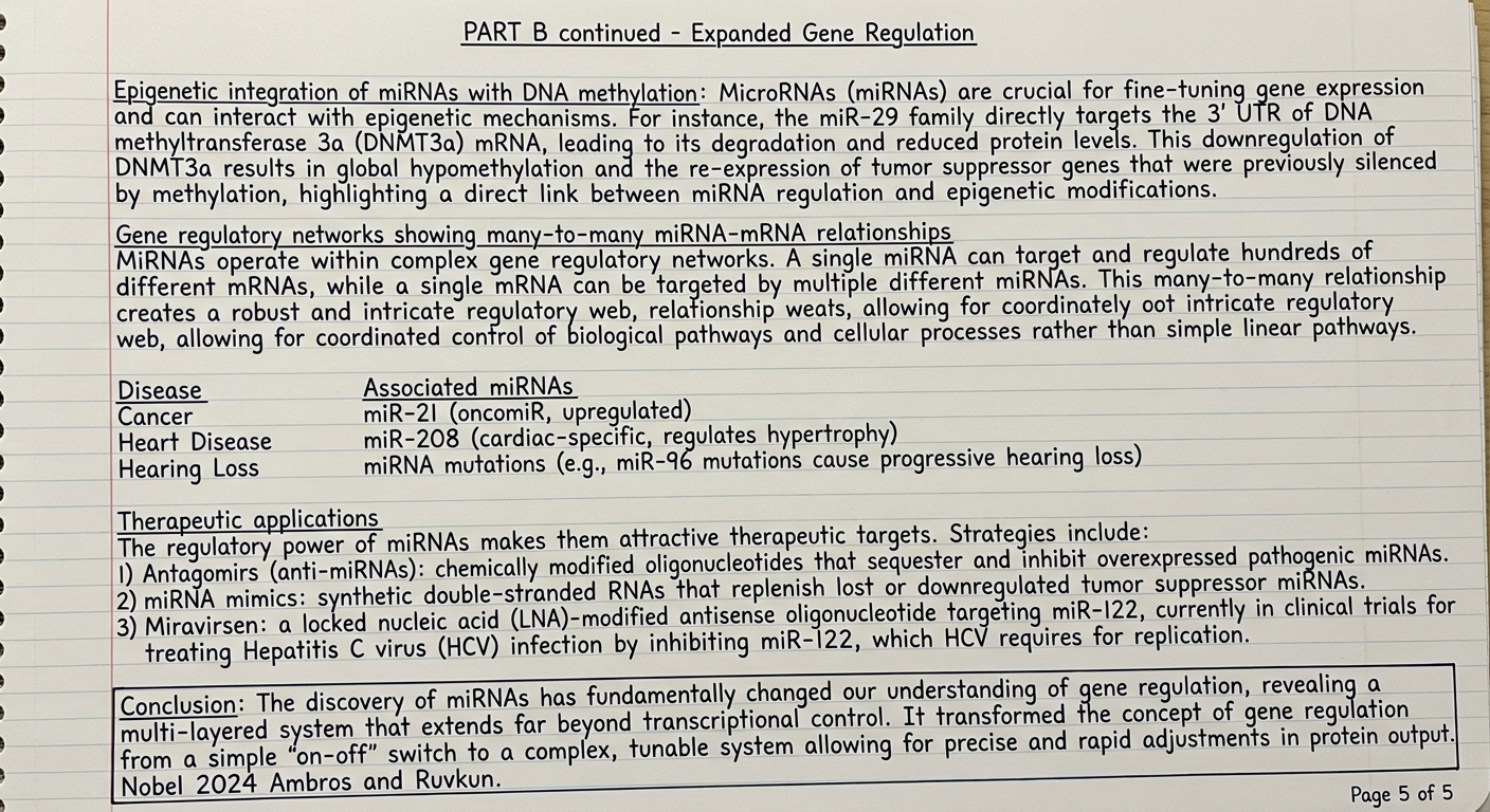
📊 Diagram — miRNA Biogenesis Pathway (Labelled)
How to use these for your exam:
| Image | Content | Marks coverage |
|---|---|---|
| Page 1 | Intro + Nobel context + Biogenesis overview | Sets up both parts |
| Pages 2-3 | Part A fully answered with 6 sub-points + summary box | 5 marks |
| Pages 4-5 | Part B fully answered with 6 sub-points + disease table + conclusion | 5 marks |
| Diagram | miRNA biogenesis pathway - draws full marks for diagrams | Bonus marks |