
hydrocephalus
hydrocephalus brain MRI CT scan enlarged ventricles
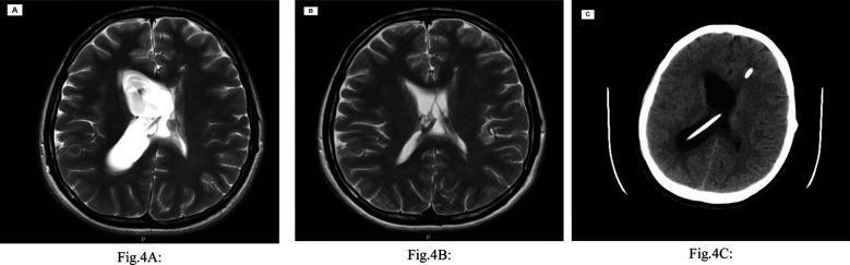
This composite figure includes radiological imaging of the brain in the axial plane, illustrating the evolution of hydrocephalus associated with ventriculoperitoneal shunt (VPS) malfunction. Panels A and B are T2-weighted MRI scans. Image A shows a significantly dilated, entrapped right lateral ventricle exhibiting a bright, hyperintense signal characteristic of cerebrospinal fluid accumulation due to right-sided shunt failure. Image B shows the same patient shortly after, displaying a reduction in ventricular volume and normalized morphology, indicating a temporary improvement in hydrocephalus. Panel C is a non-contrast axial CT scan showing a recurrence of hydrocephalus. A high-density (bright) linear structure is visible traversing the right cerebral parenchyma and terminating in the enlarged right lateral ventricle, representing the radio-opaque proximal shunt catheter. A separate circular hyperdensity within the calvarium denotes a shunt component or connector. This series highlights the diagnostic utility of MRI and CT in monitoring shunt-dependent hydrocephalus and identifying complications like entrapped ventricles.
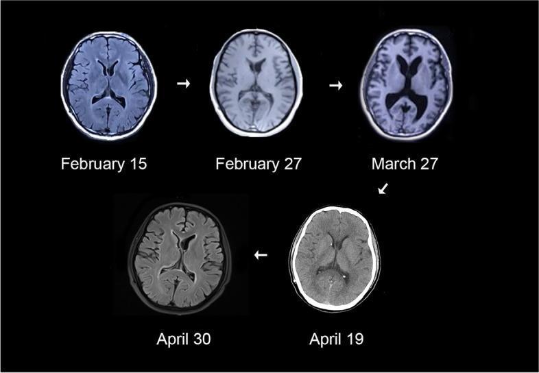
A series of five longitudinal neuroimaging studies (axial sections) demonstrating dynamic changes in ventricular size over a two-and-a-half-month period. The series primarily consists of brain MRI (T1-weighted and FLAIR sequences) and one CT scan (April 19). The images illustrate a progression from baseline ventricular morphology on February 15 to progressive ventriculomegaly by March 27, where the lateral ventricles are markedly enlarged and rounded, suggestive of hydrocephalus. Subsequent scans from April 19 and April 30 show a resolution of this enlargement, with the ventricles returning to near-baseline dimensions. This clinical progression demonstrates the fluctuation of ventricular volume, often seen in cases of intermittent hydrocephalus or following neurosurgical intervention such as shunt placement. The images provide high-contrast visualization of the cerebrospinal fluid (CSF) spaces relative to the brain parenchyma, cortical sulci, and periventricular structures, serving as a teaching tool for longitudinal monitoring of intracranial pressure-related changes.
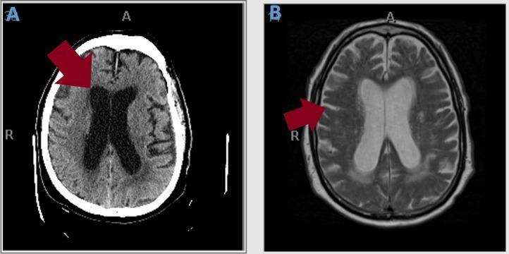
This figure presents two axial cross-sections of the brain highlighting neuroanatomical changes. Image A is a non-contrast Computed Tomography (CT) scan of the head demonstrating significant ventriculomegaly. A red arrow points to the markedly enlarged lateral and third ventricles, which occupy a disproportionate volume relative to the brain parenchyma. Image B is a T2-weighted Magnetic Resonance Image (MRI) with contrast from the same patient. This image clarifies the etiology of the ventricular enlargement, with a red arrow indicating pronounced cerebral atrophy. Key visual findings in image B include the prominent narrowing of cortical gyri and the compensatory widening of the sulci (hydrocephalus ex vacuo). The comparison between these two modalities is used to distinguish between communicating hydrocephalus and secondary ventricular enlargement due to global tissue loss. These diagnostic images are essential for evaluating geriatric patients presenting with the clinical triad of gait instability, urinary incontinence, and cognitive decline, often seen in normal pressure hydrocephalus (NPH) or neurodegenerative processes.
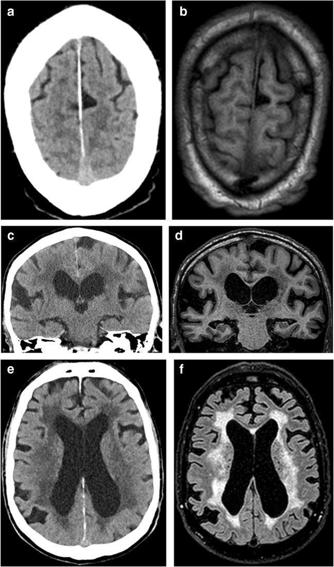
This composite diagnostic image presents a comparative study between Computed Tomography (CT) and Magnetic Resonance Imaging (MRI) of the brain in the context of Idiopathic Normal Pressure Hydrocephalus (iNPH). (a, b) Axial CT and T1-weighted MRI at the vertex level demonstrate 'disproportionately enlarged subarachnoid space hydrocephalus' (DESH), characterized by narrow sulci at the high convexity and midline, contrasting with focally enlarged sulci elsewhere. (c, d) Coronal CT and T1-weighted MRI sections highlight significant ventriculomegaly and prominently dilated Sylvian fissures, a classic diagnostic feature of iNPH. (e, f) Axial CT and T2-FLAIR MRI sequences at the level of the lateral ventricles illustrate extensive periventricular white matter changes. These appear as hypodense regions on CT and hyperintense signals on T2-FLAIR, surrounding the dilated lateral ventricles. The images serve to illustrate how both CT and MRI can identify key morphological markers of hydrocephalus, including sulcal effacement, sylvian fissural widening, and white matter signal alterations for clinical evaluation.
"hydrocephalus"[MeSH Terms] AND ("treatment" OR "shunt")
Hydrocephalus
Definition
CSF Physiology
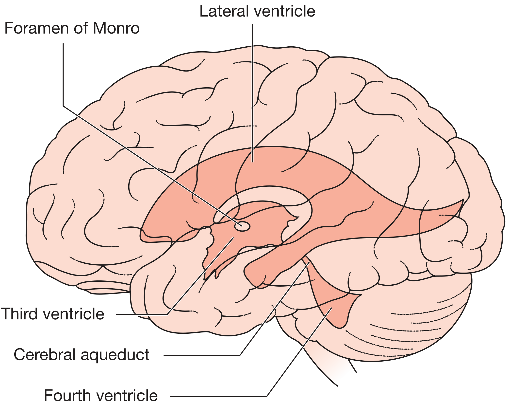
Classification
1. Noncommunicating (Obstructive / Intraventricular) Hydrocephalus
- The ventricles proximal to the obstruction enlarge; distal ventricles remain normal
- Most commonly blocked at the cerebral aqueduct (aqueductal stenosis)
- Can cause very sudden deterioration, coma, and death
- Lumbar puncture carries risk of brainstem herniation
2. Communicating (Extraventricular Obstructive) Hydrocephalus
- Causes: post-hemorrhagic, post-infectious, raised CSF protein, meningeal carcinomatosis
3. Hydrocephalus ex Vacuo
4. Overproduction (Rare)
Etiology by Age
| Age Group | Common Causes |
|---|---|
| Neonate/Infant | Posthemorrhagic (germinal matrix hemorrhage in preterms <1500g), postinfective (in utero infection → aqueductal scarring), Chiari II malformation (with myelomeningocele), Dandy-Walker malformation, aqueductal stenosis/gliosis |
| Older child | Posterior fossa neoplasms (cerebellum/brainstem), aqueductal stenosis, meningitis sequelae |
| Adult | Subarachnoid hemorrhage, colloid cyst of 3rd ventricle (ball-valve effect), ependymoma of 4th ventricle, cerebellar hemorrhage/infarction (compresses aqueduct), meningeal infections/tumors, NPH |
Clinical Features
Infants (sutures open, before age ~2 years)
- Progressive macrocephaly - head circumference crossing centile lines (the most reliable sign)
- Frontal bossing
- Tense, bulging anterior fontanelle
- Sutural diastasis, scalp vein engorgement, calvarial thinning
- "Sunsetting" eyes - failure of upward gaze (Parinaud sign due to dorsal midbrain compression)
- Lateral rectus palsy (CN VI false localizing sign)
- Leg spasticity (corticospinal tracts stretched around enlarged ventricles)
- "Cracked-pot" sound on skull percussion (McEwen sign)
Older Children (sutures fused)
- Early morning headache, nausea, vomiting (raised ICP)
- Papilledema
- Diplopia (CN VI palsy)
- Ataxia and gait disturbance
- With long-standing disease: endocrine dysfunction (short stature, menstrual irregularity, diabetes insipidus)
Adults - Acute Hydrocephalus
- Headaches, papilledema, diplopia
- Mental status changes
- Sudden death from acute pressure surge
Adults - Chronic Hydrocephalus / Normal Pressure Hydrocephalus (NPH)
- Gait disturbance - apraxic ("magnetic") gait - inability to lift feet as if stuck to the floor; wide-based; resembles Parkinsonism (most responsive to treatment)
- Cognitive impairment - subcortical dementia: psychomotor slowing, apathy, preserved language/spatial skills
- Urinary incontinence
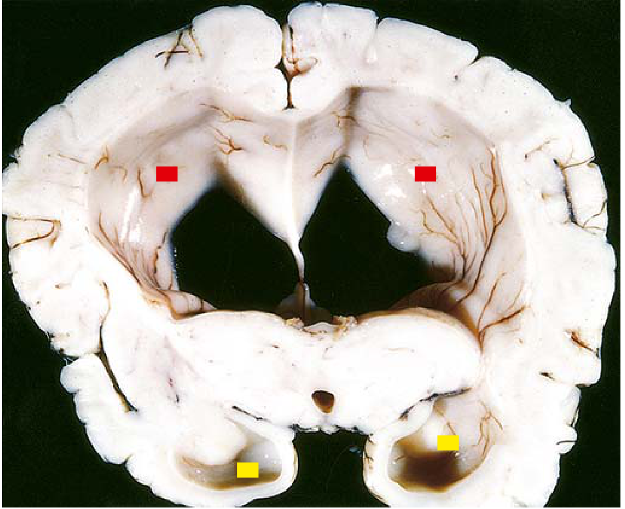
Gross Pathology
Imaging
CT
- Dilated temporal horns (early and sensitive sign of noncommunicating hydrocephalus)
- Periventricular interstitial (transependymal) edema: hypodense "halo" around ventricles on CT, hyperintense on T2/FLAIR MRI
- Small sulci and obliterated basal cisterns (distinguish from atrophic ventriculomegaly where sulci are prominent)
MRI (preferred)
- Shows all of the above plus cause of obstruction
- Aqueductal stenosis: focal narrowing on sagittal MRI, usually at level of superior colliculi; lateral and 3rd ventricles dilated, 4th ventricle normal
- NPH: DESH pattern (Disproportionately Enlarged Subarachnoid Space Hydrocephalus) - tight sulci at high convexity with enlarged Sylvian fissures
- Grainger & Allison's Diagnostic Radiology, p. 2028-2033
Pathophysiology of Ventricular Enlargement
Treatment
Surgical CSF Diversion
| Procedure | Details |
|---|---|
| External Ventricular Drain (EVD) | Acute/emergency; also allows ICP monitoring |
| Ventriculoperitoneal (VP) shunt | Most common permanent option; one-way pressure-responsive valve; drains to peritoneal cavity |
| Ventriculoatrial (VA) shunt | Used when peritoneal cavity unavailable |
| Endoscopic Third Ventriculostomy (ETV) | Creates opening in floor of 3rd ventricle → CSF drains to suprasellar cistern; preferred for aqueductal stenosis; avoids shunt hardware |
| Removal of obstructing lesion | When a resectable mass is the cause |
Shunt Complications
- Malfunction/obstruction - choroid plexus or glial tissue blocking catheter tip; presents with recurrent hydrocephalus symptoms; revision required
- Infection - requires externalization and antibiotic treatment
- Over-drainage - subdural hematomas, slit ventricle syndrome
- In children, revisions are frequently needed as the child grows
- Shunt tubing integrity is assessed by plain X-ray ("shunt series"); obstruction and fluid tracking along tubing suggest malfunction
NPH - Diagnostic Work-up Before Shunting
- Lumbar puncture (large-volume tap test) - remove 30-50 mL CSF; observe for gait improvement in the next 24-48 hours (positive tap test predicts shunt response)
- Continuous ICP monitoring or external lumbar drainage over several days
- A 2024 Cochrane review (PMID 39105473) evaluated shunting for iNPH; a 2025 systematic review (PMID 40045804) focused on standardizing the tap test protocol
Special Topics
Hydrocephalus ex Vacuo
Fetal Hydrocephalus
Post-Subarachnoid Hemorrhage
paraphimosis
paraphimosis foreskin retracted glans penis swelling
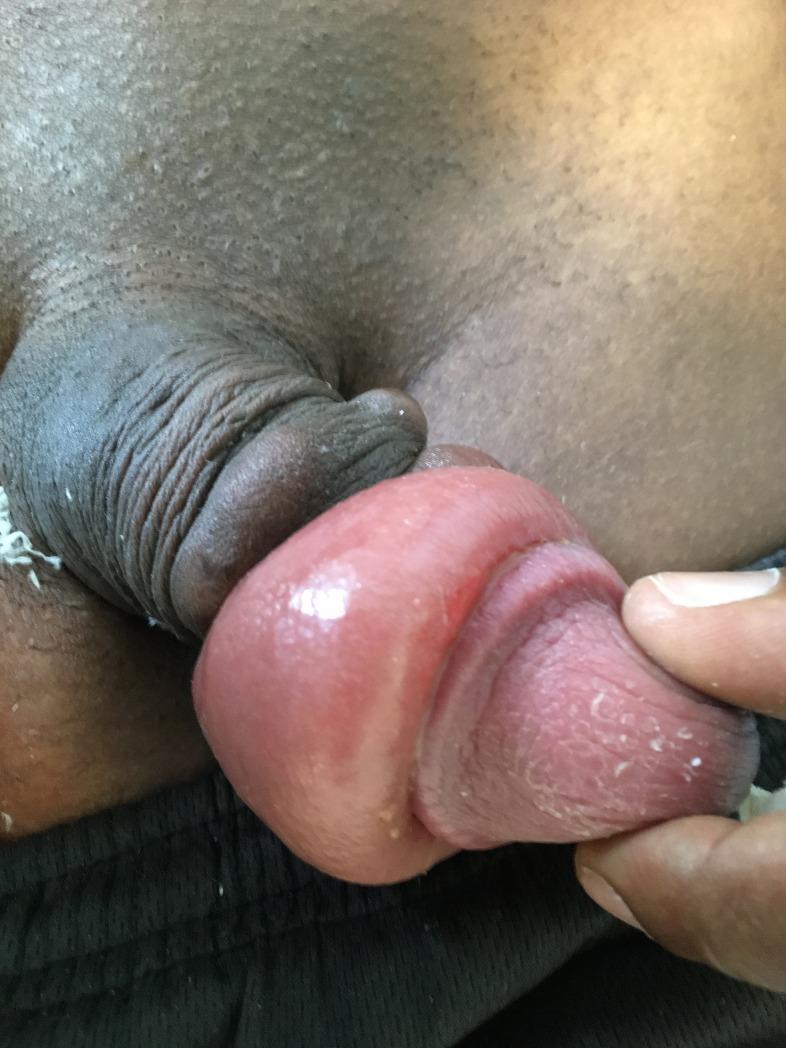
This clinical photograph displays a case of infectious paraphimosis, a urological emergency, occurring as a short-term complication following the insertion of subcutaneous penile implants. The image shows the glans penis in a state of significant edema and erythema. A constricting band of retracted foreskin is visible proximal to the glans, leading to venous and lymphatic congestion. The glans itself exhibits a shiny, tense surface texture with visible inflammation. Distally, the tissue appears somewhat desiccated with small amounts of white, flaky discharge or epithelial desquamation present. The surrounding scrotal skin and lower abdomen are visible, and the clinician's gloved hand is seen retracting the tissue to demonstrate the extent of the swelling and the characteristic ‘doughnut’ appearance of the edematous prepuce. This visual serves as an educational example of post-procedural infectious complications and the clinical presentation of paraphimosis in the context of foreign body insertion.
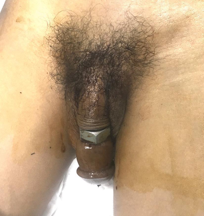
This clinical photograph displays a case of penile incarceration and strangulation caused by a metallic foreign body. A large steel hexagon nut is seen encircling the mid-shaft of the penis. Distal to the site of constriction, the penile shaft and glans exhibit significant non-pitting edema and venous congestion, appearing markedly swollen compared to the proximal shaft. The tissue distal to the nut shows a cyanotic or dark reddish-brown discoloration, suggesting compromised venous return and localized ischemia. A state of paraphimosis is visible, with the foreskin retracted and trapped behind the coronal sulcus, further contributing to the distal swelling. There are no immediate signs of overt skin necrosis or gangrene visible in this view. This image illustrates a surgical and urological emergency requiring urgent removal of the constricting band to prevent permanent tissue damage, urethral injury, or gangrene. It serves as a classic educational example of Grade III penile injury (Bhat classification) characterized by distal edema and sensory changes.
"paraphimosis" AND reduction
Paraphimosis
Definition
Pathophysiology
- The foreskin is retracted behind the coronal sulcus and becomes stuck
- The retracted prepuce forms a tight circumferential constricting band at the coronal sulcus
- This band obstructs venous and lymphatic drainage from the glans and distal foreskin
- Progressive edema accumulates in the glans, making it larger and harder to reduce
- The worsening edema tightens the constricting ring further
- If untreated: arterial compromise → ischemia → necrosis and gangrene of the glans
Clinical photograph
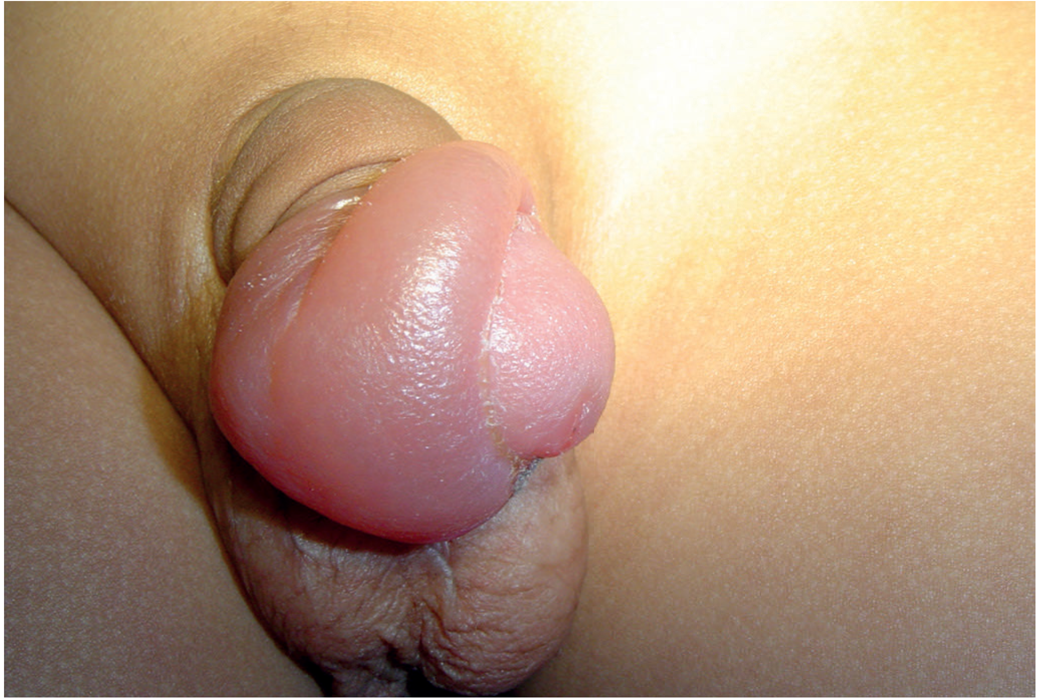
Etiology / Precipitating Causes
| Category | Examples |
|---|---|
| Iatrogenic | Failure to replace foreskin after urinary catheterization, cystoscopy, or medical examination - most common preventable cause |
| Inflammation | Balanoposthitis → chronic foreskin inflammation → contracture of preputial ring |
| Poor hygiene | Chronic smegma accumulation and recurrent infection |
| Sexual activity | Prolonged retraction |
| Physiological | Phimosis predisposes: tight preputial ring when retracted over compressible glans cannot be returned |
Key prevention rule: After any urethral catheterization, penile examination, or cystoscopy in an uncircumcised patient, always ensure the foreskin is returned to its native position covering the glans. - Roberts and Hedges', p. 1312
Differential Diagnosis
- Allergic reaction / angioedema of the foreskin
- Penile trauma
- Infection / cellulitis
- Generalized edema from systemic causes (CCF, nephrotic syndrome)
Treatment
Step 1 - Analgesia
- Topical anesthetic lubricant (e.g., EMLA cream - 2.5% lidocaine/2.5% prilocaine) applied to the foreskin and glans for mild cases
- Dorsal penile nerve block for moderate-severe cases: inject lidocaine without epinephrine at 10 o'clock and 2 o'clock positions at the penile base, just deep to Buck's fascia (3-5 mm beneath skin). Aspirate before injecting to avoid intravascular injection. Wait 5 minutes for optimal effect.
- Procedural sedation for young children or very uncooperative patients; general anesthesia for young children when necessary
Step 2 - Edema Reduction (critical for success)
| Method | Technique |
|---|---|
| Manual compression | Firmly grasp the glans + foreskin with the entire palm; apply steady circumferential pressure for several minutes |
| Elastic bandage | Wrap a 2-inch compressive bandage (ACE wrap) around the distal penis for 5 minutes |
| Iced-glove method | Half-fill a large glove with crushed ice and water; invaginate the thumb of the glove; draw it over the lubricated paraphimotic penis and hold in place for 5-10 minutes - combines cooling and compression |
| Granulated sugar | Apply to edematous foreskin for 1-2 hours (osmotic dehydration of edema) |
| Hyaluronidase injection | Inject hyaluronidase in normal saline into the edematous foreskin to enzymatically disperse the fluid |
| Multiple punctures technique | Multiple small punctures in the edematous skin to allow fluid egress before manual reduction |
Step 3 - Manual Reduction
- Place both thumbs on the urethral meatus (glans)
- Wrap index and long fingers of both hands circumferentially around the foreskin just proximal to the phimotic ring
- Apply simultaneous, slow, steady pressure: thumbs push the glans proximally while fingers pull the foreskin distally over the glans
- The key is constant, steady pressure - not sudden force
- Alternatively: grasp the shaft with one hand, use the thumb of the other to push the glans forward while sliding the foreskin distally
- Apply 6-8 Babcock (non-crushing) clamps spaced evenly around the foreskin, straddling the phimotic ring (one edge proximal, one distal to it)
- Apply simultaneous distal traction on all clamps together to pull the ring over the glans
- Inspect the foreskin carefully for injuries afterward
Step 4 - If Manual Reduction Fails: Surgical Options
| Procedure | Details |
|---|---|
| Dorsal slit | Under local anesthesia, make a superficial dorsal incision through the constricting preputial band using iris scissors (to protect underlying tissue) - allows the ring to be divided and the foreskin to be reduced. Definitive circumcision is done later once inflammation resolves. |
| Circumcision | Definitive treatment; performed after acute inflammation subsides. Also indicated for patients with recurrent paraphimosis or those who fail conservative management. |
Post-Reduction Care
- Administer antibiotics if there is evidence of underlying infection (balanoposthitis)
- Advise the patient that a phimotic foreskin remains after reduction - this predisposes to recurrence
- Plan for elective circumcision in patients with phimosis or recurrent episodes
- Counsel on the importance of always returning the foreskin after retraction
Key Points for Prevention
After every urinary catheter insertion, cystoscopy, or penile examination in an uncircumcised patient, always replace the foreskin to its native anatomical position over the glans. This single step prevents iatrogenic paraphimosis - the most common avoidable cause. - Roberts and Hedges', p. 2015
Comparison: Phimosis vs. Paraphimosis
| Feature | Phimosis | Paraphimosis |
|---|---|---|
| Problem | Foreskin cannot be retracted over the glans | Retracted foreskin cannot be returned to cover the glans |
| Emergency? | Usually not (unless urinary obstruction) | Yes - urological emergency |
| Risk | Difficulty with hygiene, UTIs, recurrent balanitis | Glans necrosis/gangrene if untreated |
| Treatment | Topical steroids (betamethasone 0.05%), preputioplasty, or circumcision | Immediate reduction; circumcision when settled |