Im a junior resident in ENT surgery Tomorrow I have a procedure Righy Tympanoplasty under General Anesthesia procedure. Make me understand necessary surgical anatomy, indications,contraindications, PreOp preparation,detailed Surgical steps, post op management. Suggest any good operative clips in Indian hospital context.
Reading File
search_medical_text
tympanoplasty surgical anatomy tympanic membrane middle ear ossicles
search_medical_text
tympanoplasty indications contraindications preoperative assessment
search_medical_image
tympanic membrane anatomy middle ear surgical landmarks
GradeResponse
GradeResponse
GradeResponse
search_medical_text
tympanoplasty surgical technique steps underlay overlay graft temporalis fascia
search_medical_text
tympanoplasty postoperative management complications hearing outcome
Here is a comprehensive pre-operative briefing for your Right Tympanoplasty under GA:
Right Tympanoplasty Under General Anesthesia
A Complete Pre-Operative Guide for the Junior Resident
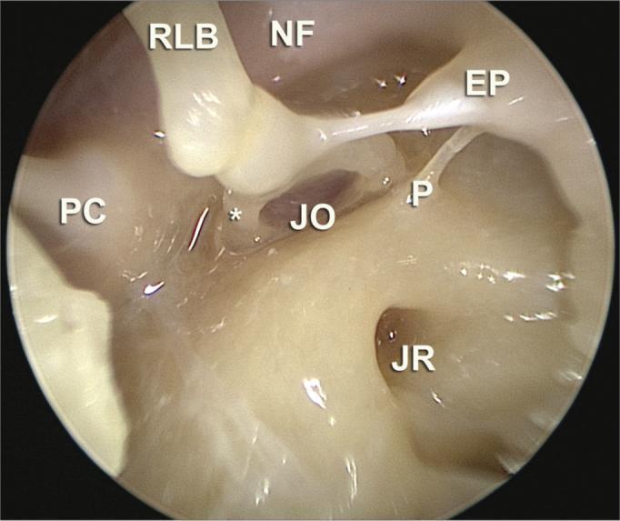
1. SURGICAL ANATOMY
The Tympanic Membrane (TM)
(Bailey & Love's Short Practice of Surgery, 28th Ed., p. 771)
The TM is a trilaminar structure — outer squamous epithelium, middle fibrous layer (pars tensa), and inner mucosal layer. It sits obliquely at ~55° to the ear canal floor.
Key landmarks you must identify intraoperatively:
| Landmark | Significance |
|---|---|
| Umbo | Tip of malleus handle — central landmark |
| Handle (manubrium) of malleus | Runs superiorly from umbo; must be preserved |
| Lateral process of malleus | Superior prominence; anterior & posterior malleolar folds arise here |
| Pars tensa | Perforation site in CSOM; your graft goes here |
| Pars flaccida | Superior/Shrapnell's membrane; site of attic perforations |
| Annulus fibrosus | Thickened rim of TM seated in tympanic sulcus |
| Light reflex | Anteroinferior cone — lost in disease |
Middle Ear (Tympanic Cavity)
Six walls to know:
- Lateral wall: TM + scutum (bony outer attic wall)
- Medial wall: Promontory (basal turn of cochlea), oval window, round window, facial nerve canal (horizontal segment runs above oval window — critical to protect)
- Anterior wall: Eustachian tube opening + carotid canal (thin bone separates them)
- Posterior wall: Aditus ad antrum, pyramidal eminence (stapedius tendon), facial nerve (descending segment)
- Roof (tegmen tympani): Thin bone separating middle ear from middle cranial fossa dura
- Floor: Thin bone over jugular bulb (may be dehiscent — aberrant jugular bulb)
Ossicular Chain
- Malleus → Incus → Stapes → oval window
- The long process of incus articulates with stapes head at incudostapedial joint
- In simple CSOM (Type I tympanoplasty), the ossicular chain is intact — your job is only to repair the TM
Facial Nerve
⚠️ Your most important structure to protect:
- Horizontal (tympanic) segment: runs above oval window in the medial wall
- Vertical (mastoid) segment: posterior to the middle ear
- May be dehiscent in 10–25% of patients — always identify before drilling
Eustachian Tube
- Opens into anterior wall of tympanic cavity
- Patency is the single most important factor for graft success
- Dysfunction → graft failure / retraction pocket
2. INDICATIONS
Absolute Indications
- Chronic Suppurative Otitis Media (CSOM) — Tubotympanic (Safe) type with central perforation
- Traumatic perforation not healed after 3 months
- Dry central perforation causing conductive hearing loss (CHL)
- Prevention of recurrent otitis media episodes
- Improvement of hearing (audiological rehabilitation)
Relative Indications
- Single hearing ear with perforation (with caution)
- Bilateral perforations (staged — worse ear first)
- Perforation in a child (debated — some prefer after age 7)
Wullstein Classification (Know This!)
| Type | What is Repaired | Indication |
|---|---|---|
| Type I | TM only (Myringoplasty) | Intact ossicular chain |
| Type II | TM + incus erosion; graft on incus | Incus long process erosion |
| Type III | Graft on stapes head (myringostapediopexy) | Incus + malleus absent |
| Type IV | Graft on stapes footplate | Only footplate remains |
| Type V | Fenestration of lateral semicircular canal | Fixed stapes |
For your case — most likely Type I unless intraoperative findings differ.
3. CONTRAINDICATIONS
Absolute
- Active wet ear / discharging ear — must treat medically first (minimum 6 weeks dry ear)
- Atticoantral (unsafe) disease / cholesteatoma — requires mastoidectomy first; tympanoplasty alone is contraindicated
- Only hearing ear with high surgical risk (relative)
- Malignancy of external/middle ear
Relative
- Eustachian tube dysfunction — graft failure rate very high; consider adenoidectomy/ET balloon dilation first
- Uncontrolled DM — poor healing, infection risk
- Immunocompromised patient
- Extremely young child — higher failure due to ET dysfunction (most centres prefer ≥5–7 years)
- Nasal/sinus disease uncontrolled — URTI, chronic sinusitis (impacts ET)
- Very poor contralateral ear — anaesthetic risk for the hearing
Practical Pre-Op Rule
"The ear must be DRY for at least 6 weeks before surgery" — this is non-negotiable in Indian practice.
4. PRE-OPERATIVE PREPARATION
Clinical Assessment
- Full otoscopic/endoscopic examination — document perforation size (small <25%, medium 25–50%, large >50%, subtotal, total), location (anterior, posterior, central), and condition of remnant TM
- Examine other ear
- Tuning fork tests: Rinne (512 Hz) and Weber — establish whether conductive loss
- Nasal examination — rhinoscopy, assess for DNS, turbinate hypertrophy, nasal polyps (ET patency)
- Eustachian tube function test (Valsalva, Toynbee)
Audiological Workup
- Pure Tone Audiometry (PTA) — mandatory; establishes baseline; air-bone gap (ABG) should correlate with perforation size
- Good prognosis if ABG < 30 dB
- Tympanometry — type B flat curve expected; check contralateral ear (As = stapes fixation, C = ET dysfunction)
- Speech discrimination score — helps counsel patient on expected post-op gain
Investigations
| Investigation | Purpose |
|---|---|
| Hb, CBC, BT/CT | Anaesthetic fitness |
| Blood glucose (FBS/RBS) | DM screening |
| Urea, creatinine | Renal function |
| ECG | If >40 yrs or cardiac risk |
| Chest X-ray | Pre-anaesthetic |
| HIV, HBsAg | Universal precautions |
| HRCT Temporal Bone (if needed) | To rule out cholesteatoma, assess ossicular status, tegmen, facial canal dehiscence |
Note: HRCT temporal bone is NOT mandatory for straightforward Type I tympanoplasty in Indian practice, but obtain it if there is any suspicion of cholesteatoma, previous failed surgery, or unusual findings.
Anaesthetic Fitness
- ENT anaesthesia joint assessment
- Standard nil-by-mouth (6 hours solid, 2 hours clear fluids)
- Consent for: tympanoplasty, possible ossiculoplasty if chain disrupted intraoperatively, possible cortical mastoidectomy if disease found
Pre-operative Aural Toilet
- Dry mopping, suction clearance under microscope
- Antibiotic ear drops (ciprofloxacin) for 1–2 weeks pre-op if any discharge
- Nasal decongestants if any nasal congestion
Medications to Stop
- Aspirin, NSAIDs — 7–10 days before
- Anticoagulants — per haematology advice
Graft Harvesting Planning
- Temporalis fascia — gold standard; harvested from same side
- Tragal perichondrium — good alternative; quicker, less morbidity
- Cartilage (tragal/conchal) — for large perforations, revision cases, or when reinforcement needed
Patient Counselling
- Success rate: 85–95% for Type I in dry ear
- Hearing improvement expected but not guaranteed
- Risk of facial nerve injury, taste disturbance (chorda tympani), dead ear (rare), tinnitus, dizziness
- Post-op water precautions for 3–6 months
5. DETAILED SURGICAL STEPS
Setup and Positioning
- GA with endotracheal intubation (RAE tube — south-facing, preferred in head and neck)
- Patient supine, head turned to left (for right ear)
- Head ring for stabilisation
- Ipsilateral shoulder slightly elevated with a roll
- Operating microscope / endoscope positioned
- Surgeon sits at head end (or at the side for endoscopic)
Injection
- Local infiltration (even under GA — reduces bleeding, aids hydrodissection):
- 1% Lignocaine with 1:200,000 adrenaline
- Inject at 4 points in posterior EAC skin (6, 9, 12 o'clock positions) and meatal incision site
Approach Options
| Approach | When Used |
|---|---|
| Permeatal (transcanal) | Adequate canal size, posterior perforation, experienced surgeon |
| Endaural | Narrow canal, moderate perforation |
| Postaural (Retroauricular) | Large perforations, revision, wide exposure needed, most common in Indian training |
For a junior resident, the postaural approach is safest and most forgiving — likely what you will assist with.
STEP-BY-STEP: Postaural Approach, Underlay Technique
Step 1 — Skin Incision (Postaural)
- Incise skin 5–7 mm behind the postauricular sulcus (stay behind sulcus to avoid visible scar)
- Deepen to periosteum
- Graft harvest at this step — identify and harvest temporalis fascia through the same incision before it becomes vascular from retraction
Step 2 — Temporalis Fascia Harvest
- Elevate skin flap anteriorly to expose temporalis muscle
- Score fascia with a scalpel, harvest adequate piece (usually 2 × 2 cm)
- Stretch on flat surface (Teflon/glass), allow to dry for 15–20 minutes (important — wet fascia is slippery, hard to handle)
Step 3 — Elevation of Periosteum + Exposure of EAC
- Separate the auricle from the bony EAC by elevating soft tissue anteriorly
- Self-retaining retractor placed (Mollison's or mastoid retractor)
- Identify the bony EAC
Step 4 — Canalplasty (if needed)
- If canal is narrow, drill posterior-superior canal wall with Skeeter/cutting burr to improve visualisation
Step 5 — EAC Skin Flap (Tympanomeatal Flap)
- Under the microscope, incisions are made in the EAC skin:
- Posterior incision: from 12 to 6 o'clock at the bony-cartilaginous junction (about 6–8 mm from the annulus)
- Radial incisions: at 12 and 6 o'clock extending to the annulus
- The tympanomeatal flap is elevated from lateral to medial using a round knife or Sickle knife
Step 6 — Elevation of the Annulus
- Identify the annulus in the tympanic sulcus
- Elevate annulus using a Fisch or Rosen elevator — posteroinferiorly first (safer, away from chorda and ossicles)
- Lift the TM remnant and annulus forward — this exposes the middle ear
Step 7 — Middle Ear Exploration
- Check ossicular chain: malleus, incus, stapes — mobility by gentle palpation
- Look for: disease (granulations, cholesteatoma — must be excluded), ossicular erosion
- Inspect: facial nerve canal, round window niche, ET orifice
- Chorda tympani — runs across the middle ear from posterior to anterior; preserve if possible; if it hinders access, can be sacrificed (causes temporary taste disturbance)
- Scutum — if needed, nibble with Citelli forceps for better visualisation of posterior epitympanum
Step 8 — Preparing the Perforation Margin
- Freshen the edges of the perforation — remove all squamous epithelium from the rim
- Use fine cup forceps, sickle knife, or picks
- Important: Any residual epithelium → pearl cholesteatoma post-op
- Also de-epithelialize the medial surface of the TM remnant for ~2 mm — this creates a raw surface for graft take
Step 9 — Graft Placement (Underlay Technique)
- Underlay = graft placed medial to the TM remnant and annulus — this is the most widely used technique
- Before placing graft:
- Pack the middle ear with Gelfoam soaked in antibiotic solution (helps hold graft)
- The Gelfoam supports the graft from below, preventing it from falling into the middle ear
- Slide the trimmed temporalis fascia graft under the TM remnant and annulus
- Ensure graft extends well beyond all margins of the perforation
- Anteriorly, slide the graft under the handle of malleus (some surgeons cut the mucosa over the handle and lay graft under it — meticulous step)
- Posteriorly and inferiorly, tuck under the annulus
Step 10 — Repositioning the Tympanomeatal Flap
- Lay the tympanomeatal flap back over the graft
- Ensure no dog-ears or folds — these cause epithelial migration
- Confirm graft position: no lateral displacement
Step 11 — Packing the EAC
- Pack the EAC with Gelfoam pieces over the repositioned flap
- This holds everything in position during healing
- Some surgeons use BIPP (Bismuth Iodoform Paraffin Paste) ribbon gauze on top
Step 12 — Wound Closure
- Close periosteum (if opened) with absorbable sutures (Vicryl 3-0)
- Subcutaneous layer closed (Vicryl 3-0)
- Skin — subcuticular Monocryl 4-0 or interrupted nylon
Step 13 — Dressing
- Mastoid pressure dressing (Jelonet + cotton wool + crepe bandage)
Overlay Technique (brief awareness)
- Graft placed lateral to the annulus and TM remnant
- Technically easier for anterior perforations
- Higher risk of lateralisation of graft and blunting of anterior angle
- Rarely used now except in specific situations
6. POST-OPERATIVE MANAGEMENT
Immediate (Recovery Room)
- Monitor vitals; watch for nausea/vomiting (common after middle ear surgery due to vestibular manipulation)
- Anti-emetics: Ondansetron 4 mg IV
- Analgesia: Paracetamol 1g IV + Diclofenac/Ketorolac
- Head end elevation (30°) to reduce oedema
Ward (Day 1–2)
| Item | Details |
|---|---|
| Antibiotics | Oral Amoxicillin-Clavulanate 625 mg TDS × 7 days (or Cefuroxime) |
| Analgesics | Paracetamol + mild NSAID |
| Anti-emetics | Ondansetron/Metoclopramide PRN |
| Nasal decongestants | Xylometazoline nasal drops TDS (keeps ET patent — critical) |
| Steroids | Some surgeons give Prednisolone 30 mg tapering × 5–7 days (reduces oedema, improves ET function) |
| Mastoid dressing | Keep intact 24–48 hours |
| Diet | Soft diet, avoid blowing nose forcefully |
Discharge (Usually Day 2–3)
- Change outer dressing; inner EAC pack left in situ
- Advise: no water in ear, no nose blowing, no strenuous activity, sneeze with mouth open
- EAC pack removal at 2 weeks (first follow-up)
Follow-Up Schedule
| Visit | Timing | What to Do |
|---|---|---|
| 1st | 2 weeks | Remove EAC Gelfoam pack under microscope, assess graft |
| 2nd | 6 weeks | Examine graft take, remove any crust |
| 3rd | 3 months | PTA to assess hearing improvement |
| 4th | 6 months | Final audiological assessment |
Criteria for Graft Uptake
- Opaque, pearlescent TM appearance
- No residual perforation
- ABG closure on PTA (aim for <20 dB)
Post-Op Complications to Watch
| Complication | Onset | Action |
|---|---|---|
| Graft failure / re-perforation | 6–8 weeks | Revision tympanoplasty after 6 months |
| Graft lateralisation | Early | May need revision |
| Anterior blunting | Weeks | Revision |
| Chorda tympani injury | Immediate | Taste disturbance — usually resolves |
| Facial nerve palsy | Immediate/delayed | Immediate — decompression; delayed — steroids + monitoring |
| Sensorineural hearing loss | Rare | Steroids urgently |
| Tinnitus / vertigo | Common, transient | Reassure; persistent → investigate |
| Wound infection | 5–7 days | Antibiotics, dressing change |
7. SUGGESTED OPERATIVE CLIPS — INDIAN CONTEXT
These are excellent resources widely used by Indian ENT trainees:
YouTube Channels to Watch Tonight
- Dr. Milind V. Kirtane (KEM Hospital, Mumbai) — detailed microscopic tympanoplasty with step-by-step narration; highly recommended for Indian trainees
- Vijay ENT Guide (Dr. Vijayendra) — practical Indian context, postaural approach well demonstrated
- ENT Surgery India — multiple cases including subtotal perforations
- Nishchint ENT — good for endoscopic tympanoplasty comparisons
- Dr. Suresh Muthusami (JIPMER, Puducherry) — academic quality, narrated for residents
Specific Search Terms for YouTube
- "Postaural tympanoplasty underlay technique India"
- "Type 1 tympanoplasty temporalis fascia India"
- "Tympanoplasty KEM Hospital Mumbai"
- "Myringoplasty under GA India resident"
PG-Level Reference Books
- Glasscock-Shambaugh: Surgery of the Ear — gold standard for operative steps
- Scott-Brown's Otorhinolaryngology — comprehensive anatomy and indications
- Stell & Maran's Operative Head and Neck Surgery
- Diseases of Ear, Nose and Throat — PL Dhingra (for quick Indian PG-level revision)
QUICK OPERATIVE CHECKLIST FOR TOMORROW
□ Patient positioned correctly, right ear up
□ LA with adrenaline injected — wait 5 min before incision
□ Temporalis fascia harvested and drying on Teflon block
□ Tympanomeatal flap elevated cleanly — annulus identified
□ Middle ear explored — ossicles intact and mobile
□ Perforation margins freshened — all epithelium removed
□ Gelfoam packing of middle ear placed
□ Graft slid under annulus and TM remnant
□ Graft tucked under malleus handle anteriorly
□ Tympanomeatal flap repositioned without folds
□ EAC packed with Gelfoam
□ Wound closed in layers — pressure dressing applied
Good luck tomorrow! The keys for a junior resident to remember intraoperatively: keep the field bloodless, handle the tympanomeatal flap gently (it tears easily), ensure all perforation epithelium is freshened, and always identify the facial nerve before any drilling. Ask your senior to point out the chorda tympani before it gets divided accidentally.
This is a shared conversation. Sign in to Orris to start your own chat.