
Atherosclerosis Pathology 10 marks answer.
Reading File
Finding Sources
Finding Sources
Reading File
Reading File
Reading File
Reading File
Reading File
Searching PubMed
atherosclerosis pathogenesis
Searching Images
atherosclerotic plaque cross section fibrous cap necrotic core diagram
Atherosclerosis - Pathology (10 Marks)
Definition
Atherosclerosis is a chronic, progressive disease of the large and medium-sized muscular arteries characterized by intimal lesions called atheromas (atherosclerotic plaques) that protrude into and narrow the vascular lumen. It is best understood as the vascular response to endothelial injury, and it underlies the pathogenesis of coronary, cerebral, and peripheral vascular disease - causing roughly half of all deaths in the Western world (Robbins & Kumar Basic Pathology, p. 313-314).
Epidemiology / Risk Factors
Constitutional (non-modifiable):
- Genetics - family history is the single most important independent risk factor; mendelian disorders like familial hypercholesterolemia are strongly associated, though they account for a small fraction of cases
- Age - clinically silent until a critical lesion threshold is reached in middle age; MI incidence rises 5-fold between ages 40-60
- Sex - premenopausal women are relatively protected compared with age-matched men; this advantage is lost after menopause
Modifiable major risk factors:
- Hyperlipidemia, especially raised LDL ("bad cholesterol") - sufficient alone to initiate lesions. HDL ("good cholesterol") is protective as it mobilizes cholesterol to the liver
- Hypertension
- Cigarette smoking
- Diabetes mellitus
- Inflammation - CRP is a useful marker of cardiovascular risk
(Robbins & Kumar Basic Pathology, p. 314-315)
Pathogenesis - "Response to Injury" Hypothesis
Atherosclerosis is currently viewed as a chronic inflammatory response of the arterial wall to endothelial injury, involving interaction among modified lipoproteins, monocyte-derived macrophages, T lymphocytes, and the cellular constituents of the arterial wall. The sequence of events is:
- Endothelial injury/dysfunction - caused mainly by hemodynamic disturbances (turbulent flow at ostia, branch points) and hypercholesterolemia; also hypertension and toxins from cigarette smoke. Dysfunctional endothelium shows increased permeability, enhanced leukocyte adhesion, and a pro-thrombotic tendency.
- Accumulation of lipoproteins (oxidized LDL and cholesterol crystals) within the intima.
- Monocyte adhesion and migration into the intima, where they transform into macrophages and engulf lipoproteins to become lipid-laden foam cells.
- Platelet adhesion to the injured/dysfunctional endothelium or exposed subendothelial matrix.
- Release of factors from activated platelets, macrophages, and vascular wall cells that induce smooth muscle cell (SMC) migration from the media (or recruitment of circulating SMC precursors) into the intima.
- SMC proliferation and extracellular matrix (collagen) production, with recruitment of T lymphocytes.
- Lipid accumulation both extracellularly and within macrophages/SMCs, enlarging the lesion.
(Robbins & Kumar Basic Pathology, p. 317)
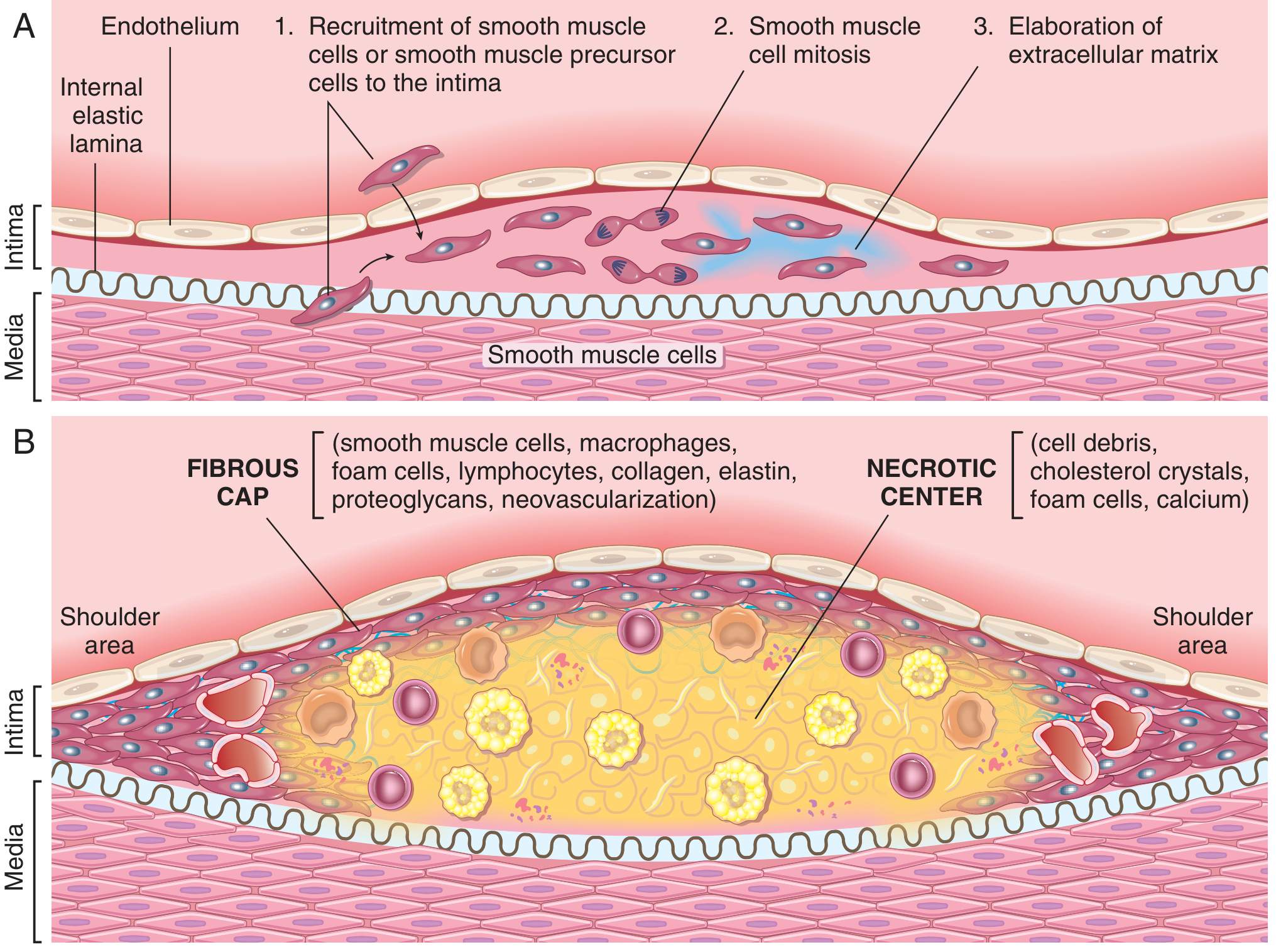
Morphology
Fatty streak - the earliest lesion, composed of lipid-filled foam cells; seen even in infants and universally present by age 10; not all progress to plaques.
Atherosclerotic plaque (atheroma):
- Gross: white-to-yellow raised lesions, 0.3-1.5 cm, may coalesce; superimposed thrombus on ulcerated plaques gives a red-brown color. Lesions are patchy and eccentric in cross-section, related to local hemodynamic turbulence.
- Distribution (descending severity): infrarenal abdominal aorta > coronary arteries > popliteal arteries > internal carotids > circle of Willis. Upper limb, mesenteric, and renal vessels are usually spared (except at ostia).
- Three principal components:
- Cells - SMCs, macrophages, T lymphocytes
- ECM - collagen, elastic fibers, proteoglycans
- Lipid - intracellular (foam cells) and extracellular (cholesterol clefts)
- Architecture: a superficial fibrous cap (SMCs + dense collagen) covers a necrotic core containing cholesterol/cholesterol esters, necrotic debris, foam cells, fibrin, and organized thrombus. The "shoulder" region (cap-media junction) is cellular, containing macrophages, T cells, and SMCs, and is often the site of neovascularization and inflammation.
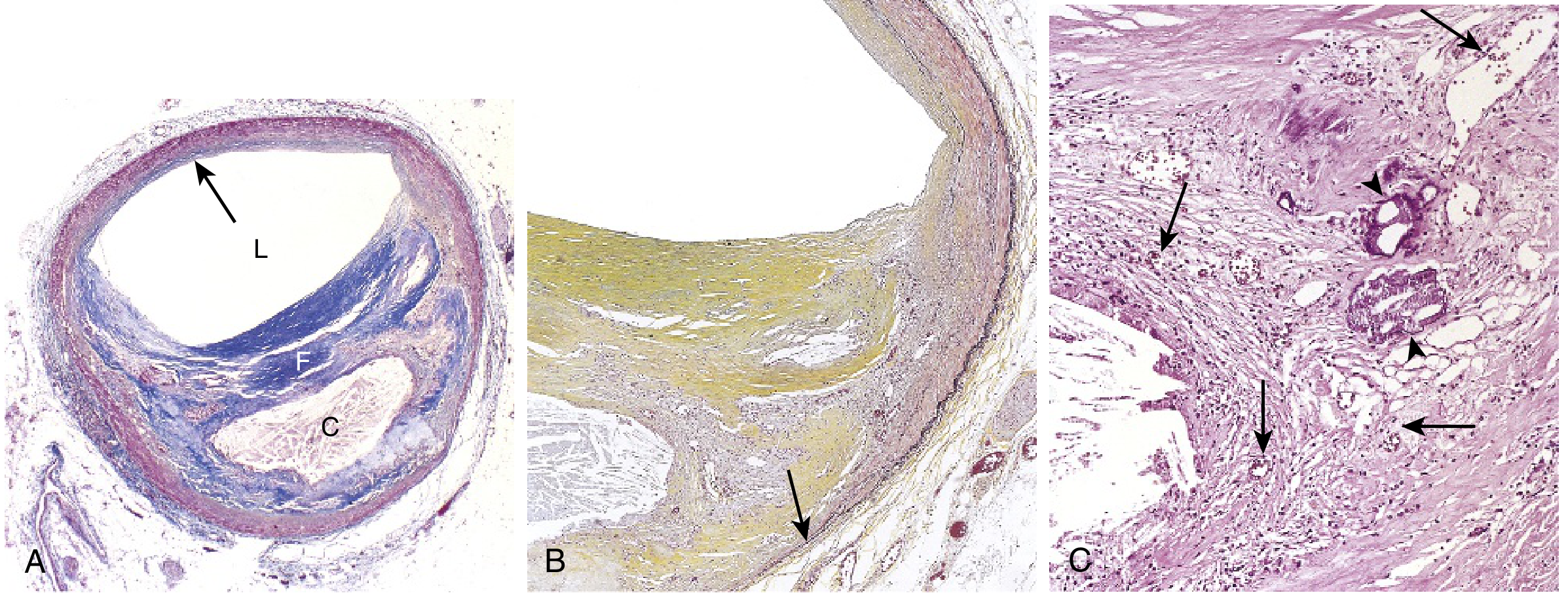
(Robbins & Kumar Basic Pathology, p. 319)
Complications / Consequences of Atherosclerosis
- Atherosclerotic stenosis - progressive luminal narrowing causing ischemia (e.g., angina, claudication).
- Acute plaque change / rupture - the central event in acute coronary syndromes. "Vulnerable plaques" have thin fibrous caps, large lipid cores, abundant foam cells, and heavy inflammatory infiltrate; inflammation increases collagen degradation and reduces synthesis, destabilizing the cap. In contrast, "stable plaques" have thick, densely collagenized caps with minimal inflammation.
- Thrombosis superimposed on a disrupted plaque - most infarctions occur on previously asymptomatic, non-flow-limiting plaques that suddenly rupture and thrombose.
- Intraplaque hemorrhage - rupture of thin-walled neovascular vessels causes a hematoma that can expand or rupture the plaque.
- Atheroembolism - discharge of plaque debris into distal circulation.
- Weakening of the media underlying the plaque (ischemia and inflammatory ECM remodeling), predisposing to aneurysm formation.
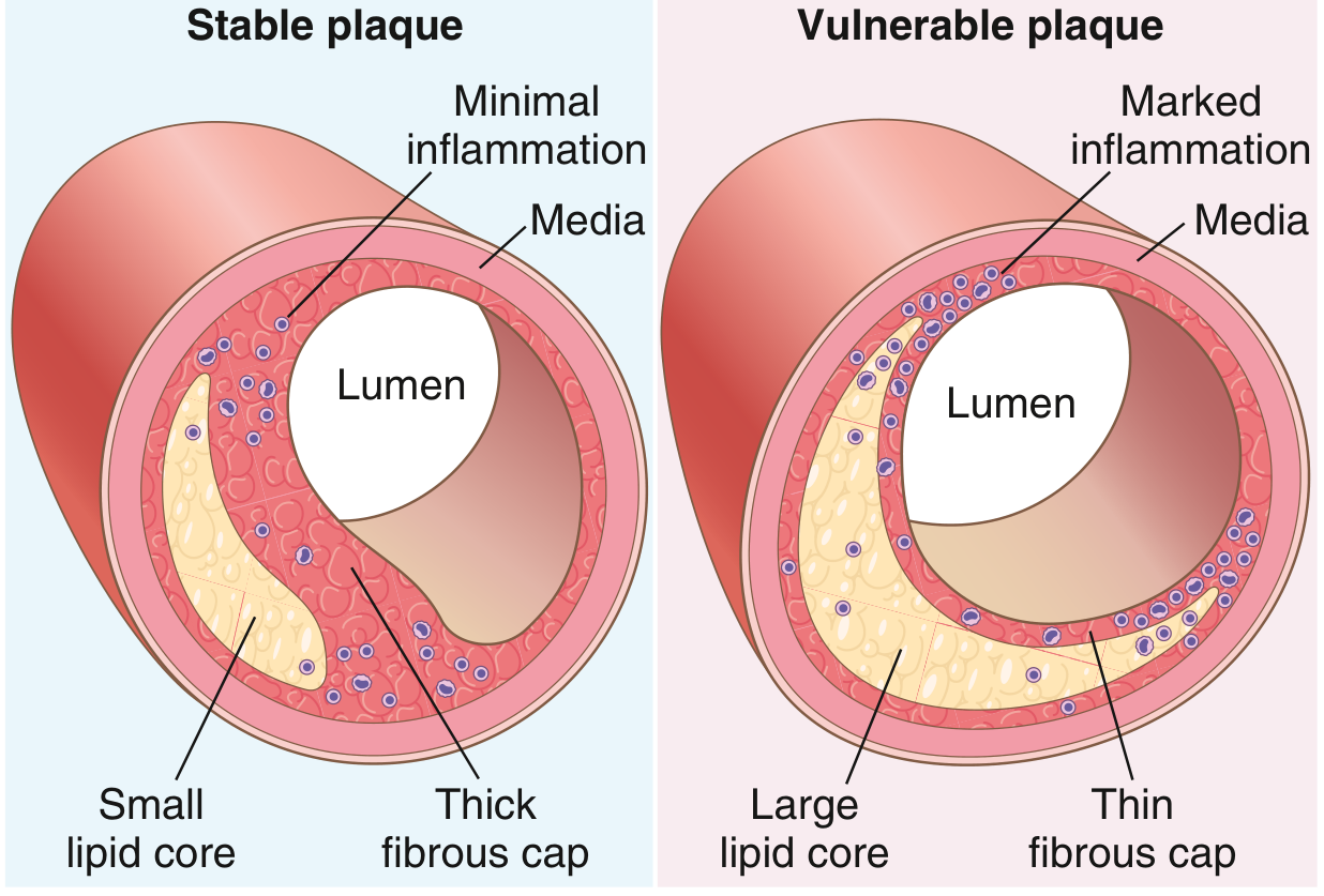
(Robbins & Kumar Basic Pathology, p. 321-322)
Summary Table
| Feature | Description |
|---|---|
| Definition | Chronic inflammatory intimal disease with atheroma formation |
| Key theory | Response-to-injury hypothesis |
| Main risk factors | Hyperlipidemia, HTN, smoking, diabetes, age, male sex, family history |
| Earliest lesion | Fatty streak |
| Plaque components | Cells (SMC, macrophage, T cell), ECM, lipid |
| Major complication | Plaque rupture -> thrombosis -> MI/stroke |
A brief literature check for recent systematic reviews (2024-2025) did not surface findings that contradict this classical response-to-injury model; recent work (e.g., PMID 37676098 on Wnt/sclerostin signaling in vascular calcification, PMID 38977083 on intraplaque neovascularization) refines molecular detail (signaling pathways, neovascularization-targeted therapies) but supports the same core mechanism described above.
This is a shared conversation. Sign in to Orris to start your own chat.