
Medicine for emphysema according to the exam of general medicine
emphysema COPD treatment management algorithm bronchodilator
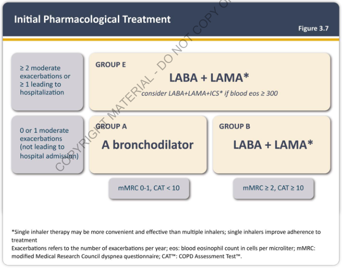
Summary : This figure presents an initial pharmacological treatment algorithm for patients with chronic obstructive pulmonary disease (COPD), stratified by exacerbation history and symptom severity, and grouped into A, B, and E for treatment recommendations. flowchart: # Nodes : • "≥ 2 moderate exacerbations or ≥ 1 leading to hospitalization" (rectangle, top left) • "0 or 1 moderate exacerbations (not leading to hospital admission)" (rectangle, bottom left) • "GROUP E: LABA + LAMA* (consider LABA+LAMA+ICS* if blood eos ≥ 300)" (rectangle, top right) • "GROUP A: A bronchodilator" (rectangle, bottom center) • "GROUP B: LABA + LAMA*" (rectangle, bottom right) • "mMRC 0-1, CAT < 10" (rectangle, below GROUP A) • "mMRC ≥ 2, CAT ≥ 10" (rectangle, below GROUP B) # Connectors : • Arrow from "≥ 2 moderate exacerbations or ≥ 1 leading to hospitalization" to "GROUP E: LABA + LAMA*" • Arrow from "0 or 1 moderate exacerbations (not leading to hospital admission)" splits to "GROUP A: A bronchodilator" and "GROUP B: LABA + LAMA*" • "GROUP A: A bronchodilator" is associated with "mMRC 0-1, CAT < 10" • "GROUP B: LABA + LAMA*" is associated with "mMRC ≥ 2, CAT ≥ 10" # Layout : • Two main branches based on exacerbation history (top left and bottom left). • Severe exacerbation branch leads directly to GROUP E (top right). • Mild exacerbation branch splits into GROUP A (center) and GROUP B (right), further differentiated by symptom scores (mMRC and CAT). • Symptom score boxes are placed below their respective treatment groups. # Treatment Recommendations : • GROUP E: LABA + LAMA*; consider LABA+LAMA+ICS* if blood eosinophils ≥ 300. • GROUP A: A bronchodilator. • GROUP B: LABA + LAMA*. • *Note: Single inhaler therapy may be more convenient and effective than multiple inhalers; single inhalers improve adherence to treatment. # Definitions & Criteria : • Exacerbations refer to the number per year. • Blood eosinophil count in cells per microliter. • mMRC: modified Medical Research Council dyspnea questionnaire. • CAT™: COPD Assessment Test™. # Analysis : • The flowchart stratifies COPD patients by exacerbation history and symptom severity (mMRC, CAT). • Patients with frequent or severe exacerbations (≥2 moderate or ≥1 hospitalization) are recommended dual bronchodilator therapy (LABA + LAMA), with possible addition of ICS if eosinophils are high. • Patients with low exacerbation risk are further divided by symptom scores: those with low symptoms (mMRC 0-1, CAT < 10) receive a single bronchodilator, while those with higher symptoms (mMRC ≥ 2, CAT ≥ 10) receive dual bronchodilator therapy. • The algorithm emphasizes tailored therapy based on risk and symptom burden, with consideration for adherence and convenience.
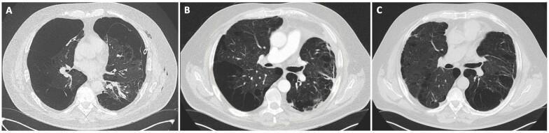
This set of three axial chest CT scans (lung window) illustrates a progression of treatment for severe emphysematous COPD and persistent pneumothorax. Image A shows a significant left-sided pneumothorax following talc pleurodesis, characterized by an absence of peripheral lung markings and compression of the left lung parenchyma against the mediastinum. Image B, captured one month after left-sided lung volume reduction surgery (LVRS), demonstrates successful resolution of the pneumothorax and re-expansion of the left lung. Although severe bullous emphysema remains visible bilaterally, the mediastinum has shifted from a rightward displacement back toward the midline. Image C shows the thorax one month after a subsequent contralateral (right-sided) LVRS. This final stage reveals improved thoracic symmetry and a further reduction in hyperinflation, with more uniform distribution of the remaining lung parenchyma. The series demonstrates the efficacy of sequential LVRS in managing refractory air leaks and optimizing lung mechanics in patients with end-stage emphysema.
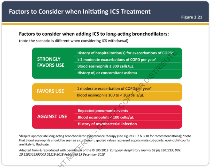
Summary : This figure presents a decision framework for clinicians on when to initiate inhaled corticosteroid (ICS) treatment in patients with COPD who are already on long-acting bronchodilators. It categorizes factors into three groups: those that strongly favor use, favor use, or argue against use of ICS. decision framework: # Strongly Favors Use : • History of hospitalization(s) for exacerbations of COPD. • ≥ 2 moderate exacerbations of COPD per year. • Blood eosinophils ≥ 300 cells/μL. • History of, or concomitant asthma. # Favors Use : • 1 moderate exacerbation of COPD per year. • Blood eosinophils 100 to < 300 cells/μL. # Against Use : • Repeated pneumonia events. • Blood eosinophils < 100 cells/μL. • History of mycobacterial infection. # Design Encodings : • Green box for "Strongly Favors Use". • Yellow box for "Favors Use". • Red box for "Against Use". • Bolded section headings and bullet points for each criterion. • Footnotes clarify that blood eosinophil values are approximate cut-points and may fluctuate, and that recommendations assume appropriate long-acting bronchodilator maintenance therapy. # Analysis : • The figure provides a clear, color-coded decision aid for clinicians, emphasizing that higher exacerbation frequency, higher eosinophil counts, and asthma history favor ICS use, while pneumonia risk, low eosinophil counts, and mycobacterial infection argue against it. The structure supports rapid clinical assessment and stratification of COPD patients for ICS therapy.
Pharmacological Treatment of Emphysema (COPD)
Emphysema is a component of COPD. Its medical management is identical to COPD management and follows the GOLD (Global Initiative for Chronic Obstructive Lung Disease) framework. Below is organized per the General Medicine exam approach.
GOLD ABE Assessment (Severity Classification)
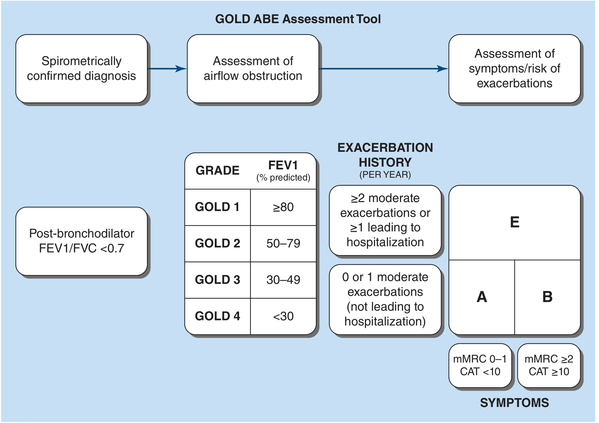
| GOLD Grade | FEV1 (% predicted) |
|---|---|
| GOLD 1 | ≥ 80% |
| GOLD 2 | 50-79% |
| GOLD 3 | 30-49% |
| GOLD 4 | < 30% |
Initial Pharmacological Treatment by Group
| Group | Criteria | First-line Treatment |
|---|---|---|
| A | Low symptoms (mMRC 0-1 / CAT < 10), 0-1 mild exacerbations | A single bronchodilator (SABA or SAMA) |
| B | More symptoms (mMRC ≥ 2 / CAT ≥ 10), 0-1 mild exacerbations | LABA + LAMA (dual long-acting bronchodilators) |
| E | ≥ 2 moderate exacerbations/year OR ≥ 1 leading to hospitalization | LABA + LAMA; add ICS if blood eosinophils ≥ 300 cells/µL |
Drug Classes in Detail
1. Bronchodilators (cornerstone of treatment)
Short-Acting (for rescue/as-needed use)
- SABA (Short-Acting Beta-2 Agonists): salbutamol (albuterol), terbutaline
- SAMA (Short-Acting Muscarinic Antagonists): ipratropium bromide - improves symptoms and acutely improves FEV1
Long-Acting Beta-2 Agonists (LABAs)
- Arformoterol, formoterol, indacaterol, olodaterol, salmeterol, vilanterol
- Provide symptomatic benefit and reduce exacerbations
- Main side effects: tremor and tachycardia
Long-Acting Muscarinic Antagonists (LAMAs)
- Aclidinium, glycopyrrolate, glycopyrronium, revefenacin, tiotropium, umeclidinium
- Improve symptoms and reduce exacerbations more than LABAs
- Side effect: dry mouth (most frequent)
LABA + LAMA Combinations
- Demonstrated superior efficacy vs. either drug alone; preferred in Group B and E
- Single-inhaler combinations improve adherence
2. Inhaled Corticosteroids (ICS)
- Hospitalization for COPD exacerbation
- ≥ 2 moderate exacerbations/year
- Blood eosinophils ≥ 300 cells/µL
- History of concomitant asthma
- Repeated pneumonia events
- Blood eosinophils < 100 cells/µL
- History of mycobacterial infection
3. Theophylline (Methylxanthine)
- Oral bronchodilator, used when inhaled therapy is unavailable or inadequate
- Narrow therapeutic window; requires serum level monitoring
- Less favored due to side effect profile (arrhythmia, seizures at toxic levels)
4. Phosphodiesterase-4 (PDE4) Inhibitor
- Roflumilast (oral): used as add-on therapy in severe COPD with chronic bronchitis phenotype and frequent exacerbations
- Reduces exacerbation frequency; not a bronchodilator
5. Biologic Therapy (newer)
- Dupilumab (anti-IL-4/IL-13 monoclonal antibody): approved for symptomatic COPD with high exacerbation risk and blood eosinophils ≥ 300 cells/µL, added to triple inhaled therapy
6. Macrolide Antibiotics (prophylactic)
- Azithromycin (daily or 3x/week): reduces exacerbation frequency in selected patients; consider risk of hearing loss and antibiotic resistance
7. Supplemental Oxygen
- Long-term oxygen therapy (LTOT): indicated when resting PaO2 ≤ 55 mmHg (or SaO2 ≤ 88%), or PaO2 56-59 mmHg with cor pulmonale/polycythemia
- One of three interventions proven to improve survival in COPD (along with smoking cessation and LVRS)
8. Smoking Cessation (most important intervention)
- Nicotine replacement therapy (gum, patch, lozenge, inhaler, nasal spray)
- Bupropion (antidepressant with dopaminergic/noradrenergic action)
- Varenicline (nicotinic acid receptor partial agonist) - most effective single agent
Non-Pharmacological / Surgical Options
| Intervention | Notes |
|---|---|
| Pulmonary Rehabilitation | Improves quality of life, dyspnea, exercise capacity; reduces hospitalizations |
| Lung Volume Reduction Surgery (LVRS) | For upper-lobe predominant emphysema + low exercise capacity after rehab; improves survival |
| Bronchoscopic Lung Volume Reduction (BLVR) | FDA-approved one-way endobronchial valves; similar outcomes to LVRS at 12 months; risk: pneumothorax |
| Lung Transplantation | Second leading indication for transplant; for very severe disease with no other surgical option |
Vaccines (mandatory)
- Influenza (annual)
- Pneumococcal
- COVID-19
- RSV
- Pertussis (if not vaccinated in adolescence)
Exam Summary Table
| Drug Class | Key Drugs | Primary Use |
|---|---|---|
| SABA | Salbutamol | Rescue/as-needed |
| SAMA | Ipratropium | Rescue or maintenance |
| LABA | Salmeterol, formoterol, indacaterol | Maintenance (Group B, E) |
| LAMA | Tiotropium, umeclidinium | Maintenance (preferred long-acting) |
| LABA+LAMA combo | Formoterol/glycopyrrolate, etc. | Group B, E first-line |
| ICS | Budesonide, fluticasone | Add-on for eosinophils ≥ 300 or asthma overlap |
| Triple therapy | LABA+LAMA+ICS | Severe disease with eosinophilia |
| PDE4 inhibitor | Roflumilast | Chronic bronchitis, frequent exacerbations |
| Biologic | Dupilumab | Eos ≥ 300 on triple therapy |
| Methylxanthine | Theophylline | Add-on (narrow window) |
| Smoking cessation | Varenicline, bupropion, NRT | All smokers |
| O2 therapy | LTOT | PaO2 ≤ 55 mmHg, proven survival benefit |
- Harrison's Principles of Internal Medicine 22E (2025), Chapter 303 - COPD, pp. 2298-2302
Pink puffer
pink puffer emphysema patient barrel chest COPD
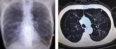
This dual-panel image displays diagnostic thoracic imaging from a patient with Chronic Obstructive Pulmonary Disease (COPD). The left panel is a posterior-anterior (PA) chest X-ray showing classic features of severe emphysema, including hyperinflated lung fields, sparse bronchovascular markings, and a widened chest diameter consistent with a barrel chest deformity. Diaphragmatic flattening and a narrowed mediastinal silhouette are also visible. The right panel is an axial CT scan of the chest (lung window) at the level of the upper lobes, revealing extensive bullous emphysema. Multiple large, thin-walled bullae are visible, particularly in the subpleural regions, along with architectural distortion of the lung parenchyma. These findings demonstrate advanced obstructive lung disease and are clinically significant for explaining symptoms of chronic shortness of breath and impaired gas exchange in elderly patients with a significant smoking history.
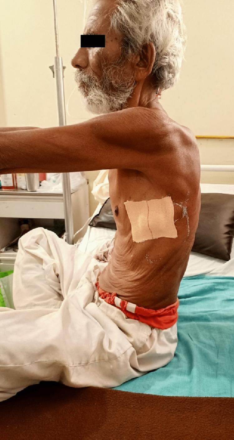
This clinical photograph provides a lateral view of an elderly male patient in a seated position, demonstrating a classic barrel-shaped chest deformity. This morphological change is characterized by a significant increase in the anterior-posterior (AP) diameter of the thoracic cage, commonly associated with chronic obstructive pulmonary disease (COPD) or severe emphysema. Visible physical findings include significant wasting of the pectoral and shoulder musculature (cachexia) and thin, wrinkled skin over the thorax. A nasal cannula is visible, indicating the need for supplemental oxygen therapy. A square medical dressing is applied to the lateral chest wall, likely at a site following the removal of an intercostal drainage (ICD) tube. The patient exhibits a 'tripod' posture or forward-leaning tendency, which is a common compensatory mechanism used to engage accessory muscles of respiration. A bedside IV pole is visible in the background, suggesting an inpatient clinical setting. This image serves as a teaching tool for physical examination findings in chronic respiratory failure and obstructive lung disease.
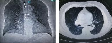
This diagnostic image pair displays thoracic radiology findings in an 80-year-old male with Chronic Obstructive Pulmonary Disease (COPD). The left panel shows a frontal chest radiograph (X-ray) exhibiting hallmark signs of hyperinflation, including a widened anteroposterior diameter and flattened diaphragmatic domes. Both lung fields demonstrate sparse bronchovascular markings and increased lucency, indicative of diffuse emphysema. The right panel shows an axial CT scan of the thorax, further characterizing the pulmonary pathology. It reveals significant emphysematous destruction of the lung parenchyma with the presence of multiple bullae and areas of decreased attenuation. Additionally, subtle interstitial lesions are visible. The imaging illustrates the classic presentation of severe COPD, characterized by permanent enlargement of airspaces distal to the terminal bronchioles and associated structural changes to the chest wall often described as a barrel chest.
Pink Puffer (Type A COPD - Pure Emphysema)
What is it?
Radiographic Basis
| Radiographic Pattern | Emphysema Type | Clinical Phenotype |
|---|---|---|
| Arterial deficiency (hyperlucent, sparse vessels) | Pan-lobular emphysema | Pink Puffer |
| Increased lung markings | Centrilobular emphysema | Blue Bloater |
- Fishman's Pulmonary Diseases and Disorders, Chapter 28
Classic Clinical Features
| Feature | Pink Puffer (Emphysema) |
|---|---|
| Appearance | Thin, cachectic, anxious |
| Skin color | Pink (relatively preserved PaO2) |
| Breathing | Labored, rapid ("puffing") |
| Dyspnea | Severe - main complaint |
| Cough/sputum | Minimal or absent |
| Chest shape | Barrel chest (increased AP diameter) |
| Breath sounds | Decreased; prolonged expiration; wheeze |
| Accessory muscles | Used prominently |
| Posture | Tripod position (leans forward on arms) |
| Pursed-lip breathing | Characteristic (auto-PEEP to keep airways open) |
| Body weight | Low (cachexia from elevated TNF-α, increased WOB) |
| Cor pulmonale | Less common (preserved PaO2 early on) |
| Blood eosinophils | May be elevated if asthma overlap |
Pathophysiology
- Protease-antiprotease imbalance (smoking → neutrophil elastase ↑, α1-antitrypsin ↓)
- Alveolar destruction → enlarged air spaces distal to terminal bronchioles
- Loss of pulmonary capillary bed → reduced DLCO
- Reduced elastic recoil → dynamic airway collapse on expiration → air trapping
- Diaphragm flattened by hyperinflation → accessory muscles recruited
- Patient compensates by hyperventilating → PaCO2 normal or low → remains "pink"
Pulmonary Function Tests (PFTs)
| Parameter | Finding | Reason |
|---|---|---|
| FEV1 | ↓ | Airflow obstruction |
| FEV1/FVC ratio | < 0.7 | Obstructive pattern |
| Total Lung Capacity (TLC) | ↑ | Air trapping / hyperinflation |
| Residual Volume (RV) | ↑↑ | Trapped air |
| Functional Residual Capacity (FRC) | ↑ | Air trapping |
| DLCO | ↓↓ (markedly) | Loss of alveolar-capillary surface |
| PaO2 | Relatively preserved (until late) | Compensatory hyperventilation |
| PaCO2 | Normal or ↓ | Hyperventilation |
The markedly reduced DLCO is the PFT hallmark that distinguishes emphysema from pure chronic bronchitis. - Frameworks for Internal Medicine, p. 273
Chest X-Ray Findings
- Hyperinflated lung fields - increased lucency
- Flattened diaphragms
- Increased AP diameter (barrel chest)
- Narrow mediastinum / vertical heart
- Sparse bronchovascular markings (arterial deficiency pattern)
- Bullae may be visible
Classic Patient Photo
Pink Puffer vs. Blue Bloater (Exam Comparison Table)
| Feature | Pink Puffer (Type A) | Blue Bloater (Type B) |
|---|---|---|
| Underlying disease | Pure emphysema | Chronic bronchitis |
| Body habitus | Thin, cachectic | Overweight, edematous |
| Skin color | Pink | Blue/cyanotic |
| Dyspnea | Severe | Mild-moderate |
| Cough/sputum | Minimal | Productive cough |
| PaO2 | Near normal | ↓ (hypoxemic) |
| PaCO2 | Normal/↓ | ↑ (hypercapnic) |
| DLCO | ↓↓ | Normal or mildly ↓ |
| Polycythemia | Absent | Present |
| Cor pulmonale | Less common | Common |
| Respiratory drive | High | Low ("won't breathe") |
| OSA risk | Lower (low BMI, high drive) | Higher |
| Emphysema type | Pan-lobular | Centrilobular |
| Vascular pattern on CXR | Arterial deficiency | Increased markings |
- Murray & Nadel's Textbook of Respiratory Medicine; Fishman's Pulmonary Diseases and Disorders
Key Exam Points
- Pink puffer = emphysema phenotype = Type A COPD
- Pink because they hyperventilate to compensate - maintains oxygenation at the cost of severe breathlessness
- DLCO is the key differentiator on PFTs from chronic bronchitis
- Associated with pan-lobular emphysema (as opposed to centrilobular in chronic bronchitis/smoking)
- Pan-lobular emphysema is also the pattern seen in α1-antitrypsin deficiency
- Relatively protected from OSA due to low BMI and high respiratory drive